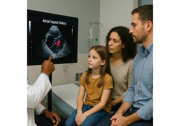
Atrial septal defect (ASD) is a congenital heart abnormality characterized by a persistent opening in the wall (septum) that separates the heart’s two upper chambers, the right and left atria. While some individuals with ASD may live symptom-free for years, this seemingly small defect can lead to significant health problems, including heart failure, arrhythmias, pulmonary hypertension, and stroke. Recognizing, understanding, and appropriately managing ASD—whether discovered in infancy, childhood, or adulthood—is essential for optimal cardiovascular health. In this comprehensive guide, we’ll explore everything you need to know about ASD from causes to lifelong care.
Table of Contents
- Thorough Exploration of Atrial Septal Defect
- Underlying Origins, Health Impacts, and Contributing Factors
- Recognizing Symptoms and Understanding Diagnosis
- Modern Treatment Approaches and Ongoing Management
- Frequently Asked Questions
Thorough Exploration of Atrial Septal Defect
Atrial septal defect, commonly known as ASD, is among the most frequently encountered congenital heart anomalies. The condition is defined by an opening in the atrial septum, allowing blood to flow from the left atrium (higher pressure) to the right atrium (lower pressure). This abnormal circulation can, over time, affect the heart and lungs.
Types of Atrial Septal Defect
There are several ASD subtypes, each with distinct characteristics:
- Ostium Secundum ASD:
The most common form (about 70% of cases), located at the center of the atrial septum where the foramen ovale typically closes after birth. - Ostium Primum ASD:
Involves the lower part of the atrial septum and is often associated with other heart defects, including mitral or tricuspid valve abnormalities. It’s more common in people with genetic syndromes such as Down syndrome. - Sinus Venosus ASD:
Occurs near the junction of the superior or inferior vena cava and the right atrium. This form is sometimes associated with abnormal drainage of pulmonary veins. - Coronary Sinus ASD:
Extremely rare, found near the coronary sinus, a vein collecting blood from the heart muscle.
Epidemiology and Lifelong Implications
- Prevalence:
ASD affects about 1 in every 1,500 live births, with a higher detection rate now due to advanced screening. - Natural History:
Many small ASDs close spontaneously in early childhood, but larger defects often persist and can cause significant complications if not addressed.
Hemodynamic Effects: How ASD Impacts the Heart
With an ASD, oxygen-rich blood from the left atrium passes into the right atrium, leading to:
- Right-sided volume overload: The right atrium and right ventricle must pump more blood than usual, potentially leading to chamber enlargement.
- Increased blood flow to the lungs: Over years, this can damage the pulmonary vessels and raise lung blood pressure (pulmonary hypertension).
- Arrhythmias: Stretched atrial tissue is more prone to irregular heart rhythms, especially atrial fibrillation.
- Risk of paradoxical embolism: Clots can pass from the right to the left atrium, entering the body’s arterial system and potentially causing a stroke.
Historical Perspective
ASD was first described in the late 19th century, and the first successful surgical closure took place in the 1950s. Over decades, minimally invasive techniques and device closures have revolutionized outcomes, allowing many people to live full, active lives.
ASD in Children vs. Adults
While most ASDs are detected and managed during childhood, some smaller defects may go unnoticed and only become problematic in adulthood, when symptoms like shortness of breath or heart rhythm disturbances develop.
Key Takeaways
- ASD is a common congenital heart defect.
- The defect’s size, type, and location influence symptoms and treatment.
- Many individuals can live healthy lives if ASD is identified and managed early.
Underlying Origins, Health Impacts, and Contributing Factors
To understand ASD, it’s essential to explore how it develops, what factors raise the risk, and what complications may arise if the defect is left untreated.
How Does an ASD Develop?
ASDs are congenital, meaning they form during fetal heart development. The atrial septum forms from two overlapping walls that should fuse completely before birth, but for various reasons, a gap may persist.
- Genetic factors:
Certain gene mutations or chromosomal abnormalities, such as those found in Down syndrome, are associated with a higher risk of ASD. - Environmental influences:
Maternal illnesses (such as diabetes), alcohol use, certain medications, or infections during pregnancy can occasionally increase the risk of congenital heart defects, including ASD.
Risk Factors for ASD
- Family history:
A parent or sibling with a congenital heart defect raises risk. - Maternal health:
Poorly controlled diabetes, rubella infection during pregnancy, or drug and alcohol exposure may increase risk. - Genetic syndromes:
ASD is more common in people with chromosomal disorders like Down syndrome, Holt-Oram syndrome, or Ellis-van Creveld syndrome.
Potential Health Consequences of Untreated ASD
- Heart failure:
Over decades, right-sided volume overload may lead to weakened heart muscles, fluid buildup, and heart failure. - Pulmonary hypertension:
High pressure in the lungs can develop, making ASD closure riskier and less effective if delayed. - Arrhythmias:
Atrial fibrillation or atrial flutter is common in adults with longstanding ASD. - Stroke risk:
Paradoxical embolism can cause a stroke if a blood clot passes through the ASD into systemic circulation. - Shortened lifespan:
Without treatment, large ASDs reduce life expectancy.
Are There Modifiable Risks?
While most risk factors are not modifiable, pregnant women can lower their risk of having a child with ASD by:
- Managing chronic conditions (especially diabetes) carefully.
- Avoiding alcohol and tobacco.
- Keeping up to date with vaccinations (especially rubella).
- Avoiding certain medications not approved during pregnancy.
Lifestyle Impacts After Diagnosis
People with ASD—especially if untreated—may need to limit intense physical activity if they develop symptoms or complications. After closure and recovery, most can lead fully active lives, though lifelong follow-up may be recommended.
Practical Advice:
If you have a family history of congenital heart disease, ask your doctor about screening for ASD and other heart anomalies, especially before pregnancy.
Recognizing Symptoms and Understanding Diagnosis
Symptoms of ASD vary widely depending on the defect’s size, type, and the patient’s age. Many cases are silent in childhood and only manifest later in life.
Typical Signs and Symptoms in Infants and Children
- Most small ASDs:
Cause no symptoms and may be discovered during routine physical examination (e.g., hearing a heart murmur). - Larger ASDs:
- Frequent respiratory infections
- Poor growth or failure to thrive
- Fatigue during feeding or activity
- Shortness of breath or easy tiring
Signs and Symptoms in Adults
ASDs often go unnoticed until adulthood, when increased strain on the heart and lungs causes:
- Shortness of breath (especially with exercise)
- Heart palpitations or irregular pulse
- Fatigue or reduced exercise tolerance
- Swelling in the legs, feet, or abdomen (signs of right-sided heart failure)
- Stroke or transient ischemic attack (TIA)
- Heart murmur detected by a healthcare provider
Physical Examination Findings
A doctor may hear a soft, blowing systolic murmur along the upper left border of the chest or notice fixed splitting of the second heart sound—a clue for ASD.
Diagnostic Tests and Imaging
1. Echocardiography (Ultrasound of the Heart):
- The gold standard for diagnosis.
- Can show the location, size, and blood flow through the defect.
- Transesophageal echocardiography (TEE) offers detailed images, especially in adults.
2. Electrocardiogram (ECG):
- May reveal right atrial enlargement, right ventricular conduction delay, or arrhythmias.
3. Chest X-ray:
- Shows heart enlargement and increased blood flow to the lungs.
4. Cardiac MRI or CT:
- Provides detailed visualization when echocardiography is inconclusive.
5. Cardiac Catheterization:
- Rarely needed solely for diagnosis, but useful for measuring pressures if pulmonary hypertension is suspected.
Screening and Early Detection
- Prenatal diagnosis:
Some ASDs are identified before birth during fetal ultrasound. - Family screening:
Recommended if there is a family history of congenital heart defects.
Key Steps for Patients:
- Report any unexplained shortness of breath, fatigue, or palpitations to your healthcare provider.
- Be proactive if you have a family history or personal risk factors.
Modern Treatment Approaches and Ongoing Management
The decision to treat ASD depends on the size and type of defect, symptoms, age at diagnosis, and presence of complications.
Watchful Waiting vs. Intervention
- Small ASDs:
Especially secundum ASDs less than 5 mm, may close on their own in early childhood. - Larger or symptomatic ASDs:
Require closure to prevent long-term complications.
Non-Surgical and Medical Management
- Observation:
Regular follow-up with echocardiography if the defect is small and asymptomatic. - Medications:
May be used to manage symptoms, such as diuretics for fluid retention or medications for arrhythmias.
Catheter-Based (Device) Closure
- Who is eligible?
Most children and adults with a secundum ASD and appropriate anatomy. - How it works:
A thin tube (catheter) is inserted through a vein and guided to the heart. A closure device is deployed to seal the defect. Over time, heart tissue grows over the device. - Benefits:
Minimally invasive, short recovery time, high success rate.
Surgical Closure
- Indications:
Large or complex ASDs, defects not suitable for device closure (e.g., primum, sinus venosus, coronary sinus types). - Procedure:
Open-heart surgery under general anesthesia. The defect is closed with sutures or a synthetic/pericardial patch. - Outcomes:
Excellent long-term survival and symptom resolution. Some require ongoing arrhythmia monitoring.
Management of Complications
- Arrhythmias:
Medications, electrical cardioversion, or ablation may be needed. - Pulmonary hypertension:
Requires specialized treatment and may impact timing or approach to ASD closure. - Stroke prevention:
In patients with paradoxical embolism, closure plus antiplatelet or anticoagulant therapy may be recommended.
Long-Term Follow-Up
- Children:
Periodic echocardiography to ensure closure and normal heart development. - Adults:
Lifelong cardiology follow-up, especially for those with repaired or unrepaired ASD and history of arrhythmias.
Lifestyle and Practical Guidance
- Physical activity:
Most can participate in normal activities after ASD closure, though restrictions may apply for those with pulmonary hypertension or arrhythmias. - Pregnancy:
Women with repaired or small ASDs can usually have successful pregnancies but should be evaluated by a cardiologist beforehand. - Endocarditis prophylaxis:
Generally not required for isolated ASD but may be advised for certain high-risk procedures or if other heart anomalies are present.
Helpful Tips:
- Keep a personal record of all heart imaging, procedures, and medications.
- Ask your doctor about activity restrictions or special precautions for travel, sports, or pregnancy.
- Join patient support groups for emotional encouragement and practical advice.
Frequently Asked Questions
What is an atrial septal defect and how serious is it?
An atrial septal defect is a congenital hole in the wall between the heart’s upper chambers. It varies in seriousness; some small defects close on their own, while larger ones can cause heart failure, arrhythmias, or stroke if untreated.
What are the signs and symptoms of atrial septal defect in adults?
Common symptoms in adults include shortness of breath, fatigue, heart palpitations, swelling in the legs, and sometimes stroke. Many adults with small defects remain asymptomatic for years.
How is an atrial septal defect diagnosed by doctors?
Doctors use echocardiography (heart ultrasound) as the primary diagnostic tool. Additional tests may include ECG, chest X-ray, cardiac MRI, or cardiac catheterization, especially if complications or other heart defects are suspected.
What are the main treatment options for atrial septal defect?
Treatment options include observation for small defects, catheter-based closure for most secundum ASDs, and surgical closure for larger or complex defects. Management of symptoms and complications may also be required.
Can an atrial septal defect go away on its own?
Small secundum ASDs in children can sometimes close spontaneously within the first years of life. Larger defects and most ASDs detected in adults will not close on their own and typically require treatment.
Is it safe to exercise or become pregnant with an atrial septal defect?
Most people with small or repaired ASDs can safely exercise and have healthy pregnancies. However, those with large or unrepaired ASDs, arrhythmias, or pulmonary hypertension should be evaluated by a cardiologist before strenuous activity or pregnancy.
Does atrial septal defect increase stroke risk?
Yes, especially if blood clots can cross the defect from the right to left atrium. Stroke prevention may involve closing the ASD and, in some cases, using anticoagulant or antiplatelet medications.
Disclaimer:
This article is for informational and educational purposes only and is not a substitute for professional medical advice, diagnosis, or treatment. Always seek the guidance of your healthcare provider with any questions regarding your health or a medical condition. Do not disregard professional advice or delay care based on information from this article.
If you found this guide helpful, please share it with others on Facebook, X (formerly Twitter), or your favorite platform. Your support enables us to continue delivering high-quality, trustworthy health content for readers worldwide. Don’t forget to follow us on social media for more updates!


