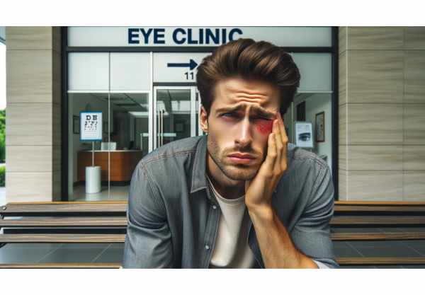
Blepharitis Basics
Blepharitis is a common and chronic inflammatory condition of the eyelids. It usually affects the area of the eyelid where the eyelashes grow, and it can affect both the anterior (front) and posterior (back) portions of the eyelid margin. Blepharitis, which is characterized by redness, irritation, and scaling of the eyelids, can be uncomfortable and impair vision if not treated properly. Several factors contribute to this condition, including bacterial infections, skin conditions such as seborrheic dermatitis, and meibomian gland dysfunction. While blepharitis is rarely sight-threatening, it can progress to more serious complications if left untreated.
In-Depth Look at Blepharitis
Blepharitis is a multifactorial condition caused by a variety of underlying causes. It is broadly divided into two types based on the location and characteristics of the inflammation: anterior blepharitis and posterior blepharitis. Understanding the unique mechanisms and contributing factors to each type is critical for effective management and treatment.
Anterior Blepharitis
Anterior blepharitis affects the outer part of the eyelid, where the eyelashes are attached. Bacterial infections, particularly Staphylococcus species, and skin conditions such as seborrheic dermatitis and rosacea are the leading causes of anterior blepharitis. Staphylococcal blepharitis is defined by the presence of bacteria at the base of the eyelashes, which causes inflammation and the formation of crusts and scales. Seborrheic blepharitis, on the other hand, is characterized by dandruff-like scaling and oily skin on both the eyelids and scalp.
Posterior Blepharitis
Posterior blepharitis, also known as meibomian gland dysfunction (MGD), is an inflammation of the meibomian glands on the inner edge of the eyelid. These glands secrete oils that form the outer layer of the tear film, preventing tears from evaporating. Dysfunction of the meibomian glands alters the lipid composition of the tear film, causing increased tear evaporation, dryness, and inflammation. Posterior blepharitis is frequently associated with acne, rosacea, and chronic blepharoconjunctivitis.
Mixed Blepharitis
Mixed blepharitis occurs when patients present with both anterior and posterior blepharitis. This overlap complicates the diagnosis and treatment of the condition because it involves multiple underlying factors and symptoms.
Pathophysiology
Blepharitis pathophysiology is complex, with microbial, inflammatory, and environmental factors all playing roles. In bacterial blepharitis, an overgrowth of Staphylococcus bacteria at the eyelid margin causes the release of toxins and enzymes, resulting in inflammation and tissue damage. These bacterial products activate the immune system, causing the recruitment of inflammatory cells and the production of pro-inflammatory cytokines.
In seborrheic blepharitis, sebaceous glands overproduce sebum, which contributes to the formation of greasy scales and crusts on the eyes. These scales can promote bacterial growth, exacerbating inflammation.
Meibomian gland dysfunction (MGD) in posterior blepharitis is defined by obstruction and inflammation of the meibomian glands. The altered lipid composition of the tear film in MGD causes increased tear evaporation, resulting in dry eye symptoms and additional inflammation of the eyelids and ocular surface.
Clinical Features
Blepharitis symptoms range in severity and can be chronic or recurring. Common clinical characteristics include:
- Redness and Swelling: The eyelids appear red, swollen, and irritated, sometimes with a burning or stinging sensation.
- Crusting and Scaling: Crusts and flakes form at the base of the eyelashes, especially after waking up in the morning.
- Itching and Discomfort: Patients frequently report itching and a gritty sensation in their eyes.
- Tearing and Dryness: Blepharitis can cause excessive tearing as well as dry eye symptoms due to tear film instability.
- Photophobia: Light sensitivity is a common symptom, especially during inflammatory flare-ups.
- Foreign Body Sensation: Patients frequently report feeling something in their eyes, which causes discomfort and irritation.
- Lash Abnormalities: Common eyelash changes include misdirection, loss (madarosis), and crusting.
Complications
Blepharitis, if left untreated, can cause a number of complications affecting ocular health and vision. The complications include:
- Chalazion and Stye: Blocked meibomian glands can cause painless lumps (chalazia) or painful, infected lumps (styes) on the eyelid.
- Conjunctivitis: Blepharitis-related inflammation can spread to the conjunctiva, causing redness, discharge, and irritation.
- Corneal Involvement: Severe or chronic blepharitis can lead to keratitis, which is an inflammation of the cornea that can cause ulceration, scarring, and vision impairment.
- Trichiasis: Inflammation can cause eyelashes to grow inward towards the eye, causing irritation and potentially damaging the cornea.
- Tear Film Instability: Persistent meibomian gland dysfunction can result in chronic dry eye syndrome, reducing visual comfort and quality of life.
Risk Factors
Several factors can increase the likelihood of developing blepharitis. The risk factors include:
- Age: Older adults are more likely to develop blepharitis due to changes in gland function and an increased risk of skin conditions such as seborrheic dermatitis.
- Skin Conditions: People with acne rosacea, seborrheic dermatitis, and eczema are more likely to develop blepharitis.
- Contact Lens Wear: Wearing contact lenses can exacerbate blepharitis symptoms, especially if proper hygiene is not followed.
- Environmental Factors: Dust, smoke, and other environmental irritants can cause eyelid inflammation.
- Hormonal Changes: Hormonal fluctuations, especially in women, can disrupt the function of the meibomian glands and contribute to blepharitis.
- Poor Eyelid Hygiene: Inadequate eyelid hygiene can result in the accumulation of oils, debris, and bacteria, increasing the risk of inflammation.
Blepharitis Prevention
- Maintain Good Eyelid Hygiene: Clean your eyelids on a regular basis with a gentle cleanser or a prescription eyelid scrub. This helps to remove excess oils, debris, and bacteria that can cause inflammation.
- Warm Compresses: Use warm compresses on your eyelids every day. This softens and loosens crusts and scales, improves circulation, and stimulates the secretion of oils by the meibomian glands.
- Avoid Eye Makeup: Limit your use of eye makeup, especially during flare-ups, to avoid further irritation and inflammation. Always thoroughly remove your makeup before going to bed.
- Practice Good Contact Lens Hygiene: If you wear contact lenses, keep them clean and stored according to the manufacturer’s instructions. Wearing contact lenses while blepharitis is active is not recommended.
- Treat and Manage Underlying Skin Conditions: Address any underlying skin conditions, such as acne, rosacea, seborrheic dermatitis, or eczema. Proper treatment of these conditions can lower the likelihood of blepharitis flare-ups.
- Avoid Environmental Irritants: Limit your exposure to environmental irritants like dust, smoke, and strong winds. Wear protective eyewear if necessary to protect your eyes from irritants.
- Use Artificial Tears: Apply preservative-free artificial tears on a regular basis to keep your eyes lubricated and reduce dryness and irritation.
- Healthy Diet: Eat a well-balanced diet rich in omega-3 fatty acids to help reduce inflammation and support the health of the meibomian gland. Omega-3-rich foods include fish, flaxseeds, and walnuts.
- Stay Hydrated: Drink plenty of water throughout the day to stay hydrated and promote good eye health.
- Regular Eye Examinations: Have regular eye exams with your ophthalmologist or optometrist to monitor your eye health and detect any early signs of blepharitis or other ocular conditions.
Blepharitis Diagnostic Methods
Blepharitis is diagnosed through a thorough clinical evaluation and, in some cases, additional diagnostic tests to determine underlying causes and the severity of the condition.
Clinical Evaluation
An ophthalmologist or optometrist conducts a thorough eye examination to diagnose blepharitis. This includes gathering a thorough patient history to determine the symptoms, their duration, and any underlying causes, such as skin conditions or contact lens use. The clinician will check for redness, swelling, and scaling of the eyelids, as well as abnormalities in the eyelashes.
Slit Lamp Examination
A slit lamp examination is an important diagnostic tool for blepharitis. This specialized microscope enables the eye doctor to closely examine the eyelids, eyelashes, and the anterior surface of the eye. The slit-lamp can reveal crusts and flakes along the eyelashes, clogged meibomian glands, and inflammation in the eyelid margin.
Meibomian Gland Evaluation
Evaluation of the function of the meibomian glands is critical, especially in cases of posterior blepharitis. The eye doctor may use techniques such as meibomian gland expression, which involves applying gentle pressure to the eyelids to assess the quality and quantity of glandular oil secretion. Poor or thickened secretions indicate that the meibomian gland is dysfunctional.
Tear Film Analysis
A variety of tests can be used to assess tear film stability. The tear break-up time (TBUT) test determines how long it takes for dry spots to appear on the cornea following a blink, indicating tear film instability. Schirmer’s test, which involves inserting a strip of paper under the lower eyelid to measure tear production, can also be used to diagnose dry eye caused by blepharitis.
Microbiological Cultures
When bacterial infection is suspected, the eye doctor may collect swabs from the eyelid margins to culture for bacteria. Identifying the specific bacterial species can aid in targeted antibiotic therapy.
Advanced Imaging Techniques
Innovative imaging techniques are increasingly being used to diagnose and treat blepharitis. Meibography, a specialized imaging technique, produces detailed images of the meibomian glands, allowing for the evaluation of gland structure and atrophy. High-definition optical coherence tomography (OCT) can be used to assess the eyelid margin and tear film dynamics, providing a non-invasive method for monitoring treatment efficacy.
Blepharitis Treatments
Blepharitis treatment aims to reduce inflammation, relieve symptoms, and address underlying causes. The condition is effectively managed through a combination of self-care measures, medications, and, in some cases, procedures.
Eyelid Hygiene
Good eyelid hygiene is the foundation of blepharitis treatment. Patients should clean their eyelids daily with warm compresses and gentle scrubbing with diluted baby shampoo or commercially available eyelid cleansers. This helps to remove crusts, debris, and excess oils, lowering the bacterial load and inflammation.
Warm Compresses
Warm compresses applied to the eyelids several times daily can help soften and loosen crusts, improve meibomian gland function, and alleviate symptoms. The heat from the compresses stimulates the flow of meibomian gland secretions, which helps to stabilize the tear film.
Topical Antibiotics
Topical antibiotics like erythromycin or bacitracin ointment are commonly used to treat bacterial blepharitis. These antibiotics help to reduce bacterial colonization and inflammation along the eyelid margins. In some cases, ointments containing both antibiotics and steroids may be used to provide additional anti-inflammatory effects.
Oral Antibiotics
Oral antibiotics such as doxycycline or azithromycin may be prescribed in cases of severe or refractory blepharitis, particularly if the blepharitis is posterior or rosacea-associated. These antibiotics have antibacterial and anti-inflammatory properties, which help to better control the condition.
Topical Steroids
Short courses of topical corticosteroids may be recommended to reduce inflammation during acute flare-ups. These medications are typically used for a limited period of time due to the risk of side effects such as increased intraocular pressure and cataract formation with prolonged use.
Artificial Tears
Preservative-free artificial tears can help patients with dry eye symptoms by lubricating their eyes and reducing irritation. These drops help to keep the tear film stable and provide comfort.
Innovative and Emerging Therapies
LipiFlow
LipiFlow is a novel treatment for meibomian gland dysfunction that employs thermal pulsation to clear blocked glands. This in-office procedure combines heat with gentle pressure to improve gland function and tear film quality. LipiFlow has shown promising results in reducing symptoms and improving gland function in blepharitis patients.
Intense Pulsed Light Therapy (IPL)
Intense Pulsed Light (IPL) therapy is a new treatment option for blepharitis and meibomian gland dysfunction. IPL reduces inflammation, kills bacteria, and stimulates the meibomian glands. This treatment has been shown to improve tear film stability and alleviate symptoms in some patients with chronic blepharitis.
Omega-3 Fatty Acid Supplements
Dietary supplements containing omega-3 fatty acids, such as fish oil or flaxseed oil, have been shown to have anti-inflammatory properties and may benefit patients with blepharitis. These supplements can improve the quality of meibomian gland secretions while also reducing inflammation.
Probiotic Therapy
Emerging research suggests that probiotics may help manage blepharitis, particularly in cases associated with rosacea. Probiotics can help balance the gut microbiome and reduce systemic inflammation, which may improve ocular symptoms.
Trusted Resources
Books
- “Blepharitis: A Comprehensive Guide” by David L. Diehl
- “Ocular Surface Disease: Cornea, Conjunctiva, and Tear Film” by Edward J. Holland and Mark J. Mannis
- “The Wills Eye Manual: Office and Emergency Room Diagnosis and Treatment of Eye Disease” by Adam T. Gerstenblith and Michael P. Rabinowitz
Online Resources
- American Academy of Ophthalmology: https://www.aao.org
- National Eye Institute: https://www.nei.nih.gov
- All About Vision: https://www.allaboutvision.com
- American Optometric Association: https://www.aoa.org


