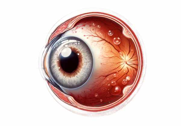
Introduction to Bullous Keratopathy
Bullous Keratopathy is an ocular condition marked by the development of fluid-filled blisters (bullae) on the cornea, the clear, dome-shaped surface that covers the front of the eye. This condition is typically caused by endothelial dysfunction, which results in corneal edema and the formation of bullae. Endothelial cells maintain corneal deturgescence by pumping excess fluid from the stroma. When these cells are damaged or lost, fluid accumulates in the cornea, causing swelling and visual impairment. Bullous Keratopathy is frequently associated with prior eye surgeries, trauma, or diseases like Fuchs’ endothelial dystrophy.
Detailed Guide to Bullous Keratopathy
Bullous Keratopathy is a chronic and debilitating eye condition that primarily affects the cornea. To fully understand its impact, one must first understand the anatomy and physiology of the cornea, as well as the pathophysiology of the condition and its various causes.
Corneal Anatomy and Physiology
The cornea has five layers: the epithelium, Bowman’s layer, stroma, Descemet’s membrane, and endothelium. The endothelium, a single layer of cells on the innermost surface, regulates fluid balance and thus maintains corneal transparency. Endothelial cells actively pump fluid from the stroma to prevent corneal swelling. Endothelial cells, unlike other cells, have a limited regenerative capacity, which means they cannot repair or regenerate themselves once damaged.
Pathophysiology
Bullous Keratopathy occurs when endothelial cells are unable to maintain the proper fluid balance, resulting in corneal edema. Excess fluid accumulates in the corneal stroma and eventually in the epithelium, resulting in painful blisters or bullae. These bullae can rupture, resulting in severe discomfort, photophobia (light sensitivity), and vision loss. Chronic edema can cause epithelial breakdown, which increases the risk of infection and complicates the condition.
Causes Of Bullous Keratopathy
- Fuchs’ Endothelial Dystrophy: This genetic condition causes progressive loss of endothelial cells. It usually appears in the fifth or sixth decade of life and can progress to severe corneal edema and Bullous Keratopathy if left untreated.
- Surgical Trauma: Eye surgeries, particularly cataract surgery, are a leading cause of endothelial damage. Intraocular procedures can damage the delicate endothelial layer, resulting in cell loss and subsequent corneal edema.
- Contact Lens Overuse: Prolonged use of contact lenses, particularly those with insufficient oxygen permeability, can cause hypoxia and endothelial stress, ultimately leading to Bullous Keratopathy.
- Glaucoma: In glaucoma, elevated intraocular pressure compresses and damages endothelial cells over time, resulting in chronic edema and bullae formation.
- Inflammatory Conditions: Chronic inflammatory conditions, such as uveitis, can result in secondary endothelial dysfunction and contribute to the development of Bullous Keratopathy.
- Trauma: Direct injury to the cornea, whether by blunt force or penetrating trauma, can damage the endothelium, causing fluid imbalance and corneal swelling.
Symptoms and Clinical Presentation
Patients with Bullous Keratopathy usually exhibit a variety of symptoms that reflect the severity of corneal edema and bullae formation. Symptoms include:
- Blurry Vision: As the cornea swells, the clarity decreases, resulting in blurred and hazy vision.
- Pain and Discomfort: Fluid-filled bullae on the corneal surface can cause severe pain, especially if they rupture.
- Photophobia: Light sensitivity is a common symptom caused by an irritated and swollen corneal surface.
- Foreign Body Sensation: Patients frequently experience the sensation that something is in their eye, which is caused by the irregular corneal surface and bullae.
- Redness and Tearing: Chronic irritation and swelling can cause redness and excessive tears.
Effects on Quality of Life
Bullous Keratopathy can significantly reduce a patient’s quality of life. Chronic pain and discomfort, combined with vision impairment, can disrupt daily activities like reading, driving, and working. The constant need to manage symptoms and seek medical attention increases the burden. Patients often experience emotional and psychological stress as they deal with the condition’s ongoing challenges.
Progress and Prognosis
The progression of Bullous Keratopathy is determined by the underlying cause and the efficacy of treatment strategies. Early intervention can slow the progression and improve outcomes, but advanced cases may necessitate more invasive procedures such as corneal transplantation. Without proper management, the condition can cause severe vision loss and permanent corneal damage.
Epidemiology
Bullous Keratopathy is a leading cause of corneal morbidity globally. It is more common in older adults because they have a higher risk of underlying conditions like Fuchs’ endothelial dystrophy and have more ocular surgeries. The condition affects both men and women equally and can appear in any ethnic group.
Understanding bullous keratopathy necessitates a comprehensive approach that takes into account the condition’s multifaceted nature. From its pathophysiological basis to its various etiologies and profound impact on patients’ lives, this ocular disorder poses a significant clinical challenge. Effective diagnosis and timely intervention are critical for managing the condition and improving the quality of life for those affected.
Essential Preventive Tips
- Regular Eye Examinations: Regular eye exams are essential, especially for people with risk factors like a history of eye surgery or a family history of corneal disease. Early detection can assist in managing conditions before they progress to Bullous Keratopathy.
- Proper Contact Lens Hygiene: Ensure that contact lenses are cleaned and handled properly. Use high-oxygen permeability lenses and adhere to the recommended wear schedule to avoid hypoxia and endothelial damage.
- Control of Underlying Conditions: Managing systemic conditions like diabetes and hypertension can help prevent secondary complications from developing into Bullous Keratopathy. Regular monitoring and treatment of glaucoma are also essential.
- Protective Eyewear: Wearing protective eyewear while participating in activities that increase the risk of eye injury can help prevent trauma-induced endothelial damage. This is especially important in occupations or sports that pose a high risk of ocular injury.
- Healthy Lifestyle: Eating a healthy, antioxidant-rich diet can benefit overall eye health. Avoid smoking because it is associated with a variety of ocular conditions, including corneal diseases.
- Early and Appropriate Treatment of Infections: Prompt treatment of ocular infections and inflammations can prevent long-term damage to the corneal endothelium.
- Post-Surgical Care: After having an eye surgery, carefully follow the post-operative care instructions. Regular check-ups with the ophthalmologist can help detect and manage complications early on.
- Educate and Inform: Raising awareness about Bullous Keratopathy and its risk factors can encourage people to seek medical attention and take preventive measures to protect their eyesight.
Diagnostic methods
Bullous Keratopathy is diagnosed using a combination of clinical and specialized diagnostic tests that assess corneal health and endothelial function. The standard and innovative diagnostic techniques used to identify and evaluate this condition are as follows:
- Slit-Lamp Examination: Ophthalmologists primarily use this clinical tool to examine the cornea. A slit-lamp exam illuminates the eye with a high-intensity light beam, allowing the clinician to see the cornea in great detail. This examination can detect corneal edema, bullae, and underlying structural abnormalities.
- Specular Microscopy: This noninvasive imaging technique is critical for evaluating the corneal endothelium. Specular microscopy provides detailed images of the endothelial cell layer, allowing for the assessment of cell density, morphology, and signs of endothelial dysfunction. Reduced cell density and abnormal cell shapes indicate poor endothelial health.
- Corneal Pachymetry: Corneal pachymetry determines the thickness of the cornea. Edema-induced corneal thickening is a common feature of Bullous Keratopathy. This measurement is useful in determining the severity of the condition and tracking its progression over time.
- Optical Coherence Tomography (OCT): OCT produces high-resolution cross-sectional images of the cornea, allowing for detailed visualization of all layers. It is especially useful for determining the degree of corneal edema and the presence of subepithelial fluid-filled bullae. Anterior segment OCT provides precise measurements that help guide treatment decisions.
- Confocal Microscopy: This advanced imaging technique enables in vivo examination of the cornea at the cellular level. Confocal microscopy provides detailed images of the corneal epithelium, stroma, and endothelium, which aids in identifying and characterizing cellular changes associated with Bullous Keratopathy.
- Anterior Segment Ultrasound Biomicroscopy (UBM): High-frequency ultrasound is used to create detailed images of the eye’s anterior segment, including the cornea. It aids in determining corneal thickness, detecting structural abnormalities, and assessing overall corneal health.
- Tear Film Analysis: Because Bullous Keratopathy can disrupt the ocular surface, examining the tear film can reveal additional information. Tear breakup time (TBUT) and osmolarity measurements are useful in determining tear film stability and ocular surface health.
Combining these diagnostic methods results in a comprehensive evaluation of Bullous Keratopathy, allowing for accurate diagnosis, disease progression monitoring, and effective management planning.
Managing Bullous Keratopathy Effectively
Bullous Keratopathy treatment aims to reduce corneal edema, alleviate symptoms, and improve visual function. The following are standard and innovative treatments for Bullous Keratopathy:
- Medications: – Hypertonic Saline: Topical hypertonic saline (5% sodium chloride) drops or ointments can reduce edema and discomfort by drawing excess fluid from the cornea. These treatments offer temporary relief but do not address the underlying endothelial dysfunction.
- Lubricative Eye Drops: Preservative-free artificial tears help to keep the ocular surface moist and reduce irritation caused by bullae. These drops offer symptom relief but do not address the underlying cause.
- Bandage Contact Lenses: Soft bandage contact lenses cover the bullae, protecting the corneal surface from mechanical trauma and reducing pain. They also help to maintain a stable tear film, which improves patient comfort. However, their use necessitates close monitoring to avoid infection.
- Corneal Transplantation: – Penetrating Keratoplasty (PK): A traditional full-thickness corneal transplant procedure that replaces the damaged cornea with a donor cornea. PK is effective, but it is not without risks, including rejection and complications.
- Endothelial Keratoplasty (EK): Descemet’s Stripping Endothelial Keratoplasty (DSEK) and Descemet’s Membrane Endothelial Keratoplasty (DMEK) are two EK techniques that selectively replace the diseased endothelial layer while preserving the rest of the cornea. These procedures have lower rejection rates and faster visual recovery than PK.
- Innovative and Emerging Therapies: – Corneal Cross-Linking (CXL): Originally developed for keratoconus, CXL is now being studied for Bullous Keratopathy to stabilize the corneal structure and reduce edema. The procedure consists of applying riboflavin drops and exposing the cornea to UV light, which strengthens the corneal collagen fibers.
- Rho Kinase Inhibitors: These medications help to regenerate endothelial cells and improve cornea clarity. Clinical trials are underway to determine their efficacy in treating Bullous Keratopathy.
- Stem Cell Therapy: Researchers are looking into stem cell therapy to regenerate damaged endothelial cells and restore corneal function. This new treatment shows promise for patients with severe endothelial dysfunction.
- Artificial Corneas: Advances in bioengineered corneas, such as the Boston Keratoprosthesis, provide alternatives for patients who are ineligible for traditional corneal transplants. These devices can help restore vision in cases of severe corneal damage.
Patients with Bullous Keratopathy can benefit significantly from timely and appropriate treatment, improving their visual outcomes and quality of life. Ongoing research and advances in corneal therapies are expanding the options for treating this difficult condition.
Trusted Resources
Books
- “Corneal Surgery: Theory, Technique and Tissue” by William J. Brunette
- “Cornea: Fundamentals, Diagnosis and Management” by Jay H. Krachmer
- “Corneal Disorders: Clinical Diagnosis and Management” by Howard M. Leibowitz


