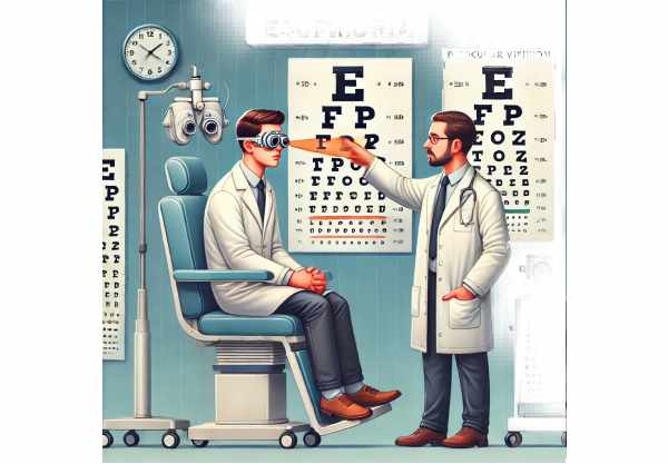
Introduction to Esophoria
Esophoria is a type of eye misalignment, or strabismus, in which one eye deviates inward towards the nose when focused on an object. Unlike esotropia, which is a constant misalignment, esophoria usually occurs intermittently and is most noticeable when a person is tired, stressed, or focuses on nearby objects for long periods of time. Symptoms of this condition include eye strain, headaches, double vision, and difficulty with binocular vision. Understanding the causes, symptoms, and diagnostic methods of esophoria is critical for its successful management and treatment.
Esophoria: Clinical Insights
When binocular vision is disrupted, one eye will deviate inward relative to the other, resulting in esophoria. This condition can be latent, which means it is not always visible but can be detected with certain diagnostic tests.
Pathophysiology
Esophoria results from an imbalance in the extraocular muscles that control eye movement. This imbalance can be caused by several factors, including:
- Muscle Imbalance: Differences in the strength and coordination of the extraocular muscles can cause one eye to turn inward.
- Refractive Errors: Large differences in refractive errors between the two eyes can result in esophoria. Hyperopia (farsightedness) is particularly associated with esophoria because the eye compensates by converging more than usual in order to see clearly.
- Neurological Factors: Abnormalities in the neural control of eye movements can also cause esophoria. This includes issues with the nerves that supply the extraocular muscles.
- Genetic Predisposition: A family history of binocular vision disorders increases the likelihood of developing esophoria.
Symptoms
Esophoria symptoms vary according to the severity and frequency of the misalignment. Common symptoms include:
- Eye Strain: Patients with esophoria frequently report eye discomfort and strain, particularly after extended periods of reading or close-up work.
- Headache: Esophoria can be characterized by frequent headaches, particularly around the eyes and on the forehead.
- Double Vision: Double vision, also known as diplopia, is caused by improper eye alignment, resulting in overlapping or doubled images.
- Blurry Vision: Intermittent blurring of vision may occur, especially when focusing on nearby objects.
- Difficulty with Binocular Vision: Difficulties maintaining proper binocular vision can cause problems with depth perception and eye coordination.
- Fatigue: Constantly maintaining proper eye alignment can cause general fatigue and difficulty concentrating.
Impact on Daily Life
Esophoria can have a significant impact on daily activities, especially those that require prolonged near vision, such as reading, computer work, and other detailed tasks. Discomfort and visual disturbances can impair academic and professional performance, resulting in decreased productivity and quality of life. Undiagnosed esophoria in children can lead to learning difficulties and a loss of interest in activities that require close visual attention.
Types of Esophoria
Esophoria can be classified according to the conditions in which it manifests:
- Accommodative Esophoria: This type is related to focusing efforts (accommodation) and is frequently associated with hyperopia. When the eyes work harder to focus, they converge more, resulting in inward deviation.
- Non-Accommodative Esophoria: This type of esophoria is unrelated to accommodation and may be caused by muscle imbalance or neurological issues.
Risk Factors
Several risk factors may increase the likelihood of developing esophoria:
- Age: Although esophoria can develop at any age, it is most commonly found in children and young adults.
- Refractive Errors: People with significant refractive errors, particularly hyperopia, are at a greater risk.
- Genetics: A family history of binocular vision disorders may predispose people to esophoria.
- Stress and Fatigue: Excessive stress and fatigue can aggravate esophageal symptoms.
- Prolonged Near Work: Activities requiring prolonged near focus, such as reading or computer use, can contribute to the development and progression of esophoria.
Complications
If left untreated, esophoria can cause a number of complications, including
- Amblyopia: Also known as “lazy eye,” amblyopia develops when one eye is consistently misaligned and not used effectively, resulting in decreased vision in that eye.
- Chronic Headaches: Consistent eye strain and misalignment can lead to chronic headaches and discomfort.
- Vision-Related Learning Difficulties: Esophoria in children can lead to difficulties with reading and learning, affecting academic performance.
- Reduced Quality of Life: The constant effort required to maintain eye alignment, as well as the associated discomfort, can have a negative impact on overall quality of life.
Patient Education and Support
Educating patients and their families about esophoria is critical for effective management. Understanding the symptoms, potential triggers, and the significance of regular eye exams can assist patients in seeking timely treatment and following prescribed management strategies. Individuals living with esophoria can benefit from support groups and educational resources.
Methods for Esophoria Diagnosis
Esophoria is diagnosed through a comprehensive eye examination and specific tests that assess eye alignment and coordination. Early and accurate diagnosis is critical to effective treatment and management.
Clinical Examination
Esophoria is first diagnosed with a thorough clinical examination by an optometrist or ophthalmologist. The key components of the examination are:
- Visual Acuity Test: This test assesses a person’s ability to see at different distances, which aids in the identification of any underlying refractive errors.
- Cover Test: This test is used to determine the presence and severity of esophoria. The examiner covers one eye and watches the movement of the uncovered eye. Esophoria occurs when the uncovered eye moves inward to fixate on a target. This test can be performed at both near and far fixation to determine the extent of the misalignment.
- Prism Test: Prism lenses are used to determine the amount of misalignment. By placing prisms of varying strengths in front of the eyes, the examiner can quantify the deviation and determine the specific degree of esophoria.
Diagnostic Tests
Several diagnostic tests are used to assess the alignment and focusing ability of the eyes.
- Maddox Rod Test: This test produces a visual distortion using a special lens with parallel red lines. When the patient looks at a light source through the Maddox rod, any misalignment of the eyes is visible. This test is used to quantify esophoria and understand its impact on binocular vision.
- Hirschberg Test: The Hirschberg test consists of shining a light into the eyes and observing the reflection on the cornea. The position of the light reflection relative to the pupil’s center indicates the presence and type of strabismus, which may include esophoria.
- Synoptophore: This instrument measures the angle of deviation and evaluates the eyes’ ability to maintain proper alignment. It provides useful information about the severity of esophoria and aids in the development of appropriate treatment plans.
Functional Tests
Functional tests assess the effects of esophoria on visual function and binocular vision.
- Convergence Insufficiency Test: This test assesses the eyes’ ability to work together when focusing on a nearby object. Esophoria patients may have difficulty maintaining proper convergence, resulting in symptoms like double vision and eye strain.
- Near Point of Convergence (NPC) Test: This test determines the closest point at which the eyes can remain aligned while focusing on a nearby object. Esophoria is characterized by difficulty maintaining convergence at close distances.
Advanced Imaging Techniques
Advanced imaging techniques may be used to assess the anatomical and functional aspects of the eye muscles and nerves.
- Magnetic Resonance Imaging (MRI): MRI can produce detailed images of the eye muscles, optic nerves, and brain structures that control eye movement. It is useful for detecting underlying neurological or anatomical abnormalities that contribute to esophoria.
- Computed Tomography (CT) Scan: CT scans can provide detailed images of the eye and surrounding structures, assisting in the diagnosis of any underlying conditions that may be causing or exacerbating esophoria.
Treatment Options for Esophoria
Esophoria treatment aims to alleviate symptoms while also improving binocular vision and overall eye alignment. The choice of treatment is determined by the severity of the condition, the presence of symptoms, and the patient’s overall visual requirements. Standard treatment options include corrective lenses, vision therapy, and, in some cases, surgery. In addition, novel and emerging therapies are being investigated in order to provide more effective esophoria treatments.
Standard Treatment Options
- Corrective lenses
- Prescription Glasses or Contact Lenses: Corrective lenses can help manage esophoria, especially if it is caused by refractive errors such as hyperopia. Correcting the underlying refractive error allows the eyes to focus more efficiently, lowering the tendency for inward deviation.
- Prism Lenses: Prism lenses can be added to glasses to help the eyes align properly. These lenses change the way light enters the eye, which helps with alignment and reduces symptoms like double vision and eye strain.
- Visual therapy
- Vision therapy is a series of structured exercises that aim to improve eye coordination and alignment. This therapy can be done under the supervision of an optometrist and may include:
- Convergence Exercises: Pencil push-ups and the use of convergence cards can help strengthen the eye muscles responsible for eye alignment during near tasks.
- Fusion Exercises: Activities that require both eyes to work together to form a single image can help improve binocular vision and alleviate esophoria symptoms.
- Computerized Vision Therapy: Specialized software programs offer interactive exercises that improve eye alignment and coordination.
- Surgery
- In severe cases of esophoria that do not respond to conservative treatments, surgery may be considered. The goal of eye muscle surgery is to better align the extraocular muscles by adjusting their tension and positioning. This procedure is typically reserved for cases where esophoria severely impairs quality of life and other treatments have proven ineffective.
Innovative and Emerging Therapies
- Botulinum toxin Injections
- Botulinum toxin (Botox) injections are being investigated as a possible treatment for esophoria. Botox can improve alignment by temporarily weakening overactive eye muscles. This method is still being investigated and is used selectively in some cases.
- Neuroplasticity-based therapies
- Research into neuroplasticity, or the brain’s ability to reorganize itself, has resulted in the development of therapies for retraining the visual system. These therapies aim to improve the brain’s ability to control eye movements and binocular vision.
- Advanced optical devices
- New optical devices, such as adjustable prism glasses and advanced contact lenses, are being developed to provide more effective and comfortable esophoria management options. These devices provide customizable correction based on individual visual needs.
- Genetic Therapy
- Gene therapy is a new field that shows promise in treating a variety of eye conditions, including esophoria. Gene therapy, which targets the underlying genetic factors that contribute to muscle imbalance and misalignment, may provide a long-term solution.
Healthcare providers can provide comprehensive and personalized management strategies for esophoria patients by combining standard treatments with innovative and emerging therapies, thereby improving their visual function and quality of life.
Esophoria Prevention Tips
- Regular Eye Examination
- Have regular eye exams to detect early signs of esophoria and other vision issues. Early detection enables timely intervention and management.
- Keep Proper Eye Hygiene
- Keep your eyes clean and free of irritations. Avoid rubbing your eyes, as this can aggravate any existing eye conditions.
- Use Corrective Lenses as prescribed
- Wear your prescription glasses or contact lenses as prescribed by your eye care professional to ensure proper vision and reduce eye strain.
- Limited Screen Time
- Take frequent breaks from screens to relieve eye strain and fatigue. Follow the 20-20-20 rule, which states that every 20 minutes, look at something 20 feet away for no less than 20 seconds.
- Practice Good Posture
- Maintain proper posture while reading, working on the computer, or doing other nearby tasks. Proper ergonomics can help to reduce strain on your eyes and neck.
- Engage in Vision Therapy
- If your eye care professional recommends it, try vision therapy exercises to strengthen your eye muscles and improve your coordination.
- Managing Stress and Fatigue
- High levels of stress and fatigue can exacerbate esophageal symptoms. Use stress-reduction techniques like meditation, deep breathing exercises, and regular physical activity.
- Stay hydrated
- Proper hydration improves overall eye health. Drink plenty of water throughout the day to keep your eyes moist and avoid dryness.
- Keep a Balanced Diet
- Eat a diet high in vitamins and minerals that promote eye health, such as omega-3 fatty acids, vitamins A, C, E, and zinc.
- Use proper lighting
- Use adequate lighting when reading or working to reduce eye strain. Reduce glare from screens and other light sources.
Trusted Resources
Books
- “Strabismus: A Decision-Making Approach” by Robert S. Lederman
- “Binocular Vision and Ocular Motility: Theory and Management of Strabismus” by Gunter K. von Noorden
- “Clinical Management of Binocular Vision: Heterophoric, Accommodative, and Eye Movement Disorders” by Mitchell Scheiman and Bruce Wick
Online Resources
- American Academy of Ophthalmology
- National Eye Institute
- American Optometric Association
- WebMD – Esophoria


