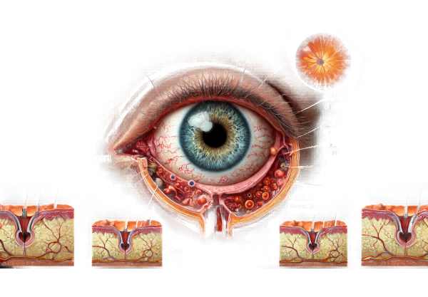
What is Graves Orbitopathy?
Graves’ Orbitopathy, also known as Thyroid Eye Disease (TED), is an autoimmune disorder that affects the orbit of the eye. It is frequently associated with Graves’ disease, a condition characterized by hyperthyroidism, or excessive thyroid activity. Graves’ Orbitopathy occurs when the immune system mistakenly targets the tissues around the eyes, causing inflammation and swelling. This can cause a wide range of symptoms, including bulging eyes (proptosis), double vision (diplopia), and discomfort. Early diagnosis and appropriate treatment are critical because the condition can have a significant impact on quality of life.
Graves’ Orbitopathy Insights
Graves’ Orbitopathy is a complex condition that primarily affects the orbit, or bony cavity that houses the eye. The underlying cause is an autoimmune reaction in which antibodies mistakenly attack the tissues around the eyes. This reaction is commonly associated with Graves’ disease-induced hyperthyroidism, but it can also occur in euthyroid and hypothyroid states.
Pathophysiology
Graves’ Orbitopathy is characterized by a number of immune-mediated processes. The immune response’s primary target is the fibroblasts in the orbital tissue. These cells express thyrotropin receptors (TSHR), which are similar to those found in the thyroid gland. When autoantibodies (TSHR antibodies) bind to these receptors, they cause a series of inflammatory responses. Cytokines like IL-1, IL-6, and TNF-α activate and promote the proliferation of orbital fibroblasts, resulting in the deposition of GAGs.
The accumulation of GAGs and the subsequent increase in osmotic pressure contribute to tissue swelling and inflammation. Furthermore, fibroblast differentiation into adipocytes causes an increase in orbital fat volume, exacerbating proptosis and compressing the ocular structures.
Graves’ Orbitopathy causes a variety of ocular symptoms, which can vary in severity. The most common symptoms are:
- Proptosis (Bulging Eyes): Proptosis, one of the hallmark features, is caused by an increase in the volume of orbital contents. It may be unilateral or bilateral.
- Lid Retraction and Lag: Upper eyelid retraction and lag are common, resulting in a staring appearance.
- Periorbital Swelling: Swelling around the eyes is caused by inflammation and fluid retention.
- Conjunctival Redness and Chemosis: Inflammation can cause redness and swelling of the conjunctiva and surrounding tissues.
- Diplopia (Double Vision): Involvement of the extraocular muscles can cause motility disturbances and double vision.
- Pain and Discomfort: Patients may feel pressure or pain in the orbit, particularly during eye movements.
- Dry Eyes and Exposure Keratopathy: Due to lid retraction and incomplete closure, the cornea may become dry, resulting in exposure keratopathy.
Risk Factors and Epidemiology
Graves’ orbitopathy is more common in people with Graves’ disease, affecting 25-50% of patients. Women are more commonly affected than men, and the condition usually appears in the fourth to sixth decades of life. Smoking is a major risk factor, with smokers experiencing a higher incidence and more severe forms of the disease.
Natural History and Phases
Graves’ Orbitopathy progresses in two phases:
- Active (Inflammatory) Phase: This phase lasts between six months and two years and is distinguished by active inflammation and progressive symptoms. During this time, the disease is most susceptible to treatment.
- Inactive (Fibrotic) Phase: Following the inflammatory phase, the disease enters a stable or quiescent phase in which the inflammation resolves but the fibrotic changes persist. Symptoms stabilize, but residual effects such as proptosis and diplopia may persist.
Effects on Quality of Life
Graves’ Orbitopathy has the potential to significantly reduce a patient’s quality of life. Visible changes in appearance can cause significant psychological distress and social embarrassment. Diplopia and visual impairment can impair daily activities such as driving, reading, and work performance. Chronic pain and discomfort increase the disease’s overall burden.
Associated Systemic Manifestations
Because of its association with Graves’ disease, systemic manifestations of hyperthyroidism frequently accompany Graves’ Orbitopathy. These may include:
Benefits include weight loss and improved heat tolerance.
- Palpitations.
- Tremors.
Symptoms include an increased appetite and fatigue. - Menstrual irregularities.
Thyroid dysfunction must be managed as part of the overall treatment plan for Graves’ Orbitopathy, as uncontrolled thyroid levels can exacerbate ocular symptoms.
Immunological Considerations
Graves’ Orbitopathy is autoimmune in nature, with a complex interplay of genetic predisposition and environmental triggers. The presence of specific HLA (human leukocyte antigen) haplotypes has been linked to an increased risk of developing Graves disease and its ocular manifestations. Environmental factors, such as stress and infection, can act as triggers in genetically susceptible people.
Research into immunological mechanisms is ongoing, with a focus on identifying specific autoantigens and immune pathways that could be therapeutic targets. Understanding the immunopathogenesis of Graves’ Orbitopathy is critical for developing new treatments that modulate the immune response and halt disease progression.
Diagnostic Techniques for Graves’ Orbitopathy
Diagnosing Graves’ Orbitopathy is diagnosed through a combination of clinical evaluation, imaging studies, and laboratory tests. A thorough examination is required to confirm the diagnosis and ascertain the extent of orbital involvement.
Clinical Examination
A thorough clinical examination is the first step in diagnosing Graves’ Orbitopathy. Key components are:
- Visual Acuity Assessment: Determine the impact on vision.
- Ocular Motility Examination: To detect and diagnose diplopia.
- Slit Lamp Examination: Examine the anterior segment for signs of inflammation, such as conjunctival redness and chemosis.
- Eyelid Position and Function: Assess lid retraction and lag.
- Exophthalmometry: Using an exophthalmometer, measure the degree of proptosis.
Imaging Studies
Imaging studies are critical for determining the extent of orbital involvement and informing treatment decisions. Common imaging modalities are:
- Computed Tomography (CT) Scan: A CT scan produces detailed images of the bony orbit and soft tissues. They are especially useful for assessing extraocular muscles and diagnosing compressive optic neuropathy.
- Magnetic Resonance Imaging(MRI): MRI provides excellent soft tissue contrast and is useful for determining the extent of muscle and fat involvement. It also aids in distinguishing active inflammation from fibrotic changes.
Lab Tests
Laboratory tests help to confirm the diagnosis and evaluate thyroid function. Important tests include:
- Thyroid Function Tests: Measurement of serum TSH, free T4, and free T3 levels to determine thyroid function.
- Thyroid Autoantibodies: The presence of TSH receptor antibodies (TRAb) and anti-thyroid peroxidase (anti-TPO) antibodies to aid in the diagnosis of Graves’ disease.
- Inflammatory Markers: High levels of inflammatory markers like erythrocyte sedimentation rate (ESR) and C-reactive protein (CRP) may indicate active inflammation.
Clinical Activity Scale (CAS)
The Clinical Activity Score is an effective tool for determining the activity of Graves’ Orbitopathy. It entails assessing several clinical signs, such as redness, swelling, and pain, and assigning a score based on their prevalence and severity. The CAS aids in determining the disease phase (active or inactive) and directing treatment decisions.
Differential Diagnosis
It is critical to distinguish Graves’ Orbitopathy from other conditions that may produce similar symptoms. Differential diagnosis includes:
- Orbital Cellulitis is an infection of the orbital tissues characterized by pain, redness, and swelling.
- Idiopathic Orbital Inflammation (Orbital Pseudotumor) is a non-specific inflammatory condition affecting the orbit.
- Orbital Tumors: These tumors, whether benign or malignant, can cause proptosis and diplopia.
Graves’ Orbitopathy Treatment Methods
Graves’ Orbitopathy treatment aims to alleviate symptoms, reduce inflammation, and prevent complications. Treatment plans are frequently tailored to the stage of the disease (active or inactive) and the severity of symptoms.
Standard Treatment Options
- Corticosteroids: High-dose corticosteroids, such as prednisone, are frequently used to reduce inflammation during the disease’s active phase. They can be given orally or intravenously.
- Orbital Radiotherapy: This treatment uses low-dose radiation to alleviate inflammation and swelling in the orbital tissues. It is usually reserved for patients who do not respond to corticosteroids or have contraindications to their use.
- Surgical Interventions: Surgery may be required for patients with severe proptosis, compressive optic neuropathy, or significant eyelid retraction. The most common surgical procedures are:
- Orbital Decompression Surgery: Removes bone and/or fat from the orbit to increase space and relieve pressure on the optic nerve.
- Strabismus Surgery: Corrects eye misalignment due to extraocular muscle involvement.
- Eyelid Surgery: Corrects lid retraction and enhances eyelid closure.
Innovative and Emerging Therapies
- Teprotumumab: Teprotumumab, a monoclonal antibody that targets the insulin-like growth factor-1 receptor (IGF-1R), has shown promising results in reducing proptosis and improving quality of life in patients with Graves’ Orbitopathy. It is administered via intravenous infusion and represents a significant advance in therapeutic options.
- Rituximab: Rituximab is an anti-CD20 monoclonal antibody that targets B cells involved in the autoimmune response. Studies have shown that it can help reduce disease activity and inflammation.
- Tocilizumab: Tocilizumab, an interleukin-6 (IL-6) receptor antagonist, is used to reduce inflammation in patients who do not respond to corticosteroids. It is administered through an intravenous infusion.
- Mycophenolate Mofetil: An immunosuppressant that prevents T and B lymphocyte proliferation. It has been used in the treatment of Graves’ Orbitopathy to reduce the need for steroids.
These emerging therapies provide new hope to patients, especially those who have not responded to conventional treatments. Ongoing research and clinical trials are underway to investigate additional therapeutic options and improve outcomes for people with Graves’ Orbitopathy.
Prevention Tips for Thyroid Eye Disease
- Stop Smoking: Smoking is a major risk factor for Graves’ Orbitopathy. Quitting smoking can help lower the risk and severity of the condition.
- Regular Thyroid Check-ups: Regular monitoring of thyroid function can aid in the early detection and management of thyroid disorders, lowering the risk of associated eye problems.
- Manage Stress: Chronic stress can cause autoimmune responses. Stress-relieving activities such as yoga, meditation, and regular exercise can help you maintain good health.
- Balanced Diet: Eating a healthy diet high in antioxidants can help the immune system function and reduce inflammation.
- Eye Protection: Wearing sunglasses and applying lubricating eye drops can help protect your eyes from environmental irritants and dryness.
- Early Medical Intervention: Seeking prompt medical attention for any symptoms of thyroid dysfunction or eye discomfort can result in early diagnosis and treatment, preventing disease progression.
- Avoid Environmental Triggers: Reducing exposure to environmental pollutants and allergens can help lower the risk of autoimmune reactions.
Individuals who follow these preventive measures can reduce their risk of developing Graves’ Orbitopathy while also maintaining good overall eye health.
Trusted Resources
Books
- “Graves’ Disease: Pathogenesis and Treatment” by Basil Rapoport and Sandra M. McLachlan
- “Thyroid Eye Disease: Understanding Graves’ Orbitopathy” by Elaine A. Moore
- “The Thyroid Eye Disease Book” by Elaine A. Moore and Lisa Marie Moore


