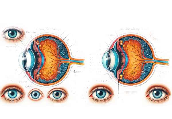
What is hyperopia?
Hyperopia, also known as farsightedness, is a refractive error in which distant objects appear more clearly than close ones. This condition develops when the eye is shorter than normal or the cornea has too little curvature, causing light to focus behind the retina rather than directly on it. Hyperopia can affect people of all ages, but it is typically congenital and becomes more noticeable with age. Symptoms may include difficulty focusing on close objects, eye strain, headaches, and, on occasion, blurred vision.
Hyperopia: Detailed Insights
One of the most common vision-related refractive errors is hyperopia, also known as farsightedness. Hyperopia primarily impairs near vision, as opposed to myopia (nearsightedness), which affects a person’s ability to see distant objects clearly. The severity of hyperopia varies greatly between individuals, and in severe cases, even distance vision can be impaired.
Anatomy and Physiology of Hyperopia
Hyperopia occurs when the eye is unable to bend light correctly, resulting in a focus point that is behind the retina. There are several anatomical variations that can cause this misalignment:
- Shortened Eyeball: The most common cause of hyperopia is a shorter-than-average eyeball. This condition causes the light rays that enter the eye to converge behind the retina, resulting in blurred near vision.
- Flattened Cornea: To properly refract light, the cornea, the eye’s outermost lens, should have a curved shape. In hyperopia, the cornea may be flatter than normal, reducing refractive power and contributing to focal point displacement.
- Lens Variations: The eye’s lens can also influence its refractive ability. A less convex lens may cause hyperopia by failing to bend light sufficiently to focus on the retina.
Symptoms of Hyperopia
Hyperopia can cause a variety of symptoms, depending on the severity of the refractive error. Common symptoms include:
- Difficulty with Near Vision: People with hyperopia frequently struggle to see objects up close, such as when reading a book or using a phone. This difficulty can cause eye strain and discomfort.
- Eye Strain: Prolonged tasks that require near vision can result in significant eye strain. Symptoms of eye strain include aching or discomfort around the eyes, particularly after prolonged reading or computer use.
- Headaches: Frequent headaches, especially after close work, may indicate uncorrected hyperopia. The strain on the eye muscles as they attempt to compensate for the refractive error can cause headaches.
- Blurred Vision: In cases of high hyperopia, distant vision may become blurred. This symptom is more noticeable in severe hyperopia, and corrective lenses are frequently required for both near and far vision.
- Squinting: To improve focus, people with hyperopia may squint, which can temporarily improve clarity by changing the shape of the eye and adjusting the focal point.
Causes and Risk Factors
Hyperopia is usually a genetic condition, so it frequently runs in families. However, a number of factors can influence its development:
- Genetics: A family history of hyperopia raises the risk of developing the condition. Genetic predisposition has a significant impact on eye shape and refractive properties.
- Age: Although hyperopia can be present from birth, it may not be noticeable until later in life. As people get older, their eyes’ lenses become less flexible, exacerbating the effects of hyperopia. This age-related change, known as presbyopia, impairs the eye’s ability to focus on nearby objects.
- Environmental Factors: Prolonged periods of near work without breaks, insufficient lighting, and poor visual ergonomics can all contribute to eye strain and affect the onset or progression of hyperopia.
Effects on Quality of Life
Hyperopia can have a significant impact on a person’s quality of life, particularly if left untreated. Difficulties with near tasks can impair daily activities such as reading, writing, and using digital devices. Children with undiagnosed hyperopia may struggle in school due to the difficulty of focusing on close-up tasks, which can lead to learning difficulties and poor academic performance.
Adults with hyperopia may also face difficulties in the workplace, especially in jobs that require prolonged near vision tasks. Eye strain and headaches can lower productivity and job satisfaction.
Hyperopia in Children
Hyperopia is fairly common in children. Many infants are born with hyperopia, but as their eyes develop, the condition often corrects itself. However, significant hyperopia that does not improve with age can result in visual issues such as strabismus (crossed eyes) or amblyopia (lazy eye). Early detection and treatment are critical in avoiding these complications.
Pediatric hyperopia can be difficult to detect because children rarely complain about their vision. Regular eye exams are essential for detecting and managing hyperopia early on, ensuring that children’s vision develops optimally.
Associated Conditions
Hyperopia can be associated with other ocular conditions, such as
- Strabismus: This condition, also known as crossed eyes, occurs when the eyes do not properly align. Hyperopic children frequently develop strabismus as their eyes work harder to focus, causing them to turn inward.
Amblyopia, also known as lazy eye, occurs when one eye is significantly more hyperopic than the other. The brain may favor the clearer eye, resulting in decreased vision in the other.
- Glaucoma: Research indicates that hyperopia may increase the risk of developing angle-closure glaucoma. Hyperopic eyes’ anatomical structure may predispose them to this type of glaucoma, which can cause increased intraocular pressure and optic nerve damage if left untreated.
Methods for Diagnosing Hyperopia
Several tests and evaluations are required to determine the extent of the refractive error and its impact on vision. Here are the main diagnostic methods used:
Comprehensive Eye Examination
A thorough eye examination is the first step in diagnosing hyperopia. During the exam, the eye care professional will perform several tests, including:
- Visual Acuity Test: This test uses a Snellen chart to determine the sharpness of vision at various distances. The patient reads letters from the chart to determine their visual acuity at different distances.
- Refraction Assessment: This test uses a phoropter and retinoscope to determine how light waves bend as they pass through the cornea and lens. The doctor will examine various lenses to determine the prescription required to correct the refractive error.
- Slit-Lamp Examination: This test allows the doctor to examine the eye’s structures with high magnification. It aids in detecting any abnormalities in the cornea, lens, and other parts of the eye that may contribute to hyperopia.
Retinoscopy
Retinoscopy is a technique for determining the refractive error of the eye. During this test, the eye doctor shines a light into the patient’s eye and looks at the reflection (reflex) off the retina. By moving the light across the eye and changing lenses in the phoropter, the doctor can estimate refractive error, including hyperopia.
Autorefractors and Aberrometers
- Autorefractors: These devices automatically determine the refractive error of the eye. The patient looks into the machine and sees an image, and the device calculates the prescription required to correct the vision.
- Aberrometers: These advanced instruments measure how light travels through the eye and can detect higher-order aberrations as well as basic refractive errors. This detailed analysis aids in the creation of more precise corrective lenses.
Cycloplegic Refraction
Cycloplegic refraction uses eye drops to temporarily paralyze the ciliary muscle, preventing the eye from shifting focus during the test. This method is especially useful for diagnosing hyperopia in children and young adults because it provides a more precise measurement of the refractive error without interference from the eye’s focusing mechanism.
Corneal Topography
Corneal topography maps the cornea’s surface curvature. This test is useful for detecting any irregularities in the corneal shape that may contribute to hyperopia. It offers detailed information about the corneal surface, which aids in the diagnosis and treatment of refractive errors.
Optical Coherence Tomography(OCT)
OCT is an imaging test that generates detailed cross-sectional images of the retina. While OCT is most commonly used to diagnose retinal conditions, it can also help assess the overall health of the eye and identify any structural issues that may be contributing to hyperopia.
Contrast Sensitivity Tests
Contrast sensitivity testing determines how well the eye distinguishes between various shades of gray. This test aids in understanding how hyperopia affects the patient’s ability to see in low-contrast environments, such as driving at night or reading faded print.
Hyperopia Treatment Insights
The treatment of hyperopia aims to correct the refractive error, improve vision, and alleviate symptoms such as eye strain and headache. Here are the standard treatment options together with innovative and emerging therapies:
- Corrective Lenses: The most common and immediate treatment for hyperopia is the use of corrective lenses. This includes prescription eyeglasses and contact lenses that change how light rays enter the eye and focus directly on the retina.
- Eyeglasses: These are the most basic and safest option, particularly for children and people with other eye conditions.
- Contact Lenses: Contact lenses offer a larger field of vision and are better suited to active lifestyles. Individual needs can dictate the type of lens prescribed, which includes soft, rigid gas-permeable, and multifocal lenses.
- Refractive Surgery: Those seeking a more permanent solution have several surgical options available:
- LASIK (Laser-Assisted in Situ Keratomileusis): This popular procedure involves reshaping the cornea to correct a refractive error. It is quick, with a short recovery period.
- PRK (Photorefractive Keratectomy): Like LASIK, PRK reshapes the cornea but does not require a corneal flap. It is suitable for people with thin corneas.
- LASEK (Laser Epithelial Keratomileusis): LASEK is a variation of PRK that preserves more of the corneal surface and is an alternative for those with thinner corneas.
- Lens Implants: For severe hyperopia, intraocular lens implants may be an option. This includes:
- Phakic Intraocular Lenses (IOLs): Implanted without removing the eye’s natural lens and suitable for severe hyperopia.
- Refractive Lens Exchange (RLE): Similar to cataract surgery, the natural lens is removed and replaced with an artificial one. This is typically reserved for people with severe refractive errors or those who have early cataracts.
Innovative and Emerging Therapies
- Corneal Inlays: These small, ring-shaped implants are inserted into the cornea to alter its shape and correct the refractive error. They are a relatively new option that can be combined with other procedures such as LASIK.
- Orthokeratology (Ortho-K): This nonsurgical procedure entails wearing specially designed rigid contact lenses overnight to temporarily reshape the cornea. It can provide clear vision throughout the day, eliminating the need for glasses or contacts.
- Wavefront-Guided LASIK: This advanced type of LASIK uses detailed measurements of how light waves travel through the eye to create a personalized treatment plan. It addresses higher-order aberrations that traditional LASIK may not correct, potentially improving visual outcomes.
- SMILE (Small Incision Lenticule Extraction): This minimally invasive surgery creates a lenticule within the cornea with a femtosecond laser before removing it through a small incision. It provides a faster recovery time and has a lower impact on corneal stability.
Individuals with hyperopia can find a solution that best suits their lifestyle and vision requirements by researching these treatment options. Consultation with an eye care professional is required to determine the best treatment option based on the severity of the condition and overall eye health.
Best Practices for Avoiding Hyperopia (Farsightedness)
- Regular Eye Exams: Have a comprehensive eye exam at least every two years, or as recommended by your eye care provider. Early detection can help to manage refractive errors before they worsen.
- Balanced Diet: Eat a diet high in vitamins A, C, and E, as well as omega-3 fatty acids, to improve overall eye health. Foods that are beneficial include leafy greens, fish, nuts, and citrus fruits.
- Proper Lighting: To reduce eye strain, use adequate lighting while reading or working. Use task lighting that is bright but not harsh, and avoid reading in low-light environments.
- Breaks During Close Work: Use the 20-20-20 rule: every 20 minutes, look 20 feet away for at least 20 seconds. This practice helps to reduce eye strain caused by prolonged near tasks.
- Protective Eyewear: Wear sunglasses with UV protection to protect your eyes from harmful ultraviolet rays. Prolonged exposure to UV light can lead to a variety of eye problems.
- Ergonomic Workspaces: Arrange your computer and workspace ergonomically to reduce eye strain. Make sure the screen is at eye level and approximately 20-30 inches away from your eyes.
- Proper Hydration: Keep your eyes hydrated by drinking plenty of water and using artificial tears as needed, particularly in dry or air-conditioned environments.
- Avoid Smoking: Smoking increases the risk of developing eye conditions such as cataracts and macular degeneration. Quitting smoking can greatly improve your eye health.
- Eye Exercises: Do regular eye exercises to strengthen your eye muscles and increase focus flexibility. Simple exercises, such as focusing on distant and nearby objects, can help.
- Monitor Screen Time: Limit screen time, particularly for children, and encourage frequent breaks to reduce digital eye strain. To reduce blue light exposure from screens, use blue light filters or glasses.
Trusted Resources
Books
- “Clinical Optics” by Troy E. Fannin and Theodore Grosvenor
- “Foundations of Clinical Ophthalmology” by Jack J. Kanski and Brad Bowling
- “Ophthalmology: A Short Textbook” by Gerhard K. Lang


