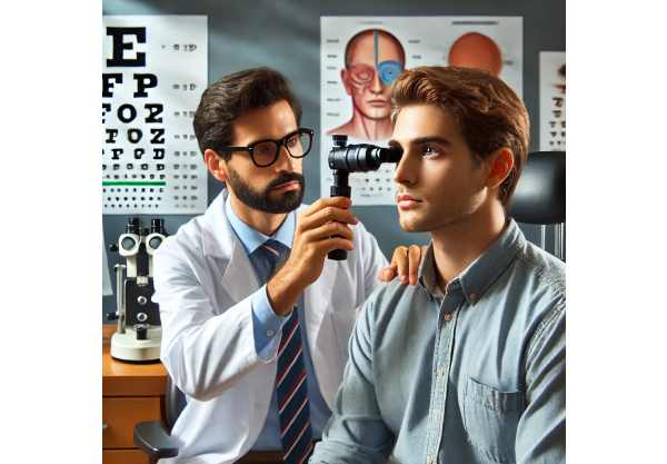
What is hypotropia?
Hypotropia is a type of strabismus in which one eye deviates downward relative to the other, resulting in misalignment. This ocular condition can impair binocular vision and depth perception, resulting in symptoms like double vision, eye strain, and headaches. Hypotropia can be congenital or acquired, and it can be caused by a variety of conditions, such as muscular imbalances, neurological disorders, or structural abnormalities in the eye. Early detection and appropriate management are critical for preventing long-term vision problems and improving those affected’s quality of life.
Thorough Examination of Hypotropia
Hypotropia, a type of vertical strabismus, poses a complex challenge for both patients and eye care professionals. It disrupts eye alignment, affecting how the brain processes visual information. This misalignment can cause a variety of symptoms and have a significant impact on daily activities as well as overall vision.
Each eye has six extraocular muscles that control its movements: superior rectus, inferior rectus, lateral rectus, medial rectus, superior oblique, and inferior oblique. Three cranial nerves (III, IV, and VI) innervate these muscles, allowing the eyes to move together.
Hypotropia is characterized by an imbalance in the function or neural control of these muscles, particularly the inferior rectus and superior oblique muscles. When the eyes are not properly aligned, the visual axis of the deviated eye is not parallel to that of the non-deviated eye, resulting in visual disturbances.
Symptoms of Hypotropia
The symptoms of hypotropia can vary greatly depending on the severity of the misalignment and the individual’s capacity to compensate. Common symptoms include:
- Double Vision (Diplopia): This is when the brain receives two different images from each eye, resulting in two images of the same object.
- Eye Strain and Fatigue: The effort to keep your eyes aligned and focused can cause significant strain and discomfort, especially during tasks that require prolonged concentration.
- Headaches: Persistent eye strain frequently causes headaches, usually around the forehead and eyes.
- Difficulty with Depth Perception: Proper depth perception requires the alignment of both eyes. Hypotropia can disrupt this, making it difficult to accurately judge distances.
- Head Tilt: People with hypotropia may use a compensatory head tilt to better align their eyes and alleviate symptoms such as double vision.
Causes and Risk Factors
Several factors can contribute to hypotropia, such as:
- Congenital Factors: Some people are born with muscle imbalances or developmental issues that cause hypotropia.
- Trauma: Injuries to the head or eye can damage the muscles or nerves that control eye movements, causing hypotropia.
- Neurological Disorders: Conditions like cranial nerve palsies, which affect the nerves that control the eye muscles, can cause vertical misalignment.
- Systemic Diseases: Conditions such as thyroid eye disease (Graves’ disease), myasthenia gravis, and diabetes can impair muscle function and contribute to hypotropia.
- Structural Abnormalities: Orbital fractures and congenital abnormalities can mechanically limit eye movement, resulting in misalignment.
Types of Hypotropia
Hypotropia is classified according to its onset, consistency, and underlying cause:
- Congenital Hypotropia: Present at birth or developing in early infancy, often necessitating early intervention to avoid amblyopia (lazy eye) and related complications.
- Acquired Hypotropia: Occurs later in life due to trauma, disease, or neurological conditions.
- Intermittent Hypotropia: The downward deviation occurs infrequently, often as a result of fatigue, illness, or other stressors.
- Constant Hypotropia: The downward deviation persists regardless of conditions.
Impact on Daily Life
Hypotropia can have a significant impact on daily life, particularly in activities that require precise vision, such as reading, writing, driving, and using digital devices. The condition can also have an impact on social interactions by making it difficult to maintain eye contact. Children with undiagnosed hypotropia may struggle in school due to visual fatigue and difficulty reading and writing. Adults may face difficulties in their professional lives, particularly in positions that require prolonged visual focus.
Psychological and Social Implications
Living with hypotropia may also have psychological and social consequences. Individuals may be self-conscious about their eye alignment, which can lower self-esteem and cause social anxiety. Visible misalignment can affect interpersonal interactions and communication, potentially leading to social isolation.
Complications
If left untreated, hypotropia can lead to a number of complications, including
- Amblyopia (Lazy Eye): In children, the brain may begin to ignore visual input from a misaligned eye, resulting in reduced vision in that eye.
- Persistent Diplopia: Chronic double vision can be debilitating, interfering with daily activities and lowering overall quality of life.
- Strain on the Non-Deviated Eye: The eye that remains aligned may become overworked, resulting in fatigue and vision issues.
Prognosis
The prognosis for hypotropia varies according to the underlying cause and the timing of the treatment. Early intervention, particularly in children, can result in significant improvements and avoid long-term complications. The success of treatment in adults is determined by the cause and severity of the condition, as well as the specific treatment method used.
Methods for Diagnosing Hypotropia
A comprehensive eye examination and several specialized tests are required to determine the extent and cause of hypotropia. Here are the main diagnostic methods used:
Comprehensive Eye Examination
A thorough eye examination by an ophthalmologist or optometrist is essential for diagnosing hypotropia. This includes:
- Visual Acuity Test: This test measures vision clarity at various distances, which aids in the identification of any vision impairment.
- Cover-Uncover Test: The doctor covers one eye and the patient focuses on a target. Observing the movement of the uncovered eye can help detect hypotropia.
- Alternate Cover Test: Similar to the cover-uncover test, this involves covering one eye first, then the other, to detect any latent deviation.
Prism Testing
Prism testing uses prisms of varying strengths to determine the degree of eye misalignment. The prism is placed in front of one eye, and the patient is instructed to focus on a target. The strength of the prism required to align the eyes is an accurate indicator of hypotropia.
Maddox Rod Test
The Maddox rod test involves inserting a series of parallel cylindrical lenses in front of one eye while the patient looks at a light source. The lenses cause a line of light to appear displaced relative to the other eye, which aids in determining the degree of misalignment.
Hirschberg Test
The Hirschberg test consists of shining a light into the patient’s eyes and observing the reflection on the cornea. The position of the reflection influences the presence and extent of hypotropia.
Neurological Examination
When neurological causes are suspected, a comprehensive neurological examination may be performed. This may include:
- Cranial Nerve Assessment: Determines the function of the cranial nerves that control eye movements.
- Imaging Studies: An MRI or CT scan may be used to detect structural abnormalities or lesions in the brain or nerves.
Sensory Testing
Sensory testing determines how well the eyes work together and the effect of hypotropia on binocular vision. This may include:
- Worth 4-Dot Test: This test evaluates fusion and suppression by having the patient look at four dots through red-green glasses.
- Stereopsis Testing: Uses various 3D tests to assess depth perception and binocular vision.
Treatment
Hypotropia treatment aims to correct the eye’s vertical misalignment, alleviate symptoms, and improve overall visual function. Here are the standard treatment options together with innovative and emerging therapies:
- Corrective Lenses: Glasses or contact lenses with prismatic correction can help realign the eyes and alleviate symptoms like double vision.
- Prism Glasses: These glasses have prisms that bend light before it enters the eye, which helps to correct vertical misalignment.
- Prescription Glasses: Corrective lenses can correct any coexisting refractive errors, resulting in less overall eye strain.
- Vision Therapy: A non-surgical approach that uses a series of eye exercises to improve coordination and strengthen the eye muscles.
- Orthoptic Exercises: Supervised exercises that improve the control and function of the eye muscles, typically performed in an eye care professional’s office.
- Home Exercises: Customized routines for patients to perform at home to reinforce the benefits of in-office therapy.
- Surgical Intervention: In severe cases where non-invasive treatments fail, surgery may be required.
- Strabismus Surgery: To correct the misalignment, adjust the length or position of the eye muscles. Techniques include:
- Muscle Resection: Shortening a muscle to increase its strength.
- Muscle Recession: Weakening a muscle by reattaching it to a different part of the eye.
Innovative and Emerging Therapies
- Botulinum Toxin Injections (Botox): Botox injections can temporarily relax overactive eye muscles and reduce misalignment. This treatment can also help determine the efficacy of surgical interventions.
- Advanced Vision Therapy Technologies: The incorporation of virtual reality and computer-based programs into vision therapy results in engaging and effective exercises tailored to the patient’s specific requirements. These technologies provide a more interactive and precise approach to improving eye coordination and muscle strength.
- Neuroplasticity Training: Studies on neuroplasticity—the brain’s ability to reorganize itself—indicate that targeted exercises and activities can improve neural pathways involved in eye movement control. This emerging field shows promise for developing new, non-invasive treatment methods.
- Wearable Technology: Devices like smart glasses with sensors and feedback mechanisms can help people with hypotropia by providing real-time data on eye alignment and guiding them through adjustments to improve visual comfort.
Trusted Resources
Books
- “Clinical Strabismus Management” by Arthur Jampolsky and Marshall M. Parks
- “Binocular Vision and Ocular Motility” by Gunter K. von Noorden and Emilio C. Campos
- “Pediatric Ophthalmology and Strabismus” by Kenneth W. Wright and Peter H. Spiegel


