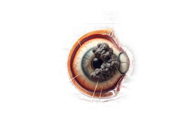
What is papilledema?
Papilledema is a swelling of the optic nerve head or disc caused by high intracranial pressure (ICP). This condition is significant in ophthalmology and neurology because it indicates elevated intracranial pressure, which can have a variety of underlying causes. Papilledema does not affect vision immediately, but if left untreated, it can cause severe visual impairment or blindness. Understanding the pathophysiology, causes, symptoms, and implications of papilledema is critical for early diagnosis and treatment.
Pathophysiology
The optic nerve transports visual information from the retina to the brain. The optic disc, which connects the optic nerve to the eye, is a critical structure. When intracranial pressure rises, it can travel to the optic nerve sheath, causing congestion and swelling of the optic disc. The pressure prevents axoplasmic flow within the optic nerve fibers, resulting in the accumulation of axoplasmic material and subsequent disc swelling.
Several mechanisms can cause increased intracranial pressure. These include increased cerebrospinal fluid (CSF) volume, intracranial mass lesions (e.g., tumors or abscesses), cerebral edema, and obstructive hydrocephalus. When CSF pressure rises, it disrupts the pressure gradient between the intracranial cavity and the optic nerve sheath, allowing fluid to leak into the optic disc, resulting in papilledema.
Causes
Papilledema is not a disease in and of itself, but rather the manifestation of an underlying condition that causes increased intracranial pressure. Several conditions can result in papilledema, including:
Intracranial Mass Lesions: Tumors, abscesses, or hematomas within the cranial cavity can raise intracranial pressure by taking up space and compressing surrounding structures. Common causes of papilledema include both malignant and benign brain tumors.
Cerebral Edema: Trauma, infection, or inflammatory processes can cause swelling of brain tissue, which can raise intracranial pressure. Meningitis, encephalitis, and traumatic brain injury are all examples of conditions in which cerebral edema can result in papilledema.
Hydrocephalus: This condition is characterized by the accumulation of cerebrospinal fluid within the brain’s ventricles, which causes increased intracranial pressure. Hydrocephalus can be congenital or acquired as a result of infection, hemorrhage, or obstruction of CSF flow pathways.
Idiopathic Intracranial Hypertension (IIH): Also known as pseudotumor cerebri, IIH is a condition characterized by elevated intracranial pressure with no apparent cause. It primarily affects overweight women of childbearing age and, if not treated properly, can progress to chronic papilledema.
Vascular Disorders: Conditions that impair cerebral blood flow, such as dural venous sinus thrombosis or arteriovenous malformations, can cause an increase in intracranial pressure and papilledema.
Systemic Conditions: Systemic diseases like severe hypertension, renal failure, and connective tissue disorders can all contribute to the development of papilledema.
Symptoms
Papilledema may not cause immediate symptoms, but the underlying cause of increased intracranial pressure and its effects on the brain and optic nerves can result in a variety of symptoms. Common symptoms associated with papilledema are:
Headache: One of the most common signs of high intracranial pressure is a headache. The headache is frequently described as diffuse and persistent, worsening in the morning or following activities that raise intracranial pressure, such as coughing or straining.
Visual Disturbances: Patients with papilledema may experience transient visual obscurations, or brief periods of vision loss or dimming. These episodes can last a few seconds and are usually triggered by changes in posture. In advanced stages, sustained vision loss or peripheral visual field defects may occur.
Nausea and Vomiting: Elevated intracranial pressure can activate the vomiting center in the brain, causing nausea and vomiting. This is more prevalent in acute or severe cases of intracranial hypertension.
Diplopia: Double vision can occur when increased intracranial pressure affects the cranial nerves that control eye movements, particularly the sixth cranial nerve (abducens nerve).
Tinnitus: Some patients experience whooshing or pulsating noises in their ears, known as pulsatile tinnitus, that correspond to their heartbeat. This symptom is frequently associated with increased intracranial pressure.
Cognitive and Behavioral Changes: Patients with chronic or severe intracranial pressure may experience changes in cognitive function, including confusion, irritability, or lethargy.
Clinical Findings
During examination, several findings can indicate the presence of papilledema:
Optic Disc Swelling: Swelling of the optic disc, visible during a fundoscopic examination, is a defining feature of papilledema. The optic disc appears elevated, with blurred margins and enlarged blood vessels. In severe cases, hemorrhages, cotton-wool spots, and exudates may appear.
Venous Congestion: The retinal veins may appear dilated and tortuous as a result of the increased pressure on venous outflow.
Paton’s Lines: These are concentric folds of the retina that form around the optic disc in some cases of chronic papilledema. They are the result of the retinal layers stretching and folding as a result of prolonged disc swelling.
Risk Factors
Certain factors can increase the risk of developing papilledema. This includes:
Obesity is an important risk factor for idiopathic intracranial hypertension.
- Female Gender: Women, particularly those of childbearing age, are more susceptible to IIH.
- Medications: Certain medications, including tetracyclines, retinoids, and growth hormone, have been linked to elevated intracranial pressure and papilledema.
- Genetics: Some genetic conditions and familial predispositions can raise the risk of intracranial hypertension and papilledema.
Complications
If left untreated, papilledema can cause serious complications, primarily due to ongoing damage to the optic nerve and the effects of the underlying cause, increased intracranial pressure. The complications include:
Permanent Vision Loss: Chronic papilledema can irreversibly damage the optic nerve fibers, resulting in permanent visual impairment or blindness. Early intervention is critical to preventing this outcome.
Brain Herniation: Severe intracranial pressure can cause brain herniation, which occurs when brain tissue is displaced due to the pressure, potentially compressing vital brain structures and posing a life-threatening risk.
Neurological Deficits: The underlying cause of papilledema, such as a brain tumor or cerebral edema, can result in a range of neurological deficits, including motor weakness, sensory disturbances, and altered consciousness.
Psychological and Social Impact
Living with papilledema and the conditions that cause it can have serious psychological and social consequences for patients. Fear of vision loss, as well as uncertainty about the underlying cause, can cause significant anxiety and stress. Furthermore, symptoms of increased intracranial pressure, such as headaches and visual disturbances, can have an impact on daily activities and overall quality of life.
Patients may struggle to maintain independence, particularly if their vision is impaired. The need for regular medical appointments, diagnostic tests, and potential treatments can also be taxing. Support from healthcare professionals, family, and support groups is critical for managing the psychological and social aspects of living with papilledema.
Prevention and Awareness
While papilledema is not preventable, being aware of the conditions that cause increased intracranial pressure and intervening early can help to reduce the risk. Regular medical check-ups, especially for people with known risk factors like obesity or systemic conditions, can help with early detection and treatment of underlying causes.
Public awareness campaigns and education about the symptoms and risks of increased intracranial pressure can encourage people to seek medical attention right away if they experience symptoms like persistent headaches, visual disturbances, or other similar signs.
Diagnostic methods
Diagnosing papilledema necessitates a thorough medical history, physical examination, and a battery of diagnostic tests to determine the underlying cause and extent of optic disc swelling.
Fundoscopy
Fundoscopy, or ophthalmoscopy, is an important diagnostic tool for detecting papilledema. An eye care professional examines the optic disc and retina using an ophthalmoscope. The presence of optic disc swelling, blurred disc margins, and venous congestion are all signs of papilledema. Fundoscopy is required for initial diagnosis and monitoring of disease progression.
Optical Coherence Tomography(OCT)
OCT is an advanced imaging technique that produces high-resolution cross-sectional images of the retina and optic nerve head. It is especially useful for determining the degree of swelling in the optic disc and detecting subtle changes over time. OCT can assess retinal nerve fiber layer thickness, which can aid in distinguishing papilledema from other conditions that cause optic disc swelling, such as optic neuritis or anterior ischemic optic neuropathy.
Visual Field Testing
Visual field testing, also known as perimetry, evaluates the patient’s peripheral vision. This test requires the patient to look into a dome-shaped instrument and respond to visual stimuli presented at various points in their field of view. Papilledema can cause specific visual field defects, such as an enlarged blind spot or peripheral constriction. Visual field testing is useful for determining the functional impact of papilledema and tracking changes over time.
Papilledema Therapy
The treatment of papilledema aims to address the underlying cause of increased intracranial pressure. Timely and effective management is critical to avoiding permanent optic nerve damage and subsequent vision loss. Here are the main approaches to treating papilledema:
Medical Treatment
Diuretics: Medications like acetazolamide or furosemide are commonly used to lower intracranial pressure by decreasing cerebrospinal fluid production. Acetazolamide, a carbonic anhydrase inhibitor, is especially effective for idiopathic intracranial hypertension (IIH).
Corticosteroids: Corticosteroids can be used to reduce inflammation and swelling that is causing increased intracranial pressure. These are frequently prescribed in cases of meningitis or autoimmune diseases affecting the central nervous system.
Antibiotics or Antivirals: If papilledema is caused by an infection, such as meningitis or encephalitis, appropriate antibiotic or antiviral treatment is required to control the infection and, as a result, lower intracranial pressure.
Surgical Treatment
Shunt Surgery: In conditions such as hydrocephalus, where cerebrospinal fluid accumulates, shunt surgery may be necessary. A shunt is a surgically implanted tube that diverts excess fluid from the brain and absorbs it in another part of the body, such as the abdominal cavity.
Optic Nerve Sheath Fenestration: This surgical procedure involves opening a small window in the sheath surrounding the optic nerve to allow excess cerebrospinal fluid to escape, thereby relieving pressure on the nerve. This procedure is particularly useful for severe or progressive papilledema that does not respond to medical treatment.
Venous Sinus Stenting: When venous sinus thrombosis or stenosis causes impaired cerebrospinal fluid drainage, stenting the affected venous sinus can help restore normal drainage and lower intracranial pressure.
Lifestyle Modifications
Weight Management: For patients with idiopathic intracranial hypertension, losing weight can significantly lower intracranial pressure and relieve symptoms. It is common to recommend a structured weight loss program under medical supervision.
Monitoring and Regular Check-Ups: Routine ophthalmologic and neurologic examinations are required to track the progression of papilledema and the efficacy of treatment. Visual field testing and imaging studies are common components of follow-up care.
Addressing the Underlying Conditions
It is critical to manage systemic conditions that cause increased intracranial pressure, such as hypertension, kidney disease, and autoimmune disorders. Effective management of these conditions can reduce the risk of recurring papilledema.
Frequently Asked Questions Regarding Papilledema
What causes Papilledema?
Papilledema is caused by elevated intracranial pressure, which can be caused by a variety of conditions such as brain tumors, hydrocephalus, idiopathic intracranial hypertension, meningitis, and cerebral edema. The elevated pressure causes the optic nerve to swell.
How is Papilledema Diagnosed?
Papilledema is diagnosed after a thorough eye exam that includes fundoscopy, optical coherence tomography (OCT), and visual field testing. Neuroimaging techniques, such as MRI or CT scans, are also used to determine the cause of elevated intracranial pressure.
Does papilledema cause blindness?
If left untreated, papilledema can permanently damage the optic nerve fibers, causing irreversible vision loss or blindness. Early diagnosis and treatment are critical for avoiding this outcome.
Is Papilledema Painful?
Papilledema is usually not painful, but the conditions that cause increased intracranial pressure, such as brain tumors or meningitis, can cause severe headaches, nausea, and other symptoms.
What are the treatments for papilledema?
The treatment focuses on addressing the underlying cause of elevated intracranial pressure. This may include medications such as diuretics and corticosteroids, surgical interventions such as shunt surgery or optic nerve sheath fenestration, and lifestyle changes such as weight management in cases of idiopathic intracranial hypertension.
Can lifestyle changes help with papilledema?
Yes, lifestyle changes, particularly weight loss in patients with idiopathic intracranial hypertension, can significantly reduce intracranial pressure and relieve symptoms. Regular monitoring and medical checkups are also required to effectively manage the condition.
Are there any support groups for people who have papilledema?
Individuals with papilledema and related conditions can find help and resources from a variety of support groups and organizations. These groups can provide helpful information, emotional support, and practical advice for managing the condition.
Trusted Resources and Support
Books and Organizations
Books:
- “Intracranial Hypertension: A Patient’s Guide” by Ian Livingstone and Deborah Friedman.
- “Neuro-Ophthalmology Illustrated”* by Valerie Biousse and Nancy J. Newman.
Organizations:
- The American Academy of Ophthalmology (AAO): Offers comprehensive information on eye health, including papilledema.
- Intracranial Hypertension Research Foundation (IHRF): Provides resources and support to patients with idiopathic intracranial hypertension.
- Brain Tumor Foundation: Offers assistance and resources to people who have brain tumors, which are a common cause of increased intracranial pressure.
Financial Aid Options
Managing and treating papilledema can be financially challenging. There are several financial aid options available to help patients manage their costs:
Insurance Coverage: The majority of health insurance plans cover diagnostic tests and treatments for papilledema. It is critical to check with your insurance provider for specific coverage details.
Patient Assistance Programs: Pharmaceutical companies frequently offer patient assistance programs that provide medications at a reduced cost or for free to qualifying patients.
Non-Profit Organizations: The HealthWell Foundation and the Patient Advocate Foundation provide financial assistance to patients for medical expenses, including treatments for conditions such as papilledema.
Government Programs: Medicaid and Medicare provide coverage to low-income individuals and those over 65, respectively. These programs can assist with the cost of medical care and treatment for papilledema.


