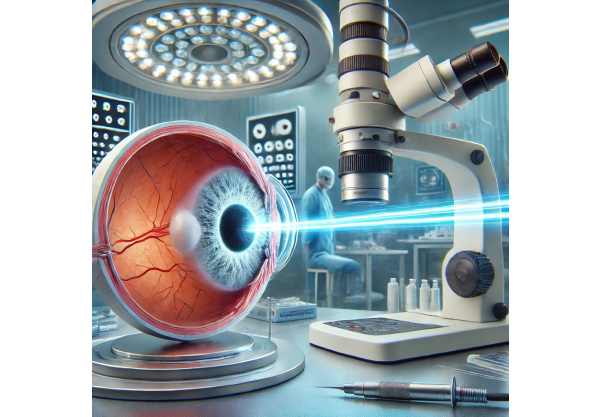
What is a Retinal Astrocytic Hamartoma?
Retinal astrocytic hamartoma is a rare, benign ocular tumor that develops from the retina’s supporting glial cells (astrocytes). These tumors are commonly associated with genetic conditions such as tuberous sclerosis complex (TSC) and neurofibromatosis type 1. Although they can occur infrequently, the vast majority of cases are associated with these genetic disorders. Retinal astrocytic hamartomas appear as yellowish or white elevated lesions on the retina and are frequently discovered during routine eye exams.
Most retinal astrocytic hamartomas are asymptomatic and do not impair vision. In some cases, however, they can cause visual disturbances if they grow significantly or if complications such as retinal detachment or macular edema develop. Retinal astrocytic hamartomas are typically diagnosed using ophthalmoscopy, which allows for the observation of their distinct appearance. Additional imaging techniques, such as optical coherence tomography (OCT) and fluorescein angiography, can help confirm the diagnosis and determine the size of the tumor.
Understanding the nature and behavior of retinal astrocytic hamartomas is critical for effectively managing the condition, especially in patients with underlying genetic disorders. Early detection and monitoring are critical for preventing potential complications and providing appropriate treatment as needed.
Standard Management and Treatment of Retinal Astrocytic Hamartoma
The management and treatment of retinal astrocytic hamartoma is primarily concerned with monitoring the condition and addressing any complications that may arise. Because these tumors are typically benign and slow-growing, a conservative approach is frequently used, particularly in asymptomatic cases.
Monitoring and Observation
Most patients with retinal astrocytic hamartoma require regular monitoring. This includes:
- Regular Eye Examinations: Follow-up visits with an ophthalmologist are required to monitor the size and appearance of the hamartoma. During these visits, a thorough eye examination, including visual acuity tests and ophthalmoscopy, is conducted.
- Imaging Studies: Advanced imaging techniques such as optical coherence tomography (OCT) and fundus photography are used to document the tumor’s characteristics and identify changes over time. OCT provides detailed cross-sectional images of the retina, which aids in the detection of complications such as macular edema and retinal detachment.
Treatment for Complications
Specific treatments are required to address vision-related complications caused by retinal astrocytic hamartomas.
- Macular Edema: If the hamartoma causes macular edema (central retina swelling), intravitreal injections of anti-VEGF (vascular endothelial growth factor) agents or corticosteroids may be used to reduce swelling and improve vision.
- Retinal Detachment: Retinal detachment is a serious complication that requires immediate surgical treatment. Pneumatic retinopexy, scleral buckle surgery, and vitrectomy are all options for reattaching the retina and preserving vision.
- Laser Photocoagulation: In some cases, laser treatment may be used to seal leaking blood vessels or to treat retinal damage caused by a hamartoma. This can help prevent further complications and keep the retina stable.
Genetic Counseling and Systems Management
Given the link between retinal astrocytic hamartomas and genetic disorders such as tuberous sclerosis complex (TSC) and neurofibromatosis type 1 (NF1), a comprehensive management plan should address the underlying systemic condition:
- Genetic Counseling: Patients with retinal astrocytic hamartoma should receive genetic counseling to determine the risk of other genetic disorders. Family members may also require testing to see if they have the same genetic mutations.
- Systemic Treatment: Managing the underlying genetic disorder is critical to preventing or treating other systemic manifestations. This may entail the use of medications, regular monitoring, and collaboration with geneticists, neurologists, and other relevant experts.
Latest Innovations in Retinal Astrocytic Hamartoma
Recent advances in medical research and technology have resulted in the development of novel therapies and techniques for treating retinal astrocytic hamartomas. These cutting-edge innovations seek to improve diagnostic accuracy, treatment outcomes, and patient support for this condition.
Advanced Diagnostic Techniques
Advanced diagnostic tools have greatly improved the detection and monitoring of retinal astrocytic hamartomas.
- Enhanced Imaging Modalities: Modern imaging techniques, such as optical coherence tomography angiography (OCTA), allow for detailed views of the retinal vasculature without the use of dye injections. OCTA can detect subtle changes in blood flow and vessel structure, allowing for the early detection of hamartoma-related complications.
- Adaptive Optics Imaging: Adaptive optics technology enables high-resolution imaging of the retinal microstructure, including detailed views of individual cells and tissue layers. This can aid in the early detection of tumor growth and the evaluation of treatment efficacy.
Pharmaceutical Advances
Innovative pharmacological treatments provide new hope for managing complications associated with retinal astrocytic hamartomas.
- Targeted Therapies: mTOR inhibitors (e.g., sirolimus and everolimus) have shown promise in treating various forms of tuberous sclerosis complex (TSC). These drugs inhibit the mTOR pathway, which is frequently dysregulated in TSC, reducing the size and activity of hamartomas. Clinical trials are currently underway to determine their efficacy for retinal astrocytic hamartomas.
- Anti-VEGF Therapy: Anti-VEGF agents, including ranibizumab and aflibercept, have been used to treat macular edema caused by retinal astrocytic hamartomas. These medications inhibit abnormal blood vessel growth and reduce fluid leakage, which improves visual outcomes in patients.
Surgical Innovations
Advances in surgical techniques have improved the safety and effectiveness of interventions for retinal astrocytic hamartomas.
- Minimally Invasive Vitrectomy: Advances in vitrectomy techniques, such as smaller gauge instruments and better visualization systems, have made this procedure safer and more effective for treating complications like retinal detachment. Minimally invasive approaches shorten recovery times and lower the risk of complications.
- Robotic-Assisted Surgery: Robotic systems are being developed to aid in precise and delicate retinal procedures. These systems allow surgeons to perform complex maneuvers with greater accuracy, potentially improving outcomes for patients with retinal astrocytic hamartomas.
Gene Therapy & Regenerative Medicine
Gene therapy and regenerative medicine show great promise for the future treatment of retinal astrocytic hamartomas.
- Gene Editing Technologies: Techniques like CRISPR-Cas9 have the potential to correct genetic mutations linked to conditions like tuberous sclerosis complex (TSC). Gene editing, which targets specific genes involved in tumor formation, could provide a long-term solution to preventing the development or progression of retinal astrocytic hamartomas.
- Stem Cell Therapy: Researchers are working to create stem cell-based treatments for retinal diseases. Stem cells have the potential to regenerate damaged retinal tissues and restore normal function, providing hope for patients who have lost significant vision due to hamartoma complications.
Personalized Medical Approaches
The personalized medicine trend is also influencing the management of retinal astrocytic hamartomas, with treatments tailored to individual patient profiles based on genetic, molecular, and clinical data.
- Genetic Profiling: Genetic testing can reveal specific mutations linked to retinal astrocytic hamartomas, guiding personalized treatment strategies. Understanding the genetic basis of the condition enables targeted treatments and preventive measures.
- Biomarker Analysis: Advances in biomarker research allow for the identification of specific molecules associated with tumor growth and activity. Biomarker analysis can aid in monitoring disease progression and treatment response, providing valuable information for personalized care plans.
Future Directions
The future of retinal astrocytic hamartoma treatment appears bright, with ongoing research and technological advancements paving the way for even more effective and minimally invasive approaches. Continued research into advanced diagnostic techniques, novel pharmacological treatments, surgical innovations, and regenerative medicine approaches is likely to yield new breakthroughs. As our understanding of the underlying mechanisms of retinal astrocytic hamartomas advances, targeted treatments that address the underlying causes of the condition will become more feasible, providing hope for long-term improvements in patient outcomes.


