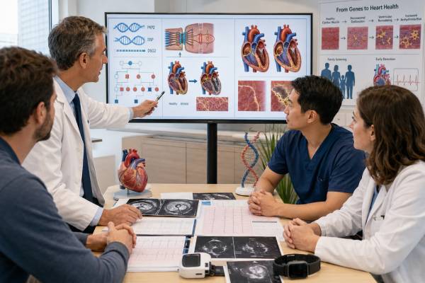
An arrhythmogenic cardiomyopathy genetic test looks for inherited variants that weaken heart-cell connections or alter other pathways involved in ventricular rhythm and muscle structure. PKP2, DSP, and DSG2 are among the most established genes, but a carefully selected panel may also include DSC2, JUP, PLN, TMEM43, DES, FLNC, and other genes supported by the person’s phenotype. A positive genetic result can strengthen a diagnosis and make screening of relatives more precise. It does not replace an electrocardiogram, rhythm monitor, echocardiogram, cardiac MRI, or clinical diagnostic criteria. Disease may involve the right ventricle, left ventricle, or both, and electrical abnormalities can appear before obvious structural change. Exercise is especially important because frequent high-intensity endurance activity can increase penetrance, arrhythmia burden, and disease progression in susceptible people. Results require cautious interpretation: a pathogenic or likely pathogenic variant may be actionable, while a variant of uncertain significance should not be used alone to label relatives, restrict activity, or justify an implantable defibrillator.
- A pathogenic variant can support arrhythmogenic cardiomyopathy and enable targeted family testing, but it does not predict a fixed severity or age of onset.
- PKP2 commonly causes right-dominant disease, while DSP often produces left-dominant or biventricular disease with myocardial injury episodes.
- A normal echocardiogram does not exclude early disease; ECG changes, ventricular ectopy, and cardiac MRI scar may appear first.
- A negative panel does not rule out arrhythmogenic cardiomyopathy because not every causal gene or variant is detectable.
- High-intensity endurance exercise can worsen disease expression in many desmosomal variant carriers and should be discussed with a specialist.
- Fainting during exercise, sustained palpitations, chest pain with arrhythmia, or sudden collapse requires urgent cardiac evaluation.
Table of Contents
- What the genetic test examines
- PKP2, DSP, DSG2, and gene-specific patterns
- Who should be tested
- How diagnosis is established beyond genetics
- Interpreting positive, negative, and uncertain results
- Exercise and risk reduction
- Treatment and surveillance
- Family screening and inheritance
What the genetic test examines
Arrhythmogenic cardiomyopathy is an inherited or acquired heart-muscle disorder characterized by ventricular arrhythmias, myocardial scar, and progressive ventricular dysfunction. Older terminology focused on arrhythmogenic right ventricular cardiomyopathy, or ARVC. Modern practice recognizes that some genetic forms predominantly affect the left ventricle or produce biventricular disease.
Many established genes encode desmosomal proteins. Desmosomes act like mechanical rivets that connect heart-muscle cells during repeated contraction. When these structures fail, mechanical stress can promote cell injury, inflammation, fibrotic replacement, and electrical instability.
A clinical panel may examine:
- PKP2, encoding plakophilin-2
- DSP, encoding desmoplakin
- DSG2, encoding desmoglein-2
- DSC2, encoding desmocollin-2
- JUP, encoding plakoglobin
- TMEM43, associated with a highly penetrant founder form in some populations
- PLN, which affects calcium handling and can cause arrhythmogenic or dilated phenotypes
- DES, FLNC, LMNA, and other genes when the phenotype overlaps with left-dominant or dilated cardiomyopathy
Panel design matters. Genes with weak or disputed evidence can create uncertain findings without improving diagnosis. The laboratory should explain gene-disease validity and the variant types it can detect, including single-nucleotide changes, small insertions or deletions, and copy-number variants.
Genetic testing works best after a cardiologist has defined the phenotype. A broad cardiovascular genetic panel may be appropriate when the findings overlap with dilated cardiomyopathy, myocarditis, conduction disease, or another inherited arrhythmia syndrome. A focused familial-variant test is preferable for relatives after a causal variant is known.
PKP2, DSP, DSG2, and gene-specific patterns
Gene names do not provide a perfect forecast, but they can explain why patients with the same broad diagnosis look different.
PKP2
PKP2 is the most frequently identified gene in classic desmosomal ARVC in many cohorts. Disease often begins with ventricular ectopy or ventricular tachycardia arising from the right ventricle. Structural right-ventricular enlargement or dysfunction may develop later. Penetrance is incomplete, and some carriers remain clinically unaffected for many years.
Exercise exposure strongly modifies PKP2-related disease. Repeated endurance activity can increase ventricular arrhythmias and accelerate structural progression. The effect is not identical in every carrier, but it is important enough to shape counseling.
DSP
DSP cardiomyopathy frequently affects the left ventricle and may resemble myocarditis. Patients can have episodes of chest pain and troponin elevation, sometimes after infection or exercise, followed by development of fibrosis visible as late gadolinium enhancement on cardiac MRI. Ventricular arrhythmias can occur even when ejection fraction is only mildly reduced.
The MRI scar pattern is often subepicardial and may form a circumferential or “ring-like” distribution. Skin or hair findings can occur with some DSP variants, especially in recessive syndromes, but many dominant cardiac cases have no obvious extracardiac signs.
DSG2
DSG2 variants can cause classic right-dominant, biventricular, or occasionally left-dominant disease. Expression varies among families. Some individuals develop prominent ventricular arrhythmias before heart failure, while others present with structural disease.
More than one variant
A person may carry pathogenic variants in more than one arrhythmogenic cardiomyopathy gene. Multiple variants can be associated with earlier or more severe disease, but laboratory classification must be rigorous. Two uncertain variants do not equal one confirmed diagnosis.
The same gene can produce different phenotypes. Conversely, similar imaging and arrhythmias can result from sarcoidosis, prior myocarditis, athlete’s heart, congenital abnormalities, or other genetic cardiomyopathies. Gene-specific clues guide evaluation but do not replace differential diagnosis.
Who should be tested
Testing is appropriate for many people with a definite or strongly suspected arrhythmogenic cardiomyopathy because the result may clarify the diagnosis and guide relatives. The most informative person to test is usually the family member with the clearest clinical disease.
Features that support testing include:
- Ventricular tachycardia with a morphology suggesting right- or left-ventricular origin
- Frequent premature ventricular complexes without another explanation
- T-wave inversion in characteristic precordial or inferolateral leads
- Epsilon waves or late potentials, though these findings require expert interpretation
- Right- or left-ventricular regional wall-motion abnormalities
- Cardiac MRI fibrosis in a pattern suspicious for arrhythmogenic cardiomyopathy
- Unexplained right-ventricular dilation or dysfunction
- Recurrent myocarditis-like episodes, particularly with persistent scar
- A family history of cardiomyopathy, ventricular arrhythmia, sudden death, or heart transplant
- Sudden unexplained death in a young relative
Testing can also be considered after autopsy-proven arrhythmogenic cardiomyopathy or sudden death with a compatible cardiac phenotype. Postmortem testing should use stored blood or tissue and be coordinated with evaluation of first-degree relatives.
Testing an unaffected person before an affected relative is generally less useful. A negative result cannot rule out familial risk if the causal variant has not been identified, and a VUS may create confusion.
Athletes with isolated right-ventricular changes require particular care because endurance training itself can enlarge the right ventricle and produce ECG changes. Diagnosis should be made by a center experienced in sports cardiology and inherited cardiomyopathy.
How diagnosis is established beyond genetics
Arrhythmogenic cardiomyopathy is a clinicogenetic diagnosis. A pathogenic variant can add major evidence, but most patients still need a combination of electrical, structural, tissue, rhythm, and family findings.
Evaluation often includes:
- Resting 12-lead ECG
- Signal-averaged ECG in selected centers
- Ambulatory rhythm monitoring for 24 hours or longer
- Exercise testing to assess ventricular ectopy and arrhythmia
- Echocardiography with right- and left-ventricular measurements
- Cardiac MRI for volumes, function, regional motion, and scar
- Review of previous myocarditis episodes and troponin results
- Coronary evaluation when ischemic disease is possible
- Endomyocardial biopsy in selected cases where inflammation, sarcoidosis, or another diagnosis must be resolved
The 2010 Task Force Criteria remain widely used for classic ARVC. Newer criteria, including the Padua framework, better account for left-dominant and biventricular forms. The 2023 European cardiomyopathy guideline organizes diagnosis around phenotype and cause rather than one rigid label.
Electrical disease can precede imaging changes. A genotype-positive relative with a normal MRI may still develop ventricular ectopy or ECG abnormalities later. Conversely, isolated nonspecific ectopy is common in the general population and does not establish disease.
Cardiac MRI interpretation requires expertise. Fat in the right ventricle is not diagnostic by itself, and overcalling normal variants can produce false diagnoses. Scar location, wall motion, chamber size, ventricular function, and the full clinical context matter.
A biopsy is not routinely required. Sampling error is common because disease is patchy, and biopsy carries procedural risk. It is most useful when another treatable inflammatory or infiltrative disorder is strongly considered.
Interpreting positive, negative, and uncertain results
| Result | Meaning | Usual action |
|---|---|---|
| Pathogenic or likely pathogenic | Supports a molecular cause when the gene fits the phenotype | Integrate with clinical criteria and offer targeted testing to relatives |
| Negative | No reportable variant was detected | Continue clinical diagnosis and family screening when suspicion remains |
| Variant of uncertain significance | Evidence is insufficient to determine causality | Do not use alone for predictive testing, activity restriction, or device decisions |
| Benign or likely benign | The variant is not considered disease-causing | Do not use it to explain the phenotype or test relatives |
A positive result does not mean that disease is inevitable in every carrier. Penetrance is age-dependent and influenced by exercise, sex, other genes, and possibly inflammatory triggers. It also does not predict a specific arrhythmia or guarantee that an implantable cardioverter-defibrillator will be needed.
A negative test does not rule out the diagnosis. The causal variant may be in a gene not yet established, a region not covered by the assay, or a structural or regulatory change that routine sequencing misses. Some cases may be predominantly exercise-induced or nonfamilial.
A VUS should remain uncertain. It may be tempting to call a rare desmosomal variant causative, especially when the phenotype is concerning, but rarity alone is not enough. Segregation in affected relatives, functional evidence, population frequency, and prior cases all matter. The laboratory should periodically reinterpret uncertain findings.
When the phenotype and genotype conflict, re-examine both. A pathogenic PKP2 variant in a person with isolated left-ventricular hypertrophy may be incidental or incompletely explanatory. A classic DSP scar pattern with a negative panel may still represent genetic disease.
Exercise and risk reduction
Exercise is a disease modifier in arrhythmogenic cardiomyopathy, not merely a trigger for symptoms. High-volume endurance training increases mechanical stress on vulnerable cell connections and is associated with greater penetrance, ventricular arrhythmias, and progression in many desmosomal variant carriers.
People with clinical arrhythmogenic cardiomyopathy are generally advised to avoid competitive endurance sports and frequent high-intensity exercise. Examples include long-distance running, intense cycling, rowing, and high-volume interval training. The exact recommendation should account for genotype, phenotype, arrhythmia history, ventricular function, and personal goals.
Complete inactivity is not usually the objective. Light-to-moderate recreational activity may be reasonable for many stable patients after specialist evaluation. A personalized exercise prescription can specify intensity, duration, warning symptoms, and monitoring.
For genotype-positive relatives without clinical disease, recommendations are more nuanced. Evidence suggests that high-intensity endurance exposure can promote disease expression, so many experts advise avoiding competitive endurance exercise. Lower-intensity activity may be acceptable through shared decision-making.
Patients should stop activity and seek assessment for exertional fainting, sustained rapid palpitations, chest pain, or an unexpected drop in exercise tolerance. Wearable devices can record heart rate or rhythm but cannot clear a person for sports or exclude dangerous ventricular arrhythmia.
Other risk-reduction measures include avoiding stimulant drugs, correcting electrolyte disturbances, treating sleep apnea, and discussing fever or myocarditis-like symptoms promptly. Family members should know cardiopulmonary resuscitation and how to use an automated external defibrillator when access and training are available.
Treatment and surveillance
Treatment aims to prevent sudden death, reduce arrhythmia symptoms, and manage ventricular dysfunction. The genetic result contributes context, but treatment decisions depend mainly on clinical risk.
Management may include:
- Beta blockers to reduce adrenergic stimulation
- Antiarrhythmic medicines such as sotalol or amiodarone in selected patients
- Catheter ablation for recurrent ventricular tachycardia
- Implantable cardioverter-defibrillator placement for secondary prevention or selected high-risk primary prevention
- Standard heart-failure therapy for ventricular dysfunction
- Advanced heart-failure or transplant evaluation when disease progresses
Ablation can reduce episodes but does not cure the underlying cardiomyopathy. Arrhythmias may arise from new scar regions over time, and epicardial ablation is sometimes needed.
An implantable defibrillator is strongly considered after cardiac arrest or sustained hemodynamically significant ventricular tachycardia. Primary-prevention decisions examine syncope, ventricular function, nonsustained ventricular tachycardia, arrhythmia burden, genotype, sex, and other risk markers. A pathogenic variant alone is usually insufficient.
Surveillance intervals depend on disease stage. Clinical patients may need regular ECG, rhythm monitoring, imaging, and exercise review. Genotype-positive relatives with normal studies need periodic reevaluation because penetrance increases with age.
DSP carriers may require attention to chest-pain episodes and troponin elevation. These episodes can be mistaken for viral myocarditis. Recurrent injury and MRI scar may affect arrhythmic risk even when ejection fraction is preserved.
Pregnancy is often possible but requires preconception review of ventricular function, arrhythmias, medicines, and device status. Hemodynamic and hormonal changes may increase symptoms in some patients, and postpartum follow-up is important.
Family screening and inheritance
Most pathogenic PKP2, DSP, and DSG2 variants causing adult arrhythmogenic cardiomyopathy are inherited in an autosomal dominant pattern. Each child of a carrier generally has a 50% chance of inheriting the variant. Penetrance is incomplete, so a positive relative may remain clinically normal for years.
Once a pathogenic familial variant is known, targeted testing can divide relatives into two groups. Carriers enter ongoing cardiac surveillance and receive exercise counseling. Noncarriers can usually stop disease-specific serial screening, provided the family has one well-established cause and the noncarrier has no independent clinical findings.
When the proband’s test is negative or shows only a VUS, first-degree relatives still need clinical screening. A typical evaluation includes history, ECG, ambulatory monitoring, echocardiography, and sometimes cardiac MRI. The starting age and interval depend on family onset and current guidelines.
Children may be tested when the result will change activity advice and surveillance. Counseling should avoid presenting a positive result as proof that the child is ill. It identifies increased susceptibility and a need for follow-up.
Some rare arrhythmogenic syndromes are recessive and include skin or hair findings, such as Naxos disease or Carvajal syndrome. When recessive inheritance is suspected, siblings may have a 25% risk and parents are usually carriers. The laboratory and genetic counselor should clarify the specific pattern.
Families should keep the complete report with the exact variant. A relative cannot receive accurate targeted testing from a gene name alone. Reclassification updates should be shared across the family because one change can affect many people.
Using gene-specific clues without replacing clinical risk assessment
Modern arrhythmogenic cardiomyopathy care increasingly recognizes that the same broad label can describe different disease pathways. PKP2-related disease often begins with electrical abnormalities and right-ventricular involvement. Ventricular ectopy or tachycardia may appear before major chamber dilation, so a normal echocardiogram does not end surveillance in a carrier with symptoms or an abnormal ECG. Cardiac MRI, ambulatory monitoring, and exercise history can reveal early disease that a resting study misses.
DSP-related cardiomyopathy often has prominent left-ventricular or biventricular fibrosis. Some carriers experience episodes of chest pain and troponin elevation that resemble viral myocarditis. These “hot phases” can precede ventricular dysfunction and may leave a characteristic subepicardial scar pattern on cardiac MRI. A pathogenic DSP result does not prove that every episode of chest pain is genetic inflammation—coronary disease, infection, pulmonary embolism, and other urgent causes still need evaluation—but it changes the threshold for specialist imaging and longitudinal follow-up.
DSG2-related disease may involve the right ventricle, both ventricles, or a broader dilated phenotype. Because imaging patterns overlap with myocarditis, sarcoidosis, athletic remodeling, and other cardiomyopathies, the variant must be classified rigorously. A rare DSG2 missense change found on a broad panel is not automatically causal. Population frequency, segregation, functional evidence, and the patient’s phenotype determine whether it can explain disease.
Genotype also informs exercise counseling, but no gene produces a universally safe workload. High-intensity endurance exercise can increase penetrance and arrhythmic events in susceptible desmosomal-variant carriers. The relevant “dose” includes intensity, duration, frequency, years of exposure, competition, and recovery. Recommendations should be individualized around symptoms, ventricular function, scar, arrhythmia burden, prior syncope, family history, and personal goals. Light or moderate activity may still provide important physical and psychological benefits when a cardiomyopathy team has assessed risk.
An implantable cardioverter-defibrillator is not prescribed because a pathogenic variant is present. ICD decisions integrate prior cardiac arrest or sustained ventricular tachycardia, unexplained arrhythmic syncope, ventricular function, scar burden, nonsustained ventricular tachycardia, sex, age, gene-associated evidence, and competing risks. The device can terminate malignant rhythms but may cause inappropriate shocks, infection, lead failure, and activity restrictions. Shared decision-making is essential, particularly for an asymptomatic carrier.
Catheter ablation can reduce recurrent ventricular tachycardia and ICD therapies, yet arrhythmogenic cardiomyopathy is progressive and arrhythmias may arise from epicardial as well as endocardial substrate. Successful ablation does not remove the inherited disease, permit unrestricted endurance exercise, or eliminate the need for surveillance. Heart-failure therapy follows ventricular phenotype, and advanced biventricular failure may eventually require transplant assessment.
The practical use of genotype is therefore directional rather than deterministic. It helps the team choose what to watch, recognize atypical presentations, counsel relatives, and interpret exercise exposure. Current symptoms, rhythm recordings, imaging, and disease trajectory remain the basis for immediate treatment decisions.
Surveillance should also be repeated after a meaningful clinical change. New palpitations, fainting, reduced exercise tolerance, chest pain with troponin elevation, pregnancy-related symptoms, or an increase in ventricular ectopy can justify earlier reassessment rather than waiting for the next routine visit. The interval that was appropriate for an unaffected carrier may no longer fit once electrical or structural findings emerge.
References
- 2023 ESC Guidelines for the management of cardiomyopathies 2023 (Guideline)
- JCS/JCC/JSPCCS 2024 Guideline on Genetic Testing and Counseling in Cardiovascular Disease 2025 (Guideline)
- Arrhythmogenic Cardiomyopathy: Current Updates and Future Challenges 2024 (Review)
- Exercise Prescription in Arrhythmogenic Cardiomyopathy: Finding the Right Balance Between Risks and Benefits 2025 (Review)
- Genetics in arrhythmogenic cardiomyopathies: where are we now and where are we going? 2025 (Review)
- Arrhythmogenic cardiomyopathy diagnosis and management: a practical review 2025 (Review)
Disclaimer
This article is educational and does not replace evaluation by a cardiologist, electrophysiologist, or cardiovascular genetics professional. Do not change exercise, medication, or device plans based only on a genetic report, especially a VUS. Exertional fainting, sustained rapid palpitations, severe chest pain, or sudden collapse requires urgent medical care.