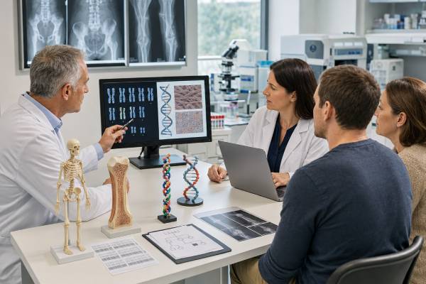
An osteogenesis imperfecta genetic test most often examines COL1A1 and COL1A2, the genes that encode type I collagen. A pathogenic variant in either gene can weaken bone and other collagen-rich tissues, causing a spectrum from mild fracture susceptibility to severe skeletal deformity or perinatal disease. Testing can confirm a clinical diagnosis, distinguish osteogenesis imperfecta from other causes of fragile bones, clarify recurrence risk, and identify relatives who may be affected. Most COL1A1- and COL1A2-related cases follow autosomal dominant inheritance, so one disease-causing variant can be enough to cause the condition. However, a negative two-gene test does not rule out osteogenesis imperfecta because many other genes can produce similar findings. Results should be interpreted with fracture history, X-rays, growth, dental findings, hearing, family history, and laboratory methods. A variant of uncertain significance does not confirm the diagnosis. Genetic counseling is especially helpful when testing a child, evaluating a pregnancy, or interpreting an apparently new variant in a family.
- A positive COL1A1 or COL1A2 result usually identifies a dominant form of osteogenesis imperfecta.
- The test can confirm the cause of bone fragility, but it cannot predict the exact number of future fractures.
- A negative result may lead to a broader bone-fragility panel because other genes also cause osteogenesis imperfecta.
- Blood, saliva, or cheek-swab samples are commonly used, and fasting is not required.
- Parental testing helps distinguish an inherited variant from a de novo change or parental mosaicism.
- Severe pain, suspected fracture, breathing difficulty, new weakness, or symptoms after head or neck injury need prompt medical assessment.
Table of Contents
- What COL1A1 and COL1A2 Testing Can Show
- How Collagen Variants Cause Bone Fragility
- Who May Benefit From Testing
- Test Methods, Samples, and Timing
- How to Read the Result
- Inheritance, Mosaicism, and Family Risk
- Limits of the Test and Next Steps
- Care After a Confirmed Diagnosis
What COL1A1 and COL1A2 Testing Can Show
COL1A1 and COL1A2 provide instructions for two chains that combine to form type I collagen. Type I collagen is a major structural protein in bone, teeth, tendons, ligaments, skin, the white part of the eye, and parts of the inner ear. A clinically significant variant can reduce the amount of normal collagen or alter its structure.
Genetic testing can answer several different questions:
- Is a person’s fracture pattern caused by COL1A1- or COL1A2-related osteogenesis imperfecta?
- Which gene and exact variant are involved?
- Did the variant come from a parent, or did it arise for the first time?
- Are siblings, children, or other relatives at risk?
- Can targeted prenatal or preimplantation testing be designed for the family?
- Does a negative result justify testing additional bone-fragility genes?
The result does not measure current bone strength, replace X-rays, or determine whether every fracture was caused by osteogenesis imperfecta. It also cannot reliably forecast the exact clinical course. Two people with the same variant may have different fracture rates, height, mobility, hearing loss, dental involvement, or need for surgery.
COL1A1 and COL1A2 explain most classic osteogenesis imperfecta, but they do not explain all cases. Other genes affect collagen folding, processing, mineralization, osteoblast function, and bone signaling. A broad genetic panel test may include IFITM5, WNT1, SERPINF1, CRTAP, P3H1, PPIB, FKBP10, SP7, BMP1, TMEM38B, and other genes. The best starting test depends on the clinical pattern and the laboratory’s coverage.
Osteogenesis imperfecta is sometimes called “brittle bone disease,” but that phrase can understate its wider effects. The same collagen defect may contribute to short stature, bone deformity, scoliosis, joint laxity, muscle weakness, easy bruising, blue or gray sclerae, dentinogenesis imperfecta, hearing loss, respiratory limitations, and chronic pain.
How Collagen Variants Cause Bone Fragility
Type I collagen is built from three protein chains: two alpha-1 chains made from COL1A1 and one alpha-2 chain made from COL1A2. These chains wind into a triple helix. The helix depends on a repeating sequence in which glycine, the smallest amino acid, appears at every third position.
Different variant mechanisms tend to produce different patterns, although exceptions are common.
| Mechanism | Effect on collagen | Typical clinical tendency |
|---|---|---|
| Loss-of-function variant, often in COL1A1 | Reduces the amount of otherwise normal type I collagen | Often associated with classic non-deforming OI and blue sclerae |
| Glycine substitution in the triple-helical region | Produces structurally abnormal collagen that can disrupt the helix | May cause moderate, severe, or perinatal disease depending on location and substitution |
| Splice-altering variant | Can remove or alter part of the collagen chain | Severity varies widely |
| In-frame deletion or duplication | Changes the chain while preserving the reading frame | Often structural collagen abnormality with variable severity |
| Exon or whole-gene deletion | May reduce collagen production or disrupt gene function | Depends on the gene, size, and exact effect |
The traditional clinical types remain useful descriptions:
- Classic non-deforming OI with blue sclerae, formerly type I, is often mild. Fractures commonly begin when a child starts walking and falling. Adult height may be near normal, and bone deformity is limited.
- Perinatally lethal OI, formerly type II, causes severe bone undermineralization, fractures, short bowed limbs, a soft skull, and a small chest. Respiratory failure is a major concern.
- Progressively deforming OI, formerly type III, is the most severe form compatible with longer survival. Recurrent fractures, marked short stature, long-bone bowing, scoliosis, and mobility limitations are common.
- Common variable OI with normal sclerae, formerly type IV, ranges from mild to moderately severe and may include dentinogenesis imperfecta.
A genetic result may support one of these patterns, but the clinical examination remains essential. Variant location can provide useful context, yet genotype-phenotype prediction is imperfect. The same broad category may contain people with very different daily function.
COL1A1 variants can also cause overlapping connective-tissue conditions, including certain Ehlers-Danlos phenotypes and Caffey disease. This is why the exact variant—not only the gene name—must be interpreted. A report stating “COL1A1 positive” without the specific change and classification is not enough.
Who May Benefit From Testing
Testing is often considered when a person has repeated fractures from minimal trauma, fractures at unusual ages or sites, low bone density without a clear acquired cause, or characteristic connective-tissue findings. It may also be ordered before birth when ultrasound shows severe skeletal abnormalities.
Children with unexplained fractures
A child may be referred after multiple long-bone fractures, vertebral compression fractures, fractures during routine handling, or a fracture pattern that does not match the reported injury. The evaluation should be careful and unbiased. Osteogenesis imperfecta can resemble non-accidental trauma, but genetic testing cannot replace a complete medical, radiologic, social, and safeguarding assessment.
Findings that may support OI include blue or gray sclerae, dentinogenesis imperfecta, joint hypermobility, short stature, wormian bones in the skull, osteopenia, bowing, hearing loss in relatives, or a family history of fractures. None is required. Blue sclerae can occur in healthy infants, and some people with OI have normal scleral color.
Adults with mild or previously unrecognized disease
Mild OI may not be diagnosed until adulthood. Clues include recurrent fractures since childhood, hearing loss, unusually low bone mass, early tooth wear, family members with similar features, or persistent joint and connective-tissue findings. An adult may seek testing after a child receives a diagnosis.
Prenatal findings
Ultrasound may detect shortened or bowed limbs, reduced skull mineralization, rib fractures, a small chest, or multiple fractures. These findings can result from several skeletal dysplasias. A targeted COL1A1/COL1A2 test may be appropriate when OI is strongly suspected, while a broader fetal skeletal-dysplasia panel or exome sequencing may be better when the pattern is unclear.
Known family variant
When a pathogenic variant has already been identified in a relative, targeted familial-variant testing is usually the most direct approach. Testing the exact family change provides clearer risk information than a general bone-health DNA screen. It also reduces the chance of unrelated uncertain findings.
Testing may be diagnostic, predictive, or prenatal, and those purposes should not be confused. A person with symptoms needs diagnostic evaluation. An unaffected relative may be undergoing predictive genetic testing. A pregnancy at known risk requires a separate consent and counseling process.
Test Methods, Samples, and Timing
Most laboratories use blood, saliva, or a cheek swab. Blood is often preferred when mosaicism is a concern or when other tests are being collected. Saliva may contain a mixture of white blood cells and cheek cells, which can sometimes help detect mosaicism, but sample quality and laboratory validation matter more than sample type alone.
No fasting or medication changes are usually needed. A recent bone fracture, bisphosphonate treatment, calcium intake, or exercise does not change the person’s DNA result. These factors can affect clinical measurements such as bone turnover markers or bone density, but not the inherited sequence.
Sequence analysis
Next-generation sequencing reads COL1A1 and COL1A2 for substitutions and small insertions or deletions. Laboratories generally examine coding exons and nearby splice boundaries. Sanger sequencing may confirm a detected variant or perform targeted family testing.
Deletion and duplication analysis
Sequence testing may miss one or more deleted or duplicated exons. Copy-number methods such as multiplex ligation-dependent probe amplification, quantitative PCR, or validated sequencing-based algorithms can detect these changes. The report should state whether deletion/duplication analysis was included.
Panel, exome, and genome testing
A multigene panel is often the strongest first choice when the phenotype could reflect another hereditary bone disorder. Panels usually offer high coverage of relevant genes and fewer unrelated findings than exome or genome sequencing.
Whole-exome sequencing may be used after a negative panel or in a complex presentation. Genome sequencing can assess coding and noncoding regions more broadly and may identify structural variants that are difficult for exome testing. Neither approach detects every possible mechanism.
Prenatal sampling
Targeted prenatal diagnosis requires fetal DNA from chorionic villus sampling, usually performed around 10 to 13 weeks, or amniocentesis, commonly performed from about 15 weeks onward. Exact timing depends on local practice. These are diagnostic procedures, not screening blood tests. A known familial variant makes interpretation much more direct.
Turnaround time ranges from roughly two to eight weeks for many standard tests. Urgent fetal or neonatal testing may be completed faster. Complex variant analysis, parental studies, or reclassification can extend the timeline.
How to Read the Result
The report should list the gene, transcript, DNA-level change, protein-level change, zygosity, classification, testing method, and limitations. The most important distinction is between a pathogenic or likely pathogenic variant and a variant of uncertain significance.
Pathogenic or likely pathogenic variant
A heterozygous pathogenic or likely pathogenic variant in COL1A1 or COL1A2 can establish the molecular diagnosis when the person’s clinical and radiographic findings fit. “Heterozygous” means the variant is present in one of the two gene copies.
A positive result does not automatically define severity. A variant associated with reduced collagen quantity often suggests a milder pattern, while certain structural variants are associated with more severe disease. However, individual outcomes can differ, and the report should not be used as a precise prognosis calculator.
Negative result
A negative result means no reportable pathogenic variant was found with the methods used. It may reduce the likelihood of COL1A1/COL1A2-related OI, but it does not exclude:
- a variant in another OI gene;
- a copy-number or structural change not assessed;
- a deep intronic or regulatory variant;
- low-level mosaicism;
- poor coverage of a relevant region;
- a newly recognized mechanism; or
- a non-genetic cause of bone fragility.
The report’s technical limitations and the pretest probability determine how reassuring a negative result is.
Variant of uncertain significance
A VUS means the evidence is insufficient to decide whether the change causes disease. It should not be used alone to diagnose OI, label a child as affected, excuse or prove a fracture mechanism, or make irreversible reproductive decisions.
Family studies may help when the variant tracks with disease across several relatives, but segregation is only one form of evidence. A VUS found in a healthy parent does not always prove benignity because expression can be mild or variable. Conversely, a de novo VUS is not automatically pathogenic. The principles described for a variant of uncertain significance are especially important in large skeletal-dysplasia panels.
Benign or likely benign variant
These variants are not considered the cause of OI. They may appear in a technical appendix or not be reported at all. Common polymorphisms do not explain recurrent fractures simply because they occur in a collagen gene.
| Result | Meaning | Usual follow-up |
|---|---|---|
| Pathogenic variant with matching features | Molecular diagnosis supported | OI specialist care, parental testing, family counseling |
| Pathogenic variant in an atypical presentation | May still be causal, but phenotype should be reviewed | Variant-specific review and assessment for allelic disorders |
| Negative COL1A1/COL1A2 test | Classic two-gene OI less likely, not excluded | Check methods and consider broader testing |
| VUS | Unresolved | Clinical correlation, selected family studies, periodic reinterpretation |
Inheritance, Mosaicism, and Family Risk
Most COL1A1- and COL1A2-related OI is autosomal dominant. An affected person has a 50% chance of passing the variant to each child. The probability is the same in every pregnancy and does not depend on the child’s sex.
A family history may be absent because the variant arose de novo in the affected person. De novo means it was not detected in either parent’s tested blood sample. Severe forms are frequently caused by new variants, while mild forms may be inherited from a parent whose features were overlooked.
Parental testing is important even when both parents appear unaffected. A parent may have mild OI, such as a few childhood fractures, subtle blue sclerae, hearing loss, or reduced height. Another possibility is mosaicism, in which the variant is present in only some cells. Blood testing can miss low-level gonadal mosaicism, so a negative parental result usually lowers—but does not reduce to zero—the chance of recurrence in another pregnancy.
The recurrence estimate for an apparently de novo variant is therefore not simply zero. A genetics specialist may discuss a small residual risk from parental gonadal mosaicism and whether testing other tissues could be useful.
When an affected parent has mosaicism, transmission risk may be lower than 50%, but it is difficult to calculate from the percentage found in blood. The level in blood does not necessarily match the level in eggs or sperm.
Reproductive options include natural conception, prenatal diagnosis, in vitro fertilization with preimplantation genetic testing for a monogenic condition, donor gametes, donor embryos, or adoption. Prenatal genetic testing is most reliable when the exact familial variant is known before pregnancy.
Families should receive the full laboratory report, not only a verbal result. Exact variant notation is necessary for testing relatives and designing prenatal assays. A family letter can summarize who may benefit from targeted testing without disclosing more medical detail than needed.
Limits of the Test and Next Steps
Genetic testing is powerful, but it answers a molecular question rather than every clinical question. It cannot determine whether a new pain represents a fracture, whether a child’s injury was accidental, whether bone density will improve with treatment, or whether a person will need surgery.
After a negative or unclear result, the clinician should review:
- whether both COL1A1 and COL1A2 were fully sequenced;
- whether exon-level deletion and duplication analysis was performed;
- whether the phenotype fits another OI gene or skeletal dysplasia;
- whether mosaicism testing needs a higher-depth assay or another tissue;
- whether vitamin D deficiency, celiac disease, endocrine disease, immobility, medication exposure, or another acquired cause contributes to fragility; and
- whether reanalysis or broader sequencing is appropriate.
A negative genetic test should not stop appropriate fracture care or rehabilitation when clinical OI remains likely. Conversely, a positive result does not mean every future symptom is caused by OI. Common medical problems still need ordinary evaluation.
Reanalysis can be valuable because variant databases, gene-disease knowledge, and classification criteria change. A reasonable interval is often one to three years, or sooner if new clinical features appear or another relative receives a more informative result.
Consumer DNA services are not substitutes for clinical testing. They may assay only selected variants, omit copy-number changes, and use methods not validated for rare collagen disorders. Any medically important finding should be confirmed in a certified diagnostic laboratory.
Care After a Confirmed Diagnosis
A confirmed diagnosis supports a coordinated plan rather than a single treatment. Care is usually shared among a metabolic bone specialist, clinical geneticist, orthopedic surgeon, physical and occupational therapists, dentist, audiologist, rehabilitation team, and other specialists according to severity.
Initial assessment may include:
- a detailed fracture and pain history;
- growth, limb alignment, spine, joint, and mobility examination;
- review of skull-base symptoms and neurologic signs;
- dental evaluation for dentinogenesis imperfecta;
- hearing assessment, especially from adolescence onward;
- respiratory review in people with severe chest or spinal deformity;
- vitamin D, calcium, kidney, liver, thyroid, and other laboratory testing when clinically indicated; and
- bone density imaging interpreted with age, body size, growth, and vertebral shape in mind.
Bisphosphonates are commonly used in children and adults with vertebral compression fractures, frequent long-bone fractures, or more severe disease. They can increase bone mineral density and may improve vertebral shape and pain, but they do not correct the collagen variant. Choice of drug, dose, interval, duration, and monitoring should be individualized.
Physical activity should build strength and function without unnecessary impact risk. Swimming and adapted exercise can be useful, but complete avoidance of activity can worsen weakness and bone health. Therapists familiar with OI can teach safe transfers, positioning, fall reduction, and return to movement after fractures.
Fractures are generally immobilized for the shortest practical period because prolonged casting can worsen osteopenia and muscle loss. Lightweight casts, early rehabilitation, and intramedullary rodding may be appropriate. Surgery and anesthesia require careful positioning, airway planning, and awareness of fragile bones and possible cervical spine abnormalities.
Dental care should begin early. Dentinogenesis imperfecta can cause translucent gray-brown teeth, rapid wear, fractures, and bite problems. Hearing testing matters because conductive, sensorineural, or mixed loss may develop, often in adolescence or adulthood.
Seek urgent care for suspected fracture with deformity, severe or uncontrolled pain, breathing difficulty, new numbness or weakness, loss of bladder or bowel control, severe headache or neurologic symptoms after trauma, or neck pain after a fall. People with severe OI may need specialized handling during emergency transport and imaging.
The genetic diagnosis can guide relatives and reproductive planning, but long-term health depends on regular clinical monitoring. The most useful care plan combines molecular information with fracture prevention, mobility, pain control, dental and hearing care, orthopedic judgment, and the person’s goals for school, work, family, and independence.
References
- COL1A1- and COL1A2-Related Osteogenesis Imperfecta 2025 (Review)
- Comprehensive Review of Osteogenesis Imperfecta 2025 (Review)
- Osteogenesis imperfecta: shifting paradigms in pathophysiology and care in children 2024 (Review)
- Cardiovascular disease in adults with osteogenesis imperfecta: clinical characteristics, care recommendations, and research priorities 2025 (Position Statement)
- Efficacy and safety of intravenous Zoledronic acid in the treatment of pediatric osteogenesis imperfecta: A systematic review 2024 (Systematic Review)
Disclaimer
This article is for general education and does not diagnose osteogenesis imperfecta or replace care from a geneticist, metabolic bone specialist, or orthopedic team. Genetic results must be interpreted with clinical and radiographic findings, and variant classifications may change. Seek urgent medical care for suspected serious fracture, breathing difficulty, neurologic symptoms, or significant head or neck trauma.