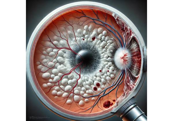
Purtscher’s Retinopathy is a rare but severe retinal vascular disorder that causes sudden, painless vision loss, usually after trauma, acute pancreatitis, or other systemic conditions. This condition, first described by Dr. Otmar Purtscher in 1910, was initially seen in patients who had suffered head trauma. It has long been associated with a variety of non-traumatic systemic conditions, making it an important diagnosis to consider in cases of sudden vision loss.
Pathophysiology
Purtscher’s Retinopathy is primarily associated with microvascular damage to the retina, which results in the occlusion of small retinal arterioles and venules. This vascular injury causes retinal hemorrhages, cotton wool spots (areas of retinal nerve fiber layer infarction), and Purtscher’s flecken, which are distinctive white retinal lesions located around the optic disc and macula.
Purtscher’s Retinopathy is thought to be caused by the embolization of leukocytes, fibrin, or fat, as well as the activation of complement pathways, which results in vascular occlusion. In the event of trauma, such as head injuries or chest compressions, a sudden increase in intravascular pressure can propel air, fat, or other emboli into the retinal circulation. This process causes microvascular occlusion, ischemia, and subsequent retinal infarction.
Purtscher’s Retinopathy has been associated with systemic conditions that cause microvascular damage in non-traumatic cases, including acute pancreatitis, connective tissue disorders (such as systemic lupus erythematosus and scleroderma), and renal failure. The exact mechanism in these cases is unknown, but it is thought that similar embolic or complement-mediated processes are involved.
Epidemiology
Purtscher’s Retinopathy is a rare condition with a low incidence rate. The condition affects both genders equally, though the specific incidence associated with trauma may differ depending on the nature of the injury. The age range of affected individuals varies greatly, as the condition can affect both children and adults, depending on the underlying cause.
Purtscher’s Retinopathy is more common in patients who have sustained significant physical injuries, such as those from car accidents, falls, or crush injuries, due to its association with trauma. However, it is critical to understand that the condition can also occur in patients with no history of trauma, particularly those with acute systemic illnesses.
Clinical Presentation
Purtscher’s Retinopathy is characterized by sudden, painless vision loss that can range from mild to severe. Vision loss is usually bilateral, but it can be asymmetrical, with one eye more severely affected than the other. The severity of the underlying systemic condition and the extent of retinal involvement are frequently associated with the degree of vision loss.
Patients with Purtscher’s Retinopathy may experience a variety of visual disturbances, including:
- Decreased Visual Acuity: Patients frequently experience a sudden loss of visual sharpness, with vision dropping to counting fingers or worse in severe cases.
- Central Scotoma: A central scotoma, or a blind spot in the central vision, is common and associated with macular disease.
- Photopsia: Some patients may report seeing flashes of light or experiencing transient visual phenomena, especially in the early stages of the disease.
- Metamorphopsia: This vision distortion in which straight lines appear wavy can occur if the macula is involved.
Fundoscopic Findings
Purtscher’s Retinopathy has a classic fundoscopic appearance that includes a number of retinal findings that are essential for diagnosis. These findings are typically observed within hours to days after the onset of vision loss, and include:
- Purtscher’s Flecken: These are multiple, white or yellow-white, polygonal retinal lesions that usually appear in the peripapillary region and the macula. They are considered pathognomonic for Purtscher’s Retinopathy and result from precapillary arteriole occlusion, resulting in localized retinal ischemia.
- Cotton Wool Spots: These white, fluffy lesions indicate retinal nerve fiber layer infarction caused by microvascular occlusion. They are most commonly found in the posterior pole, near the optic disc and macula.
- Retinal Hemorrhages: Purtscher’s Retinopathy is characterized by small, flame-shaped hemorrhages, especially in the posterior pole. These hemorrhages occur when capillaries and venules rupture due to increased intravascular pressure or ischemic injury.
- Optic Disc Edema: Swelling of the optic disc may occur, especially in severe cases or those with extensive retinal involvement. Optic disc edema can lead to further visual impairment and is a significant prognostic factor.
- Macular Edema: In some cases, the macula may become edematous, causing additional visual distortion and central vision loss. Macular involvement is a significant predictor of visual prognosis in Purtscher’s Retinopathy.
Systematic Associations
Purtscher’s Retinopathy is not a single ocular condition; it is frequently associated with systemic diseases, particularly those involving microvascular injury or embolization. Some of the most common systemic associations are:
- Trauma: Head trauma, chest trauma, long bone fractures, and crush injuries are some of the most common causes of Purtscher’s Retinopathy. The sudden increase in intravascular pressure caused by these injuries is believed to contribute to retinal vascular occlusion.
- Acute Pancreatitis: Purtscher’s Retinopathy has been associated with acute pancreatitis, particularly in severe cases. The precise mechanism is unknown, but it could involve fat embolism, complement activation, or other inflammatory processes that harm retinal vessels.
- Connective Tissue Disorders: Purtscher’s Retinopathy has been associated with autoimmune diseases such as systemic lupus erythematosus, scleroderma, and dermatomyositis. Systemic inflammation and microvascular injury are present in these conditions, which may predispose patients to retinal vascular occlusion.
- Renal Failure: Patients with chronic or acute renal failure may develop Purtscher’s Retinopathy, possibly as a result of uremia-related endothelial damage or other metabolic disturbances affecting the retinal vasculature.
- Childbirth: Rare cases of Purtscher’s Retinopathy have been reported in women after giving birth, particularly after complicated deliveries with significant blood loss or other systemic complications.
- Vasculitis: Systemic vasculitides, such as polyarteritis nodosa or granulomatosis with polyangiitis, can cause widespread inflammation and vessel occlusion in the retina, including Purtscher’s Retinopathy.
Prognosis
The visual prognosis in Purtscher’s Retinopathy is variable and dependent on a number of factors, including the extent of retinal involvement, the presence of macular involvement, and the underlying systemic condition. In some cases, spontaneous visual recovery can take weeks or months, especially if the macula is unharmed and there is little retinal damage.
However, in more severe cases with significant retinal ischemia, macular involvement, or optic disc edema, the prognosis can be bleak, with permanent vision loss. Early detection and management of the underlying systemic condition are critical for improving outcomes in patients with Purtscher’s Retinopathy.
Differential Diagnosis
Purtscher’s Retinopathy must be distinguished from other conditions that can produce similar fundoscopic appearances due to its unique retinal findings. Some of the key differential diagnoses are:
- Central Retinal Vein Occlusion (CRVO): CRVO can cause retinal hemorrhages and cotton wool spots, but it usually results in more diffuse retinal hemorrhage and optic disc swelling. CRVO is typically associated with underlying vascular risk factors like hypertension or diabetes.
- Diabetic Retinopathy: Patients with advanced diabetic retinopathy may exhibit cotton wool spots, retinal hemorrhages, or macular edema. Diabetic retinopathy, on the other hand, is associated with a history of chronic diabetes and more extensive vascular changes, such as neovascularization.
- Hypertensive Retinopathy: Severe hypertension can cause cotton wool spots, retinal hemorrhages, and optic disc edema, which are similar to Purtscher’s Retinopathy. However, hypertensive retinopathy is frequently associated with other signs of hypertensive damage, such as arteriolar narrowing and arteriovenous nicking.
- Terson Syndrome: Terson syndrome is defined by intraocular hemorrhage and intracranial hemorrhage. Patients may present with sudden vision loss and fundoscopic evidence of retinal or vitreous hemorrhage, but without the distinctive Purtscher’s flecken.
- Retinal Vasculitis: Inflammatory conditions of the retinal vessels, such as Behçet’s disease or sarcoidosis, can result in retinal hemorrhages and cotton wool spots. However, these conditions are often associated with uveitis or vitreous inflammation.
Diagnostic Techniques for Purtscher’s Retinopathy
Purtscher’s Retinopathy is diagnosed through a combination of clinical examination, fundoscopic evaluation, and imaging tests. Given the link to systemic conditions, a comprehensive patient history and systemic workup are also required.
Fundoscopy
Fundoscopy remains the primary method of diagnosing Purtscher’s Retinopathy. A dilated fundus examination allows for direct visualization of the retina, revealing the characteristic features of Purtscher’s flecken, cotton wool spots, retinal hemorrhages, and optic disc edema. These findings, when combined with a history of trauma or an associated systemic condition, strongly support the diagnosis of Purtscher’s Retinopathy.
Optical Coherence Tomography(OCT)
Optical Coherence Tomography (OCT) is a non-invasive imaging technique that generates high-resolution cross-sections of the retina and optic nerve head. OCT is especially useful in the diagnosis and monitoring of Purtscher’s Retinopathy because it allows for detailed visualization of the retinal layers, which can detect subtle changes that are not visible during fundoscopic examination.
In patients with Purtscher’s Retinopathy, OCT typically reveals the following:
- Inner Retinal Thickening: This is caused by ischemia and swelling of the retinal nerve fiber layer, and it is commonly seen around cotton wool spots.
- Disruption of the Outer Retinal Layers: Damage to the photoreceptor layers and retinal pigment epithelium (RPE), particularly in the macula, can lead to visual acuity loss.
- Subretinal Fluid: When macular edema or serous retinal detachment is present, OCT will reveal an accumulation of fluid beneath the retina.
OCT can also help monitor the resolution of retinal edema and guide treatment decisions, particularly when macular involvement is significant.
Fluorescein Angiography(FA)
Fluorescein Angiography (FA) is another useful diagnostic tool for Purtscher’s Retinopathy. FA is the injection of fluorescein dye into the bloodstream, which allows for the visualization of retinal circulation and aids in the detection of retinal ischemia and vascular leakage.
In Purtscher’s Retinopathy, FA usually shows:
- Non-Perfusion Areas: These are zones in which the dye does not fill the retinal capillaries, indicating capillary dropout or occlusion.
- Delayed Arteriovenous Transit Time: This delay indicates vascular occlusion or decreased retinal perfusion, which is consistent with the pathophysiology of Purtscher’s Retinopathy.
- Leakage from Retinal Vessels: In cases of severe retinal involvement, dye leakage from damaged retinal vessels can be seen, contributing to retinal edema.
FA can also help differentiate Purtscher’s Retinopathy from other conditions with similar fundoscopic findings by revealing distinct patterns of vascular involvement.
Indocyanine green angiography (ICGA)
Indocyanine Green Angiography (ICGA) is a technique for evaluating choroidal circulation that can be useful when choroid involvement is suspected. ICGA is less commonly used than FA in the diagnosis of Purtscher’s Retinopathy, but it may provide additional information in complex cases or when choroidal pathology is suspect.
Visual Field Testing
Visual field testing, such as automated perimetry, can help determine the extent of visual field loss in patients with Purtscher’s Retinopathy. This is especially true for patients who present with central scotomata or other field defects. Visual field testing can quantify the disease’s impact on vision and track changes over time, providing useful information for managing the condition.
Laboratory Workup and Imaging
Given the systemic nature of Purtscher’s Retinopathy, a thorough laboratory workup is frequently required to rule out any underlying conditions. This can include:
- Blood Tests: To detect systemic inflammation (e.g., erythrocyte sedimentation rate, C-reactive protein), pancreatic enzymes in suspected pancreatitis, or autoimmune markers in cases of connective tissue disease.
- Imaging Studies: A CT scan or MRI may be required to assess for trauma or other systemic causes. In cases of suspected pancreatitis, abdominal imaging may be used to confirm the diagnosis.
Differential Diagnosis
As part of the diagnostic process, it is critical to distinguish Purtscher’s Retinopathy from other retinal conditions with similar symptoms. This entails a thorough review of clinical findings, imaging studies, and systemic evaluation. Based on the clinical and imaging features of Purtscher’s Retinopathy, it is necessary to consider and rule out conditions such as central retinal vein occlusion, hypertensive retinopathy, and other causes of retinal vasculopathy.
Purtscher’s Retinopathy
Best Practices for Purtscher’s Retinopathy Management
Purtscher’s Retinopathy is difficult to manage because of its rarity and the variety of underlying causes. There are no specific therapies that directly target retinal pathology, so treatment focuses on addressing the underlying systemic condition. Systemic management, ocular management, and supportive care are the three main areas of management.
Systematic Management
Because Purtscher’s Retinopathy is frequently the result of trauma or systemic disease, addressing the underlying cause is crucial. For example:
- Trauma: When Purtscher’s Retinopathy develops as a result of head or chest trauma, the primary goal should be to treat the physical injuries. Depending on the type and severity of the trauma, this could include surgical intervention, fracture stabilization, or intracranial pressure management. Addressing the trauma can sometimes lead to stabilization or improvement in retinal symptoms, but this is not always the case.
- Acute Pancreatitis: When Purtscher’s Retinopathy is coexisting with acute pancreatitis, the treatment focuses on the pancreatitis. This includes supportive care like fluid resuscitation, pain management, and nutritional support. Early and aggressive pancreatitis treatment can help reduce systemic inflammation, potentially preventing additional retinal damage.
- Connective Tissue Disorders: In cases involving autoimmune or connective tissue diseases, immunosuppressive therapy may be required. High-dose corticosteroids, immunomodulatory agents (such as azathioprine or methotrexate), or biologic agents (such as rituximab) may be used to treat the underlying disease and reduce the risk of further retinal damage.
- Renal Failure: For patients with renal failure, treating the underlying uremia or addressing the causes of acute renal injury can help alleviate the systemic conditions that contribute to Purtscher’s Retinopathy. Dialysis or alternative renal replacement therapies may be required.
Eye Management
Although direct treatment for the retinal manifestations of Purtscher’s Retinopathy is limited, certain measures can be considered to support retinal health and address complications:
- Corticosteroids: Some clinicians recommend the use of systemic corticosteroids to reduce retinal inflammation, especially in cases of significant retinal edema or an inflammatory systemic condition. The use of corticosteroids in trauma-related Purtscher’s Retinopathy is more contentious, with conflicting reports on their effectiveness.
- Anti-VEGF Therapy: If choroidal neovascularization (CNV) develops as a complication of Purtscher’s Retinopathy, intravitreal injections of anti-VEGF agents such as bevacizumab (Avastin), ranibizumab (Lucentis), or aflibercept (Eylea) may be required. These treatments can help reduce neovascularization and associated macular edema, potentially saving or improving vision.
- Observation: In many cases, Purtscher’s Retinopathy may improve on its own over time, eliminating the need for direct ocular intervention. Regular monitoring, including fundus examinations, optical coherence tomography (OCT), and visual field testing, is required to track the progression of retinal changes and determine whether or not intervention is necessary.
Supportive Care
Supportive care is essential in managing patients with Purtscher’s Retinopathy, especially those who have significant vision loss:
- Visual Aids: Patients with residual visual deficits may benefit from low-vision aids like magnifying lenses, electronic reading devices, and specialized software that improves contrast and readability.
- Occupational Therapy: Referring patients to an occupational therapist who specializes in low vision can help them adapt to their vision loss and maintain independence in everyday activities.
- Psychological Support: Sudden and severe vision loss can be emotionally distressing. Counseling or therapy may be beneficial, especially for patients dealing with the psychological effects of their condition.
Prognosis and Follow-up
The prognosis for Purtscher’s Retinopathy is variable, depending on the severity of retinal involvement and the efficacy of systemic management. While some patients see significant vision improvement in weeks or months, others may have permanent visual deficits, especially if the macula or optic nerve is involved.
Regular follow-up is required to monitor the progression of the condition and the efficacy of treatment. This usually includes periodic fundus examinations, OCT, and visual field testing, especially in the first year after diagnosis.
Trusted Resources and Support
Books
- “Retinal Vascular Disease” by A.M. Joussen, U. Kirchhof, et al. – This book provides a comprehensive overview of retinal vascular conditions, including Purtscher’s Retinopathy, with a focus on pathophysiology, diagnosis, and management.
- “Duane’s Ophthalmology” by William Tasman and Edward A. Jaeger – A thorough reference that covers a wide range of ocular conditions, including detailed chapters on retinal diseases like Purtscher’s Retinopathy.
Organizations
- American Academy of Ophthalmology (AAO) – The AAO offers a wealth of resources on various eye conditions, including educational materials on retinal diseases. AAO Website
- National Eye Institute (NEI) – As part of the NIH, the NEI provides extensive information on eye diseases and ongoing research, making it a valuable resource for patients and healthcare professionals. NEI Website


