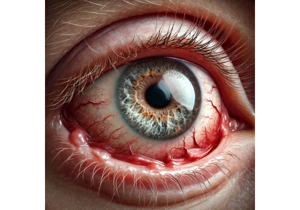
What is uveitic glaucoma?
Uveitic glaucoma is a complex and potentially blinding ocular condition that develops as a result of uveitis, an inflammatory disease that affects the uveal tract of the eye. The uveal tract contains the iris, ciliary body, and choroid, all of which play important roles in eye function. Uveitis can cause inflammation in one or more of these structures, triggering a series of pathological changes that can impair intraocular pressure (IOP) regulation and ultimately lead to glaucoma. Uveitic glaucoma is especially difficult to treat because of the combined effects of inflammation and high IOP, which increase the risk of optic nerve damage and subsequent vision loss.
Understanding Uveitis
To fully comprehend uveitic glaucoma, you must first understand uveitis. Uveitis is a broad term for inflammation of the uveal tract, but it can also affect other structures such as the retina, vitreous, and optic nerve. The location of the inflammation determines the type of uveitis.
- Anterior Uveitis: Inflammation primarily affects the eye’s anterior segment, which includes the iris and the anterior chamber. Anterior uveitis is the most common type of uveitis, and it is frequently associated with ankylosing spondylitis, juvenile idiopathic arthritis, and sarcoidosis.
- Intermediate Uveitis: Inflammation primarily affects the vitreous and peripheral retina, and is frequently associated with systemic conditions such as multiple sclerosis and sarcoidosis.
- Posterior Uveitis: This type of uveitis affects the retina and choroid and is associated with infectious agents such as toxoplasmosis and autoimmune diseases like Behçet’s disease.
- Panuveitis is an inflammation of the entire uveal tract that affects the anterior, intermediate, and posterior segments of the eye. This form is common in conditions such as Vogt-Koyanagi-Harada disease and sympathetic ophthalmia.
A variety of factors can contribute to uveitis, including autoimmune diseases, infections, trauma, and toxic exposure. Uveitis-related inflammation can disrupt the eye’s normal function, particularly the aqueous humor drainage system, resulting in increased IOP and secondary glaucoma.
Pathogenesis of Uveitic Glaucoma
The pathophysiology of uveitic glaucoma is multifactorial, with both inflammatory and mechanical mechanisms contributing to elevated IOP and optic nerve injury.
- Inflammatory Mechanisms: Uveitis can cause several changes in the eye that predispose it to glaucoma. Inflammatory cells and proteins can accumulate in the trabecular meshwork, the primary drainage pathway for aqueous humor, clogging it and reducing outflow. Chronic inflammation can also cause scarring and fibrosis of the trabecular meshwork and surrounding tissues, which impedes aqueous humor drainage.
- Mechanical Mechanisms: Uveitis can cause structural changes within the eye, which contribute to high IOP. For example, posterior synechiae (adhesions between the iris and lens) can cause pupillary block, which is characterized by an obstruction in the flow of aqueous humor from the posterior chamber to the anterior chamber, resulting in increased pressure. Furthermore, peripheral anterior synechiae (adhesions between the iris and the trabecular meshwork) can physically block the outflow of aqueous humor, resulting in angle-closure glaucoma.
- Corticosteroid-Induced Glaucoma: Corticosteroids are commonly prescribed to treat the inflammation caused by uveitis. However, in some patients, corticosteroid therapy can cause or worsen glaucoma by increasing resistance to aqueous humor outflow. This corticosteroid-induced IOP elevation is especially difficult to manage in patients with uveitic glaucoma, as it requires balancing the need for anti-inflammatory treatment with the risk of glaucoma progression.
- Secondary Angle Closure: When uveitis causes significant inflammation, it can progress to secondary angle-closure glaucoma. This happens when the angle of the anterior chamber, which contains the trabecular meshwork, closes or narrows as a result of peripheral anterior synechiae or other inflammatory changes. Angle closure prevents proper drainage of the aqueous humor, resulting in a rapid increase in IOP.
- Neovascularization: Chronic uveitis can cause neovascularization, which is the formation of new, abnormal blood vessels in the iris and angle of the eye. These vessels are fragile and prone to bleeding, which further obstructs the trabecular meshwork and raises IOP.
Clinical Presentation
Patients with uveitic glaucoma may experience a variety of symptoms, which differ depending on the severity of the inflammation, the extent of IOP elevation, and the underlying cause of the uveitis. Common symptoms include:
- Redness and Pain: Patients with uveitic glaucoma frequently report a red, painful eye. Redness is usually caused by underlying uveitis, whereas pain can be caused by both inflammation and elevated IOP.
- Blurred Vision: Blurred vision is a common symptom of uveitic glaucoma, caused by a combination of inflammation, corneal edema, cataract formation, and optic nerve damage due to elevated IOP.
- Photophobia: Patients with uveitic glaucoma frequently suffer from photophobia, or light sensitivity. It is frequently associated with anterior uveitis, in which inflammation of the iris and ciliary body causes increased light sensitivity.
- Visual Field Loss: As uveitic glaucoma progresses, patients may gradually lose peripheral vision. This visual field loss is caused by optic nerve damage caused by chronically elevated IOP, which can progress to central vision loss if not treated properly.
- Halos Around Lights: Patients may notice halos around lights as a result of corneal edema (swelling of the cornea) or the presence of cataracts.
Risk Factors
Several risk factors for developing uveitic glaucoma include:
- Chronic or Recurrent Uveitis: Patients with chronic or recurrent uveitis are more likely to develop uveitic glaucoma as a result of ongoing inflammation and the cumulative effects of multiple episodes of elevated IOP.
- Corticosteroid Use: Corticosteroids, especially in high doses or for long periods of time, are a known risk factor for secondary glaucoma in patients with uveitis.
- Systemic Autoimmune Diseases: Patients with systemic autoimmune diseases like sarcoidosis, lupus, and rheumatoid arthritis have a higher risk of developing uveitis and, as a result, uveitic glaucoma.
- History of Ocular Surgery: Patients who have had ocular surgery, particularly in the anterior segment, may be more likely to develop uveitic glaucoma due to postoperative inflammation and scarring.
- Genetic Predisposition: Genetic factors may influence the development of uveitic glaucoma, especially in patients with a family history of glaucoma or other ocular conditions.
Complications Of Uveitic Glaucoma
If not properly managed, uveitic glaucoma can cause a number of serious complications, including:
- Optic Nerve Damage: The most serious complication of uveitic glaucoma is damage to the optic nerve, which can cause irreversible visual loss. Elevated IOP damages the optic nerve fibers, resulting in progressive visual field loss and, eventually, blindness if not treated.
- Cataract Formation: Chronic inflammation and long-term corticosteroid use in patients with uveitic glaucoma can hasten the development of cataracts, further impairing vision.
- Secondary Angle-Closure Glaucoma: In some cases, the inflammation associated with uveitis can cause the formation of peripheral anterior synechiae, resulting in angle-closure glaucoma, which is more difficult to manage and has a higher risk of rapid vision loss.
- Retinal Detachment: Severe uveitis and elevated IOP can result in retinal detachment, a sight-threatening condition requiring immediate surgical intervention.
- Macular Edema: Chronic inflammation in uveitic glaucoma can cause fluid accumulation in the macula, the central part of the retina responsible for detailed vision, resulting in macular edema and severe visual impairment.
Techniques for Diagnosing Uveitic Glaucoma
To diagnose uveitic glaucoma, a comprehensive and systematic approach is required to assess both the underlying uveitis and the associated elevated intraocular pressure. The diagnostic process combines clinical evaluation, imaging studies, and laboratory tests to accurately identify the condition and guide appropriate treatment.
Clinical Evaluation
- Tonometry: Measuring intraocular pressure (IOP) is critical in detecting uveitic glaucoma. Tonometry-detected elevated IOP is a key indicator of glaucoma in the presence of uveitis. The Goldmann applanation tonometer is the industry standard for measuring IOP and provides accurate results. Other methods, such as non-contact tonometry (also known as the “air puff” test), are available, but they are generally less accurate than Goldmann tonometry. Consistently elevated IOP levels, especially when combined with other signs of uveitis, strongly indicate uveitic glaucoma.
- Gonioscopy: Gonioscopy is a technique for visualizing the anterior chamber angle, which is where aqueous humor drains from the eye via the trabecular meshwork. In uveitic glaucoma, gonioscopy can reveal abnormalities such as peripheral anterior synechiae (PAS), angle neovascularization, or angle closure, all of which can contribute to high IOP. Gonioscopy is critical for distinguishing between open-angle and angle-closure mechanisms in uveitic glaucoma, which has a direct impact on the management strategy.
- Fundoscopy: A thorough examination of the optic nerve head with a fundoscope is essential for determining optic nerve health. Uveitic glaucoma causes optic nerve damage, which manifests as increased cupping (enlargement of the central depression of the optic nerve head) and changes in the neuroretinal rim. These findings suggest glaucomatous optic neuropathy, which can cause permanent vision loss if not treated promptly. Fundoscopy can also detect other complications, such as posterior synechiae and choroidal inflammation, which are common in uveitic eyes.
- Visual Field Testing: Visual field testing, which is usually done with automated perimetry, is used to determine the functional impact of glaucoma on the patient’s vision. Uveitic glaucoma can produce distinct patterns of visual field loss, such as arcuate scotomas or nasal steps. Regular visual field testing is critical for monitoring disease progression and assessing treatment efficacy.
Imaging Studies
- Optical Coherence Tomography (OCT): OCT is a non-invasive imaging technique for obtaining detailed cross-sectional images of the retina and optic nerve. In uveitic glaucoma, OCT can detect early signs of optic nerve damage, such as retinal nerve fiber layer thinning (RNFL). OCT is also useful for monitoring macular edema, a common complication of uveitis that can worsen vision. OCT, which provides high-resolution images of retinal structures, aids in treatment decision-making and monitoring the effectiveness of therapeutic interventions.
- Ultrasound Biomicroscopy (UBM): UBM is a cutting-edge imaging technique that uses high-frequency ultrasound to examine the anterior segment of the eye. UBM can detect structural abnormalities in uveitic glaucoma such as ciliary body detachment, iris bombe (anterior bowing of the iris), and PAS that standard slit-lamp biomicroscopy cannot detect. UBM is especially useful in cases of secondary angle-closure glaucoma, as it can aid in determining the underlying cause of angle closure.
- Anterior Segment Optical Coherence Tomography (AS-OCT): AS-OCT is a type of OCT that images the anterior segment of the eye, which includes the cornea, iris, and anterior chamber angle. AS-OCT generates detailed images of angle structures, allowing for the detection of PAS, iris thickening, and angle closure. AS-OCT is an effective tool for assessing the anatomical changes associated with uveitic glaucoma and planning surgical interventions, if necessary.
Lab Tests
- Blood Tests and Serological Studies: Because uveitic glaucoma is frequently associated with systemic diseases, laboratory tests are required to identify any underlying conditions that may be causing uveitis. Common tests for systemic inflammation include erythrocyte sedimentation rate (ESR) and C-reactive protein (CRP), as well as specific serological markers like antinuclear antibodies (ANA), rheumatoid factor (RF), and HLA-B27. Identifying an underlying autoimmune or infectious etiology can aid in the treatment of both uveitis and associated glaucoma.
- Aqueous or Vitreous Sampling: If the cause of uveitis is unknown or there is a suspicion of an infectious etiology, an aqueous or vitreous tap may be performed to collect fluid samples for microbiological and cytological examination. Polymerase chain reaction (PCR) testing can detect the presence of specific viral, bacterial, or fungal pathogens, whereas cytology can assist in identifying malignant cells in masquerade syndromes like lymphoma.
- Imaging for Systemic Disease: In patients with uveitic glaucoma and systemic disease, imaging studies such as chest X-rays, computed tomography (CT) scans, or magnetic resonance imaging (MRI) may be required to rule out systemic involvement. For example, chest X-rays can detect sarcoidosis, which is characterized by bilateral hilar lymphadenopathy, whereas MRI can assess neurological involvement in conditions such as multiple sclerosis.
Treatment Approaches for Uveitic Glaucoma
Uveitic glaucoma is especially difficult to manage because it requires simultaneous control of intraocular pressure (IOP) and intraocular inflammation. Because of the condition’s dual nature, treatments must address both underlying uveitis and secondary glaucoma in order to prevent optic nerve damage and preserve vision. The treatment strategy is often tailored to the individual patient, taking into account the severity of the glaucoma, the cause of the uveitis, and the patient’s overall health.
Medical Management
- Anti-inflammatory Therapy: Controlling the underlying inflammation is critical for treating uveitic glaucoma. Corticosteroids are commonly used for this purpose, and they can be administered topically (eye drops), systemically (oral or intravenous), or locally (periocular or intravitreal injections). Topical corticosteroids, such as prednisolone acetate, are frequently the first line of treatment for anterior uveitis. Systemic corticosteroids or immunosuppressive agents such as methotrexate, azathioprine, or cyclosporine may be required for severe or chronic uveitis. However, long-term corticosteroid use, particularly systemic use, can have serious side effects, including exacerbation of glaucoma.
- IOP-Lowering Medications: The next critical aspect of treating uveitic glaucoma is lowering intraocular pressure to avoid optic nerve damage. Several types of medications are used to lower IOP:
- Prostaglandin Analogues: Drugs such as latanoprost, travoprost, and bimatoprost promote aqueous humor outflow via the uveoscleral pathway. However, their use in uveitic glaucoma is controversial because they may exacerbate inflammation in some patients.
- Beta-Blockers: Timolol and betaxolol reduce aqueous humor production and are frequently used as first-line agents because of their efficacy and low side-effect profile.
- Alpha Agonists: Brimonidine and apraclonidine reduce aqueous humor production and can be used as an adjunctive therapy in conjunction with other IOP-lowering medications.
- Carbonic Anhydrase Inhibitors: Topical dorzolamide and brinzolamide, as well as systemic acetazolamide, reduce aqueous humor production. These drugs are especially useful for treating acute IOP spikes.
- Rho Kinase Inhibitors: Newer agents, such as netarsudil, lower IOP by increasing aqueous humor outflow via the trabecular meshwork, providing an alternative mechanism of action.
- Mydriatics and Cycloplegics: In cases of anterior uveitis, mydriatic and cycloplegic agents, such as atropine or cyclopentolate, are used to prevent the formation of posterior synechiae and relieve pain by paralyzing the ciliary muscle. These agents help to stabilize the blood-aqueous barrier, which reduces protein leakage and cellular infiltration into the anterior chamber.
Surgical Management
When medical therapy fails to control IOP or the side effects of medications become unbearable, surgical intervention may be required.
- Laser Therapy: Due to the inflammatory nature of uveitic glaucoma, laser trabeculoplasty is generally less effective than it is in primary open-angle glaucoma. However, selective laser trabeculoplasty (SLT) may be considered in some cases because it is less likely to cause postoperative inflammation.
- Filtration Surgery: Trabeculectomy is a common surgical procedure that creates a new drainage pathway for aqueous humor, thereby lowering IOP. However, the increased risk of scarring and closure of the surgical site due to ongoing inflammation often limits the success of trabeculectomy in uveitic glaucoma. Anti-fibrotic agents such as mitomycin C or 5-fluorouracil are commonly used during surgery to reduce the risk of scarring.
- Glaucoma Drainage Devices: If trabeculectomy is ineffective or deemed inappropriate, glaucoma drainage devices (also known as tube shunts) such as the Ahmed, Baerveldt, or Molteno implants may be used. These devices provide an alternative route for aqueous humor to exit the eye, avoiding the trabecular meshwork and aiding in IOP control. However, the presence of these implants necessitates meticulous postoperative care to avoid infection and other complications.
- Cyclodestructive Procedures: Patients with refractory uveitic glaucoma who are not candidates for conventional surgery may benefit from cyclodestructive procedures such as cyclophotocoagulation. This technique involves using a laser to destroy a portion of the ciliary body, thereby reducing aqueous humor production and IOP. While effective, this procedure has a higher risk of complications, such as hypotony (an abnormally low IOP) and vision loss.
Management of Complications
- Cataract Surgery: Cataract formation is a common complication of uveitic glaucoma, especially in patients receiving long-term corticosteroid therapy. Cataract surgery in these patients is difficult due to the higher risk of postoperative inflammation and glaucoma progression. Careful preoperative planning, including the use of perioperative steroids and anti-inflammatory medications, is critical for successful outcomes.
- Management of Macular Edema: Chronic inflammation causes macular edema, which is a significant complication of uveitic glaucoma. To reduce inflammation and fluid accumulation in the macula, patients are typically treated with intravitreal corticosteroids or anti-VEGF agents.
- Monitoring for Disease Progression: Regular follow-up with comprehensive eye examinations, including IOP measurements, optic nerve evaluation, and visual field testing, is critical for tracking disease progression and adjusting treatment as necessary. Uveitic glaucoma is a chronic condition that requires long-term management and vigilance to prevent vision loss.
Trusted Resources and Support
Books
- “Uveitis: Fundamentals and Clinical Practice” by Robert B. Nussenblatt and Scott M. Whitcup: This comprehensive book provides an in-depth understanding of uveitis, including the complications such as uveitic glaucoma. It is an essential resource for both clinicians and researchers focused on ocular inflammation.
- “Glaucoma: Clinical, Surgical, and Imaging Aspects” edited by R.N. Weinreb and P.A. Netland: This book covers the full spectrum of glaucoma management, including specific challenges and strategies for treating secondary glaucomas like uveitic glaucoma.
Organizations
- American Academy of Ophthalmology (AAO): The AAO offers a wide range of resources for patients and professionals, including guidelines on the management of uveitis and associated glaucoma. Their website provides access to the latest research, clinical guidelines, and educational materials.
- The Glaucoma Foundation: This organization provides support and information for patients with all forms of glaucoma, including uveitic glaucoma. They offer resources for understanding the condition, exploring treatment options, and finding support networks.


