What is xanthelasma?
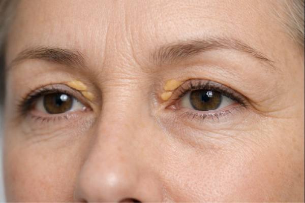
Xanthelasma, also known as xanthelasma palpebrarum, is a common, harmless condition in which yellowish plaques or nodules appear on the eyelids, particularly around the inner canthus (the area near the nose). Cholesterol deposits accumulate within the skin, forming soft, flat, or slightly elevated plaques. Xanthelasma primarily affects the upper eyelids, but it can also appear on the lower eyelid. Although xanthelasma is not harmful or causes discomfort, it can be a cosmetic concern for many people due to its visible and often symmetrical nature.
Epidemiology and Demographics
Xanthelasma is one of the most common types of xanthomas, which are lipid-rich deposits that can appear in a variety of body locations. Xanthelasma refers to xanthomas that appear on the eyelids. This condition can affect people of all ages, but it is most common in middle-aged and older adults, with a higher incidence in those over the age of 40. Women are more commonly affected than men, though the reason for this gender disparity is unclear.
The prevalence of xanthelasma varies by population and ethnic group. According to studies, it may be more common in people of Mediterranean and Asian descent, but it can affect any ethnic group. While xanthelasma can appear in people with normal lipid levels, it is frequently associated with hyperlipidemia, a condition defined by elevated levels of lipids in the blood, such as cholesterol and triglycerides. It is important to note that, while xanthelasma is associated with lipid disorders, it can also occur in people who have no underlying lipid abnormalities.
Pathophysiology
The development of xanthelasma is closely related to lipid accumulation, particularly cholesterol, in the dermis—the layer of skin beneath the epidermis. These lipid deposits are primarily made up of low-density lipoprotein (LDL) cholesterol, which is absorbed by macrophages, a type of immune cell in the skin. When macrophages engulf and store large amounts of cholesterol, they transform into “foam cells,” which are characteristic of xanthoma development.
The precise mechanism by which xanthelasma develops is unknown, but it is thought to be influenced by a number of factors:
- Lipid Metabolism: Abnormal lipid metabolism, such as hyperlipidemia, is thought to play an important role in the development of xanthelasma. Elevated LDL cholesterol levels in the blood may cause cholesterol deposition in the skin, especially in areas with a high blood supply, such as the eyelids.
- Genetic Factors: There is evidence that genetic factors may influence the development of xanthelasma. People with a family history of the disease are more likely to develop it themselves. Furthermore, certain genetic disorders affecting lipid metabolism, such as familial hypercholesterolemia, can raise the risk of xanthelasma.
- Inflammatory Processes: Inflammatory processes within the skin can also contribute to the development of xanthelasma. Chronic inflammation can increase the permeability of blood vessels, allowing lipids to leak into the surrounding tissue and accumulate in the dermis.
- Aging: The risk of developing xanthelasma rises with age. This could be due to age-related changes in lipid metabolism, as well as a decrease in the skin’s ability to remove accumulated lipids.
- Local Factors: The eyelids’ unique anatomy, including their thin skin and abundant vascular supply, may make them more prone to the development of xanthelasma. The repetitive motion of blinking, as well as the high exposure to environmental factors, may contribute to the condition.
Clinical Features
Xanthelasma appears as soft, yellowish plaques or nodules that are usually symmetrical and located on the upper and lower eyelids. The lesions are usually flat or slightly raised, with clearly defined borders. They can range in size from a few millimetres to several centimetres in diameter. In some cases, multiple plaques can combine to form larger, irregularly shaped lesions.
- Location: Xanthelasma is most commonly found on the medial aspects of the upper eyelids, near the inner canthus, but it can also occur on the lower eyelid. The condition is often bilateral, which means it affects both eyes.
- Texture: The plaques are typically soft to the touch and may have a slightly granular appearance. They are usually non-tender and do not cause discomfort or itching.
- Color: The lipid content within the lesions causes xanthelasma’s characteristic yellow color. The color may vary slightly, appearing more whitish or orange, depending on the individual’s skin tone and lipid concentration.
- Progression: Xanthelasma is a slow-progressing condition. Once formed, the plaques tend to stay stable or grow slowly over time. In some people, the lesions remain small and asymptomatic, while in others, they grow larger and more visible.
- Cosmetic Concerns: Although xanthelasma rarely causes physical symptoms, it can be a cosmetic issue for many people. The visible nature of the lesions, especially if they are large or numerous, can cause self-consciousness or dissatisfaction with their appearance.
Associated Conditions
Although xanthelasma is commonly regarded as a benign and isolated condition, it can be associated with underlying systemic conditions, particularly those involving lipid metabolism. The presence of xanthelasma should prompt a comprehensive examination of the patient’s lipid profile and cardiovascular risk factors.
- Hyperlipidemia: A significant proportion of people with xanthelasma have hyperlipidemia, which is defined by high levels of LDL cholesterol, triglycerides, or both. Hyperlipidemia is a significant risk factor for atherosclerosis and cardiovascular disease, and xanthelasma may be an early warning sign of these conditions.
- Cardiovascular Disease: Some studies indicate that xanthelasma may be an independent risk factor for cardiovascular disease, even in people with normal lipid levels. The presence of xanthelasma could indicate underlying atherosclerosis or other vascular abnormalities.
- Diabetes Mellitus: Xanthelasma has been linked to diabetes mellitus, specifically type 2. Individuals with diabetes frequently have lipid abnormalities, such as elevated triglycerides and low HDL cholesterol, which may contribute to the development of xanthelasma.
- Familial Hypercholesterolemia: Familial hypercholesterolemia is a genetic disorder characterized by abnormally high LDL cholesterol. Individuals with this condition are more likely to develop xanthelasma and other xanthomas throughout the body.
- Hypothyroidism: Hypothyroidism, which occurs when the thyroid gland fails to produce enough thyroid hormone, can cause changes in lipid metabolism and may be associated with the development of xanthelasma.
Differential Diagnosis
When evaluating a patient with xanthelasma, it is critical to consider other conditions that may present with similar clinical characteristics. The differential diagnosis of xanthelasma includes the following:
- Sebaceous Hyperplasia: Sebaceous hyperplasia is a condition characterized by the enlargement of sebaceous glands, which cause yellowish or skin-colored papules on the face, including the eyelids. These lesions are typically smaller and have a central depression, making them distinct from xanthelasma.
- Syringomas: Syringomas are benign sweat gland tumors that can manifest as small, flesh-colored or yellowish papules on the eyelids. They are generally smaller and firmer than xanthelasma plaques.
- Basal Cell Carcinoma: Basal cell carcinoma (BCC) is a common type of skin cancer that sometimes appears as a yellowish plaque on the eyelids. BCC lesions are typically more irregular in shape and may be associated with ulceration or bleeding.
- Xanthogranulomas: Xanthogranulomas are benign lesions with lipid-laden histiocytes, similar to xanthomas. They can appear as yellowish nodules on the skin, but they are typically solitary and larger than xanthelasma.
- Other Xanthomas: Although xanthelasma is a type of xanthoma, other types of xanthomas, such as eruptive xanthomas (small, red-yellow papules) and tuberous xanthomas (firm, yellow nodules), can also appear on the skin. These other xanthomas are frequently associated with severe lipid abnormalities.
Finally, xanthelasma is a common, benign condition that causes yellowish plaques to appear on the eyelids. While the condition is not inherently dangerous, it may be associated with underlying lipid abnormalities and cardiovascular risk factors. Proper diagnosis and evaluation are required to identify any underlying systemic conditions and guide appropriate management.
Diagnosing Xanthelasma: Key Approaches
Diagnosing xanthelasma is primarily based on clinical examination, with the distinctive appearance of the lesions frequently sufficing for an accurate diagnosis. However, additional diagnostic tests may be required to assess underlying lipid abnormalities and associated conditions, especially in people who have risk factors for cardiovascular disease or other systemic disorders.
Clinical Examination
A thorough clinical examination of the affected area is usually required to diagnose xanthelasma. Xanthelasma’s distinguishing features—yellowish plaques on the eyelids, frequently located near the inner canthus—are usually visible upon visual inspection. The lesions are soft, flat, or slightly raised, with well-defined edges. Detailed examination should include:
- Assessment of Lesion Characteristics: Examine the lesions’ size, shape, texture, and color carefully. Xant
Helasma plaques are typically soft to the touch, with a yellowish hue that varies according to the individual’s skin tone and the lipid content of the lesions. Plaques are typically well-defined, with smooth edges, and range in size from a few millimeters to several centimeters. They are frequently symmetrical and bilateral, affecting primarily the upper eyelids, though the lower eyelids may also be involved. - Examination of the Surrounding Skin: The clinician should examine the skin around the lesions to rule out any other dermatological conditions that may exhibit similar characteristics. This includes looking for signs of inflammation, ulceration, or other abnormalities that may indicate a different diagnosis.
- Palpation: Touching the lesions can confirm their soft, lipid-rich texture. Unlike some other skin conditions, which can present with firm or cystic nodules, xanthelasma lesions are usually soft and pliable.
Lipid Profile Testing
Given the strong link between xanthelasma and lipid disorders, a lipid profile test is an important part of the diagnostic process. This blood test determines the levels of several lipids in the bloodstream, including:
Elevated levels of low-density lipoprotein (LDL) cholesterol, also known as “bad” cholesterol, are common in people with xanthelasma. High LDL levels can cause cholesterol deposits in the skin and arteries, raising the risk of cardiovascular disease.
- High-Density Lipoprotein (HDL) Cholesterol: HDL, also known as “good” cholesterol, aids in the removal of cholesterol from the blood. Low levels of HDL cholesterol may increase the risk of cardiovascular disease.
- Total Cholesterol: This test determines the total cholesterol level in the blood, which includes both LDL and HDL cholesterol. Elevated total cholesterol is frequently indicative of hyperlipidemia.
- Triglycerides: Triglycerides are another type of blood lipid. High triglyceride levels can cause xanthelasma and increase the risk of cardiovascular disease.
The results of the lipid profile can aid in determining whether the patient has hyperlipidemia or other lipid abnormalities that may require treatment. It is also critical to assess these levels in light of the patient’s overall cardiovascular risk.
Biopsy (if needed)
In most cases, a biopsy is not required to diagnose xanthelasma because the clinical appearance of the lesions is sufficient for an accurate diagnosis. If there is any doubt about the diagnosis, or if the lesions have an unusual appearance (e.g., irregular borders, rapid growth, or ulceration), a biopsy may be performed to rule out basal cell carcinoma or other skin cancers.
- Histopathological Examination: If a biopsy is performed, the tissue sample will be examined using a microscope. The presence of foam cells—lipid-laden macrophages—in the dermis is a typical histopathological finding in xanthelasma. These cells give the lesions their distinctive yellowish appearance. A biopsy may also reveal cholesterol clefts and a mild inflammatory infiltrate.
- Differential Diagnosis: A biopsy can help distinguish xanthelasma from other conditions that share similar characteristics, such as sebaceous hyperplasia, syringomas, or xanthogranulomas. The histopathological characteristics of these conditions differ from those of xanthelasma, allowing for a precise diagnosis.
Additional Diagnostic Tests
Other tests, in addition to a lipid profile, may be required depending on the patient’s clinical presentation and risk factors. These tests can aid in determining underlying conditions that may be contributing to the development of xanthelasma:
- Fasting Blood Glucose and Hemoglobin A1c (HbA1c): Because xanthelasma is associated with diabetes, particularly type 2 diabetes, it is critical to monitor the patient’s blood glucose levels. Elevated fasting blood glucose or HbA1c levels may indicate diabetes or prediabetes, requiring additional treatment.
- Thyroid Function Tests: Hypothyroidism can cause lipid abnormalities and may be associated with xanthelasma. Thyroid function tests, such as thyroid-stimulating hormone (TSH) and free thyroxine (T4), can help doctors determine whether a patient has hypothyroidism.
- Cardiovascular Risk Assessment: Patients with xanthelasma, especially those with hyperlipidemia, should have their cardiovascular risk assessed. This may include measuring blood pressure, performing an electrocardiogram (ECG), and looking for other risk factors like smoking, a family history of heart disease, and obesity.
Best Practices for Xanthelasma Management
The treatment of xanthelasma entails addressing both the cosmetic concerns associated with the condition and any underlying systemic issues, particularly lipid abnormalities. The treatment options differ depending on the size, location, and number of lesions, as well as the patient’s preferences and overall health. While xanthelasma is a benign condition, its association with hyperlipidemia and cardiovascular risk factors necessitates a multifaceted approach to treatment.
Lifestyle and Medical Management
Managing underlying lipid abnormalities is an important part of xanthelasma treatment for patients. Addressing hyperlipidemia not only slows the progression of xanthelasma, but also lowers the risk of cardiovascular disease.
- Diet and Exercise: Lifestyle changes are frequently the first line of therapy for hyperlipidemia. A diet low in saturated fats, trans fats, and cholesterol, combined with regular physical activity, can help reduce LDL cholesterol and triglyceride levels. Increased intake of omega-3 fatty acids (found in fish and flaxseed) and fiber (found in fruits, vegetables, and whole grains) can also improve lipid profiles.
- Lipid-Lowering Medications: Patients with significant hyperlipidemia may require lipid-lowering medications. Statins are the most commonly used drugs to lower LDL cholesterol. Fibrates, niacin, and omega-3 fatty acid supplements are also options for treating high triglycerides and low HDL cholesterol. It is critical for patients to collaborate with their healthcare provider to determine the most effective and tolerable treatment regimen.
- Management of Associated Conditions: In addition to lipid abnormalities, it is critical to manage other xanthelasma-related conditions such as diabetes and hypothyroidism. Controlling blood glucose levels and thyroid function can help reduce the risk of xanthelasma recurrence while also improving overall health.
Cosmetic and Surgical Treatments
For patients concerned about the cosmetic appearance of xanthelasma, there are several treatment options available to remove or shrink the lesions. Depending on the location and complexity of the lesions, a dermatologist, plastic surgeon, or ophthalmologist will usually perform these procedures.
- Chemical Peels: Chemical peels containing trichloroacetic acid (TCA) or other exfoliants can help reduce the appearance of xanthelasma. The peel works by removing the top layers of skin, allowing new skin to regenerate and reducing the appearance of lesions. Multiple treatments may be required to achieve the desired outcome.
- Laser Therapy: Laser therapy, especially with CO2 or Erbium:YAG lasers, is a popular method of treating xanthelasma. The laser precisely targets and vaporizes the lipid deposits, removing the lesions while causing minimal damage to the surrounding skin. Laser therapy is generally well-tolerated and can produce excellent cosmetic results, though there is a risk of scarring or pigment changes.
- Cryotherapy: Xanthelasma plaques are frozen using liquid nitrogen or another cryogenic agent. The extreme cold degrades the lipid deposits, and the lesions eventually fall off as the skin heals. Cryotherapy works well for smaller lesions, but it may not be appropriate for larger or deeper plaques due to the risk of scarring and skin depigmentation.
- Surgical Excision: Larger or more persistent xanthelasma lesions may benefit from surgical excision. The plaques are removed with a scalpel, and the skin is sutured afterwards. While surgical excision can provide permanent removal of the lesions, it has a higher risk of scarring than other treatment options. To reduce scarring and achieve a good cosmetic outcome, precise surgical technique is required.
- Radiofrequency Ablation is a minimally invasive procedure that uses radiofrequency energy to heat and destroy xanthelasma lesions. This method is effective for treating small to medium-sized plaques and has the advantage of being quick with little downtime. Scarring is minimal, making it an appealing option for a wide range of patients.
Recurrence and Follow-up
Despite successful removal of xanthelasma lesions, there is a risk of recurrence, especially if the underlying lipid abnormalities are not treated. Patients should be informed about the possibility of recurrence and the importance of continuing to manage their lipid levels and cardiovascular risk factors.
- Regular Follow-Up: Patients with xanthelasma should have regular follow-up appointments to monitor their lipid levels and check for recurrence of lesions. Ongoing management of lipid disorders, including lifestyle changes and medication, is critical for lowering the risk of recurrence.
- Patient Education: Educating patients on the importance of living a healthy lifestyle and following their treatment plan is critical for long-term success. Patients should be encouraged to continue engaging in regular physical activity, eating a heart-healthy diet, and taking their lipid-lowering medications as prescribed.
Trusted Resources and Support
Books
- “Lipid Disorders: A Comprehensive Guide” by Scott M. Grundy
- This book provides an in-depth overview of lipid disorders, including hyperlipidemia and its associated conditions, such as xanthelasma. It offers practical information on the diagnosis, management, and treatment of lipid abnormalities.
- “Cosmetic Dermatology: Principles and Practice” by Leslie Baumann
- This book is an excellent resource for understanding the various cosmetic treatments available for skin conditions, including xanthelasma. It covers a wide range of procedures, from chemical peels to laser therapy, and offers guidance on selecting the most appropriate treatment options.
Organizations
- American Academy of Dermatology (AAD)
- The AAD provides extensive resources on skin conditions, including xanthelasma. Their website offers information on diagnosis, treatment options, and patient education, making it a valuable resource for both patients and healthcare providers.
- National Lipid Association (NLA)
- The NLA is dedicated to the prevention and treatment of lipid disorders. Their website offers resources on managing hyperlipidemia, including guidelines for lipid-lowering therapies and lifestyle modifications. They also provide educational materials for patients and healthcare professionals.


