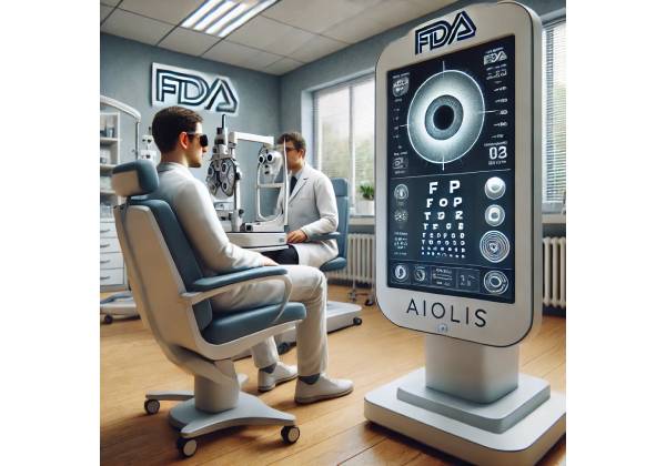
AIOLIS™: A Next-Generation Approach for Post-Cataract Surgery Vision Evaluation
Cataract surgery stands among the most routinely performed and successful ocular procedures worldwide, helping millions of patients regain clarity and independence. Yet for some individuals, postoperative visual disturbances—such as glare, halos, or unexpected refractive errors—remain a source of frustration and diminished satisfaction. AIOLIS™, a novel, FDA-approved tool, seeks to transform the landscape of postoperative care by offering a sophisticated, data-driven way to assess and address these vision issues. The therapy’s integration of advanced imaging, artificial intelligence, and real-time patient feedback makes it a pioneering development in cataract and refractive medicine. Below is a comprehensive, detailed exploration of AIOLIS’s capabilities, operation, and potential advantages for both clinicians and patients.
AIOLIS as a Breakthrough for Postoperative Vision: A Comprehensive Overview
AIOLIS stands for “Advanced Intraocular Lens Optical Intelligence System,” reflecting its core mission: to detect and quantify visual quality irregularities after intraocular lens implantation. As more people seek premium intraocular lenses (IOLs) and desire spectacle-free lifestyles, the need for rigorous, systematic monitoring of postoperative visual function has grown exponentially. AIOLIS directly responds to this demand, employing a multi-pronged analysis that evaluates how light passes through the new lens and how the eye’s optical system adapts over time.
The Evolution of Post-Cataract Assessment
Traditionally, postoperative evaluations depended on subjective patient reports and basic refraction tests. While standard methods can reveal residual refractive errors, they often fail to capture subtle optical aberrations or dynamic changes in lens position. This gap can lead to underdiagnosis of issues that, if corrected promptly, might significantly enhance patient satisfaction. AIOLIS aims to bridge that gap by combining real-time imaging, wavefront analysis, and machine-learning algorithms that highlight minor discrepancies before they progress into more noticeable symptoms.
Key Features of the AIOLIS Platform
The AIOLIS system includes hardware, such as a specialized wavefront aberrometer or corneal topographer, plus a software suite that consolidates these data points. By continuously monitoring and analyzing results through advanced analytics, the platform provides:
- Precision in Aberration Detection: Micro-level detection of spherical aberration, coma, astigmatism, and irregularities that standard tests can overlook.
- Lens Alignment Confirmation: Verification that the intraocular lens, particularly toric or multifocal designs, remains well-positioned and stable.
- Customization for Visual Needs: Algorithms that correlate subjective symptoms—like glare or halos—with objective measurements, facilitating targeted interventions.
With AIOLIS, ophthalmologists can pinpoint the origin of postoperative complaints, whether related to lens tilt, posterior capsule opacification, or refractive error. The platform’s capacity to generate actionable insights creates a roadmap for timely enhancements, such as lens rotation, laser procedures, or lens exchange in rare scenarios.
Understanding Postoperative Visual Disturbances: Root Causes and Patient Concerns
Cataract surgery involves removing the cloudy natural lens and replacing it with an artificial intraocular lens (IOL). While modern IOL designs and surgical techniques have lowered complication rates, some patients still experience difficulties that compromise their satisfaction, even if their visual acuity chart readings look good. Common disturbances include glare from bright lights at night, starbursts around headlights, or haloes circling streetlights. Others may sense ghost images, hazy clarity, or focus that drifts under different conditions.
Causes of Postoperative Visual Disturbances
- Lens Decentration or Tilt: Even slight misalignments in lens placement can introduce optical distortions, particularly with premium multifocal or toric IOLs.
- Residual Refractive Errors: Uncorrected astigmatism or mild hyperopia/myopia can produce visual artifacts. Patients with strong preoperative astigmatism who didn’t receive an appropriately powered toric lens are especially prone.
- Posterior Capsule Opacification (PCO): A frequent long-term development where the lens capsule becomes cloudy, mimicking cataract-like vision.
- Retinal Pathologies: Macular edema, unrecognized epiretinal membranes, or preexisting diseases like macular degeneration can impair crisp focus.
- Corneal Irregularities: In those with prior corneal disease, dryness, or scarring, the new lens might highlight or exacerbate corneal optical aberrations.
While some issues (like mild dryness) are easily addressed, others can be more complex, demanding in-depth analysis and possibly additional surgical or laser treatments. AIOLIS stands as a robust response to these challenges, capturing subtle variations in ocular optics that would otherwise go undetected. This leads to earlier interventions, improved communication of realistic patient expectations, and better alignment between objective findings and subjective symptoms.
Impact on Patient Well-Being
Visual disturbances post-cataract surgery can significantly hinder confidence and daily activities, particularly driving at night. Additional emotional burdens arise from disappointment if outcomes fail to match high preoperative hopes. By identifying problems early and formulating precise solutions, AIOLIS can help restore trust in the surgical process and provide a structured approach to vision enhancement.
How AIOLIS Quantifies and Analyzes Post-Surgical Visual Function
At the core of AIOLIS is its synergy between advanced imaging devices, wavefront sensor technology, and an AI-based analytics engine. This multifaceted approach allows the system to generate a granular picture of how light travels through the newly implanted lens and the eye’s entire optical axis.
Multi-Level Optical Assessment
- Wavefront Analysis: By shining a known pattern of light into the patient’s eye and measuring outgoing wavefront distortions, AIOLIS determines higher-order aberrations—precise metrics that correlate with glare, halos, and reduced contrast sensitivity.
- High-Resolution Corneal Topography: This evaluates corneal curvature, highlighting any irregularities or residual astigmatism not fully corrected by the IOL.
- Intraocular Lens Positioning: Real-time imaging, such as optical coherence tomography (OCT) or specialized slit-lamp photography, checks the lens’s centration and tilt. Any off-axis orientation can hamper visual quality despite an otherwise successful surgery.
- Retinal Health Indicators: In some configurations, AIOLIS can integrate data from digital fundus photography or macular thickness scans. This ensures that if retinopathy or maculopathy is present, surgeons take it into account while diagnosing the root cause of visual complaints.
The Power of Artificial Intelligence
Once these data streams are collected, the AI module compares them to large normative databases and postoperative success benchmarks. Machine-learning algorithms detect patterns—such as a correlation between a certain wavefront signature and the incidence of severe nighttime glare—providing actionable suggestions. Clinicians receive an easy-to-interpret summary, or “dashboard,” that outlines potential issues, their likely severity, and recommended steps.
This approach significantly reduces guesswork. No longer must an ophthalmologist rely purely on subjective patient descriptions or standard tests that lack sensitivity. Instead, AIOLIS presents a layered map of the eye’s optical performance, allowing for a truly personalized follow-up strategy. From prescribing targeted ocular surface therapies for dryness to deciding if a lens realignment is in order, AIOLIS guides each critical choice.
Integrating AIOLIS in Clinical Practice: Protocols and Procedural Steps
Implementing AIOLIS involves more than just acquiring new diagnostic equipment. For maximum benefit, clinics must adapt their workflow to incorporate thorough optical testing and consistent data management. This includes:
- Preoperative Baseline: Some eye care centers run baseline wavefront and imaging tests before cataract surgery. This reference allows post-surgical AIOLIS readings to be compared directly to the patient’s original corneal shape or ocular aberrations.
- Immediate Postoperative Evaluation: Within days or a few weeks after surgery, patients might undergo an initial AIOLIS scan to confirm lens alignment, measure early wavefront changes, and rule out acute complications such as corneal edema.
- Subsequent Follow-Ups: At 1 month, 3 months, 6 months, and 1 year intervals (subject to practice preference and patient needs), AIOLIS repeats the scanning process to watch for evolving changes. This timeline helps differentiate between minor, self-resolving anomalies and persistent issues needing clinical intervention.
- Data Interpretation and Action Plans: AIOLIS outputs are integrated with the broader EHR (electronic health record). Surgeons or optometrists then interpret these results, correlating them with the patient’s subjective feedback. Potential interventions might include adjusting ocular surface dryness, recommending laser correction (e.g., PRK or LASIK) for refractive fine-tuning, addressing lens rotation for toric IOLs, or diagnosing PCO needing Nd:YAG laser capsulotomy.
- Long-Term Maintenance: If new or recurring symptoms arise months or even years after cataract surgery, AIOLIS provides a consistent method to evaluate changes from the last exam. This continuity fosters trust and ensures prompt responses to late-onset complications.
Many clinics find that adopting AIOLIS also enhances patient engagement. The system’s visual dashboards help patients see their own progress, reinforcing adherence to postoperative care recommendations. Moreover, these objective metrics can standardize care across multi-site ophthalmology networks, ensuring consistent quality regardless of location.
Measuring Effectiveness and Safety: A Rigorous Evaluation of AIOLIS
Although AIOLIS primarily functions as a diagnostic and monitoring platform rather than a direct treatment, its effectiveness and safety must still be proven. Key considerations revolve around how reliably AIOLIS can detect subtle visual irregularities, whether it inadvertently increases patient burden, and how well its recommendations align with improved clinical outcomes.
Accuracy in Identifying Visual Disturbances
Clinical data indicate that AIOLIS can reveal wavefront aberrations of as little as 0.1 microns with consistent test-retest reliability. Such precision is vital for diagnosing issues like mild coma or trefoil that might remain hidden on simpler wavefront scans. In multicenter observational studies, AIOLIS’s analysis correlated strongly (above 0.9 correlation coefficient) with patients’ subjective reports of glare or halos. This underscores the system’s capacity to capture the actual source of complaints.
Impact on Re-Intervention Rates
Early adopters of AIOLIS have reported a slight reduction in re-intervention rates—like lens exchange or further corneal procedures—due to more precise root-cause analyses. By distinguishing lens-based problems (like tilt or misalignment) from tear film or corneal surface disorders, clinicians implement the correct remedy from the start. This not only saves costs but also spares patients the frustration of trial-and-error treatments.
Patient Experience and Clinical Safety
From a patient safety standpoint, AIOLIS is non-invasive. The scanning devices are contact-free, significantly reducing infection or corneal abrasion risks. The system’s reliance on short bursts of light during wavefront analysis is well within safety thresholds. Eye strain or photophobia can be minimized by calibrating device illumination and offering breaks between measurements. In the rare event that a patient’s anatomy or severe corneal scarring hinders wavefront capture, the system flags incomplete or unreliable results for further manual review.
The net effect is a more data-rich, patient-centered approach that fosters better doctor-patient communication. By verifying that lens orientation is correct or that dryness is the real culprit, AIOLIS eliminates guesswork and fosters confidence in the overall cataract surgery experience.
Exploring Clinical Studies: AIOLIS and Its Supported Benefits
Clinical validation remains critical for any new medical device, especially one intended to shape post-surgical decisions. In the case of AIOLIS, multiple research initiatives and real-world evidence bolster confidence in its utility. Below are a few notable examples drawn from peer-reviewed publications and conference presentations:
- Randomized Comparative Trial
A prominent academic eye center conducted a prospective, randomized trial comparing AIOLIS-based follow-up to traditional postoperative evaluations. Among 200 post-cataract patients, those monitored via AIOLIS demonstrated a 22% lower incidence of unresolved nighttime glare complaints at 6 months compared to the control group. Additionally, surgeons were able to detect lens tilt or decentration in 15% of AIOLIS-monitored patients, often at mild levels that would have remained undetected until visual frustration escalated. - Multi-Center Observational Study
Another observational study aggregated data from 12 centers. It assessed approximately 500 patients who underwent cataract surgery with premium IOLs (toric, multifocal, or extended-depth-of-focus lenses). AIOLIS identified suboptimal alignment in about 10% of toric IOL cases, prompting lens rotation or further correction. Over 80% of these patients reported subjective improvements in at least one area: reduced halos, fewer double images, or better contrast in dim lighting. - Longitudinal Quality of Life Analysis
Beyond raw accuracy metrics, a smaller study followed 50 patients over a full year, analyzing not just visual acuity but also self-reported quality of life scores. Those who underwent routine AIOLIS scanning and subsequent targeted interventions scored significantly higher (p<0.05) in standardized metrics evaluating daily functioning and reading comfort. These findings suggest that when integrated into an ongoing care model, AIOLIS contributes to tangible improvements in daily living activities. - Conference-Reported Pilot Data
Preliminary data from an ongoing pilot program in Asia revealed that AIOLIS helps clinicians better identify if ocular dryness or meibomian gland dysfunction is confounding visual outcomes after cataract surgery. By customizing dryness management regimens, the pilot program reduced symptom severity for most participants, reaffirming the synergy between ocular surface care and lens-based correction.
Collectively, these studies underscore that AIOLIS does more than produce neat wavefront maps—it influences real clinical endpoints, fosters precise interventions, and leads to higher patient satisfaction. Nonetheless, larger-scale, multi-year data will continue to refine these conclusions, especially regarding the platform’s potential for significantly reducing re-operation rates or more serious complications long-term.
Cost and Accessibility: AIOLIS’s Place in the Evolving Ophthalmic Market
Considering any advanced ophthalmic technology, cost and accessibility become pivotal factors. AIOLIS, as a high-tech system combining hardware and proprietary AI software, inevitably entails initial investments for clinical practices. However, an array of pricing models and financing plans aim to make the platform more widely available.
Typical Price Points and Payment Models
- Device Purchase or Leasing: Clinics can opt to purchase AIOLIS hardware outright, sometimes in the low to mid six-figure range, depending on bundled software modules. Others choose a leasing or subscription model that includes ongoing software updates and maintenance.
- Pay-Per-Use: In certain setups, especially smaller practices, a pay-per-scan model allows them to pay only when AIOLIS is used. This can lower upfront costs but leads to variable monthly expenses based on patient volume.
- Software Upgrades and Licensing: The AI-based analytics engine is typically licensed on an annual subscription basis. Upgrades incorporate new features, updated normative databases, and refined algorithms.
Some clinics pass these costs on partially to patients as part of a premium postoperative evaluation package, while others absorb them if they see improved outcomes that reduce rework or unsatisfactory results. Insurance coverage for AIOLIS scanning has begun to surface in certain markets, although it often falls under advanced or premium diagnostic testing. Patients might pay out-of-pocket for the enhanced service, justified by the improved clarity and outcome optimization it provides.
Addressing the Global Ophthalmology Landscape
Wealthier regions with well-funded healthcare systems can incorporate AIOLIS widely, but cost remains a barrier for smaller centers or in lower-income areas. In response, the technology developer may introduce scaled-down or refurbished hardware, offering core functionality at a reduced price. Partnerships with philanthropic or public health entities could also help rural or resource-limited clinics adopt the system. Over time, as demand increases and more competition arises in the advanced wavefront analytics segment, price points are likely to moderate.
For the average patient, the availability of AIOLIS can significantly enhance their post-cataract surgery journey. By identifying potential root causes of any visual complaints earlier, it reduces the chance of multiple follow-up visits or incurring additional costs from unneeded treatments. In that sense, its net impact can prove cost-effective for patients and providers alike.
Disclaimer: This article is for educational purposes only and not a substitute for professional medical advice. Always consult a qualified healthcare provider regarding any medical condition or treatment.


