
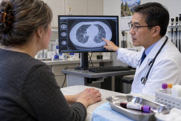
The anti-MDA5 antibody test identifies autoantibodies against melanoma differentiation-associated protein 5, an intracellular sensor of viral RNA. A convincing positive result strongly supports an anti-MDA5 dermatomyositis spectrum when it occurs with characteristic skin disease, inflammatory arthritis, hand lesions, or interstitial lung disease. Muscle weakness may be mild or absent, so the condition is sometimes called clinically amyopathic dermatomyositis. The most important complication is rapidly progressive interstitial lung disease, which can worsen over days to weeks and requires urgent specialist care.
A positive result does not mean that severe lung disease is inevitable. Risk varies by symptoms, oxygenation, CT findings, ferritin, inflammatory markers, coexisting anti-Ro52, geography, and treatment timing. Test quality also matters: weak line-blot positives can be false, especially when the phenotype is absent or several myositis antibodies are reported together. Anti-MDA5 should therefore trigger prompt clinical and lung assessment, not prognosis from the antibody number alone. A negative test does not exclude dermatomyositis or other myositis-associated lung disease.
- Anti-MDA5 is linked to a dermatomyositis phenotype that may have striking skin disease with little muscle weakness.
- Interstitial lung disease can be absent, chronic, or rapidly progressive; the antibody alone cannot determine which course will occur.
- New breathlessness, falling oxygen, fever, or rapidly changing chest imaging requires urgent evaluation.
- Weak isolated line-blot results should be confirmed when clinical features do not fit.
- Monitoring relies on symptoms, oxygen, pulmonary function, high-resolution CT, ferritin, and other clinical data—not antibody titers alone.
Table of Contents
- MDA5 and the Dermatomyositis Spectrum
- Skin, Joint, and Muscle Clues
- Lung Risk and Warning Signs
- Interpreting the Antibody Test
- Risk Assessment and Monitoring
- Treatment Principles
- What to Do After a Positive Result
MDA5 and the Dermatomyositis Spectrum
MDA5 is encoded by the IFIH1 gene and helps the innate immune system detect viral double-stranded RNA. Activation leads to interferon signaling and antiviral responses. In anti-MDA5 disease, autoimmunity targets this sensor and is accompanied by a distinctive interferon-rich inflammatory phenotype.
Anti-MDA5 was initially described in patients with clinically amyopathic dermatomyositis, meaning typical dermatomyositis skin findings with no clinically significant muscle weakness for a prolonged period. Broader experience has shown a spectrum: some patients remain amyopathic, some have mild enzyme elevation, and others develop definite inflammatory myopathy.
The diagnosis is not based on the antibody alone. A compatible phenotype may include Gottron papules, a heliotrope rash, ulceration, palmar papules, painful hand lesions, alopecia, oral ulceration, arthritis, fever, and lung disease. Different populations show different frequencies and outcomes, so statistics from one region cannot be applied mechanically to every individual.
Anti-MDA5 is a myositis-specific antibody. It is generally mutually exclusive with other major myositis-specific antibodies, although commercial panels sometimes report multiple weak bands. Anti-Ro52 can coexist because it is a myositis-associated antibody rather than a mutually exclusive defining specificity.
The antibody may be present before the complete syndrome is obvious. A patient can first appear in dermatology with ulcerative hand lesions, in rheumatology with arthritis, or in pulmonology with unexplained ILD. Recognizing the cross-organ pattern matters because lung disease can progress before substantial muscle symptoms develop.
Anti-MDA5 titers may fall with treatment and have prognostic associations in research cohorts, but assays are not standardized sufficiently to use a universal cutoff. Clinical response and lung physiology remain more actionable.
Skin, Joint, and Muscle Clues
The skin phenotype can provide an early warning. Classic dermatomyositis findings include violet or erythematous papules over the knuckles, eyelid discoloration and swelling, a photosensitive shawl or V-sign rash, and nailfold changes. Anti-MDA5 disease also has several enriched features.
Painful palmar papules or macules can occur over the finger joints and creases. Ulcers may affect Gottron papules, the nailfolds, elbows, or other pressure sites. These lesions may reflect vasculopathy and can be tender or slow to heal. Oral ulcers, diffuse hair loss, and panniculitis are reported in some patients.
Mechanic’s hands—rough, cracked skin on the sides of the fingers—can appear but are not specific. Raynaud phenomenon may coexist. Skin severity and lung severity do not always move together; dramatic skin disease can occur without severe ILD, while dangerous lung progression can occur with modest rash.
Inflammatory arthritis is common and can resemble rheumatoid arthritis. Morning stiffness, swollen small joints, and difficulty making a fist may precede the dermatomyositis diagnosis. Rheumatoid factor or anti-CCP can occasionally be present, so clinicians must integrate the skin and lung picture.
Muscle involvement may be subtle. Questions should cover difficulty climbing stairs, rising from a chair, lifting the arms, washing hair, holding the head upright, swallowing, and sustaining activity. Creatine kinase may be normal or only mildly elevated. Aldolase, MRI, electromyography, and examination can detect disease missed by CK alone.
| System | Possible findings | Important caution |
|---|---|---|
| Skin | Gottron papules, ulceration, palmar papules, heliotrope rash, alopecia | Skin severity does not predict lung severity perfectly |
| Joints | Inflammatory small-joint pain and swelling | Can resemble rheumatoid arthritis |
| Muscle | No weakness, mild weakness, or definite myositis | Normal CK does not exclude the syndrome |
| Lungs | Dry cough, breathlessness, low oxygen, ILD | Progression may be rapid and life-threatening |
Skin biopsy may support dermatomyositis but usually cannot identify anti-MDA5 specifically. Infection, vasculitis, pressure injury, and medication reactions may need exclusion when ulcers are present.
Lung Risk and Warning Signs
Interstitial lung disease is the complication that gives anti-MDA5 its urgency. It can develop gradually, remain stable, or progress rapidly. Rapidly progressive ILD generally means substantial worsening over weeks to a few months, with increasing oxygen need, expanding CT abnormalities, or declining respiratory function.
Symptoms include dry cough, breathlessness with activity, chest tightness, fatigue, and reduced exercise tolerance. Fever can reflect autoimmune inflammation but must also raise concern for infection. Home pulse oximetry may detect change, but a normal reading at rest does not exclude exertional desaturation or early ILD.
High-resolution CT often shows ground-glass opacity, consolidation, organizing pneumonia, nonspecific interstitial pneumonia, or diffuse alveolar damage. Lower-lung and peripheral changes are common, but no single pattern proves anti-MDA5 disease. Infection, pulmonary edema, hemorrhage, aspiration, and drug toxicity can look similar.
Pulmonary function tests measure forced vital capacity and diffusing capacity, but a patient with rapidly progressing disease may be too ill for reliable testing. Serial CT, oxygen requirement, respiratory rate, blood gases, and clinical trajectory can be more important in an acute setting.
Warning signs requiring urgent same-day assessment include:
- Breathlessness worsening over days or a few weeks.
- New oxygen saturation below the person’s usual level.
- Shortness of breath at rest or inability to speak comfortably.
- New diffuse opacities on chest imaging.
- Fever with respiratory symptoms during immunosuppression.
- Coughing blood, chest pain, confusion, or marked weakness.
Anti-Ro52 co-positivity has been associated with worse lung outcomes in some cohorts. High or rising ferritin, elevated inflammatory markers, lymphopenia, and extensive CT disease may also indicate higher risk. None of these markers is sufficient alone; a low ferritin does not guarantee safety.
The anti-Ro52 test can add context, while high-resolution CT and oxygenation show the organ directly. Clinicians may involve rheumatology, pulmonology, critical care, radiology, infectious disease, and transplant services early when disease is severe.
Interpreting the Antibody Test
Anti-MDA5 can be detected by enzyme immunoassay, line immunoblot, dot blot, immunoprecipitation, or other specialist methods. Immunoprecipitation has historically been a reference technique but is not routinely available. Commercial enzyme assays may provide a numeric value; line blots often report weak, moderate, or strong bands.
A strong positive result in a patient with characteristic rash, arthritis, or ILD has high clinical significance. A weak positive in someone with no dermatomyositis features, normal lungs, and several other weak myositis bands has a lower positive predictive value.
| Result pattern | Likely meaning | Reasonable next step |
|---|---|---|
| Strong positive with typical skin disease or ILD | Strong support for anti-MDA5 dermatomyositis spectrum | Urgent baseline lung and multisystem assessment |
| Weak line-blot positive without a compatible phenotype | Possible false-positive or low-specificity result | Confirm before assigning diagnosis |
| Negative with classic dermatomyositis | Another dermatomyositis subtype remains likely | Review the broader myositis panel and organ findings |
| Falling titer during treatment | May support response | Do not substitute for lung and clinical monitoring |
Line-blot testing has known limitations across myositis antibodies. Weak multiple positives are especially suspicious because true myositis-specific antibodies are usually mutually exclusive. A clinician may request confirmation with an enzyme assay, immunoprecipitation, or another validated method when the result will drive intensive treatment.
The numerical value is method-specific. A result of 40 units on one platform cannot be mapped to another laboratory’s risk threshold. Serial testing, if used, should stay on the same platform.
Pretest probability has a large effect on interpretation. A strong result in a patient with ulcerative Gottron lesions and new bilateral ground-glass opacities is clinically coherent. A weak result in a person tested for nonspecific fatigue, with normal skin examination, CK, oxygen, and CT, is far more likely to be misleading. Clinicians should not begin aggressive immunosuppression solely to treat a laboratory band.
When confirmation is needed, the goal is not to delay urgent care. A patient with rapidly worsening respiratory disease may need treatment while a reference assay is pending. In a stable patient with no phenotype, confirmation can prevent an incorrect lifelong diagnosis and unnecessary surveillance.
A negative test does not exclude ILD, dermatomyositis, antisynthetase syndrome, or another autoimmune process. It only means anti-MDA5 was not detected by that assay. The broader myositis antibody panel can identify other phenotypes, but clinical evaluation remains necessary even when the panel is negative.
Risk Assessment and Monitoring
Every clinically credible anti-MDA5-positive patient needs a baseline respiratory assessment, even if muscle disease is minimal. This commonly includes symptoms, oxygen saturation, pulmonary function testing, and high-resolution CT when indicated. The urgency depends on current respiratory status and how quickly symptoms are changing.
Ferritin is often checked because very high levels have been associated with severe inflammation and poorer outcomes. CRP, ESR, LDH, blood counts, liver enzymes, CK, aldolase, and markers of tissue injury may also be followed. Ferritin is nonspecific and can rise with infection, liver disease, malignancy, and macrophage activation.
Monitoring intervals are individualized. Stable outpatient disease may be followed with repeated symptoms, examination, PFTs, and selected imaging. Rapidly progressive disease may require daily or more frequent assessment in hospital. ACR/CHEST guidance supports PFTs and HRCT for screening and monitoring ILD in idiopathic inflammatory myopathies, with testing matched to clinical need.
The goal is to detect trajectory. A modest FVC decline, increasing oxygen need, expanding ground-glass changes, and rising ferritin together are more concerning than a single isolated abnormality. Conversely, CT abnormalities can persist after inflammation is controlled and may represent residual fibrosis.
A personal baseline is valuable. Recording resting and exertional oxygen, respiratory rate, FVC, DLCO, CT extent, ferritin, weight, skin photographs, strength, and functional capacity creates a comparison point. The same marker can mean different things in different patients, so changes should be interpreted against that baseline and the treatment timeline.
Infection monitoring is essential because the treatment itself can be intensive. Pneumocystis pneumonia, viral infection, bacterial pneumonia, and fungal disease can mimic autoimmune ILD progression. Bronchoscopy or other microbiologic testing may be necessary when the diagnosis is uncertain.
Muscle and skin monitoring should continue even when lungs dominate care. Photographs can document rash and ulcers. Strength testing, swallowing assessment, weight, and functional measures show whether other organs are improving or deteriorating.
Cancer screening should be age-appropriate and symptom-guided. Anti-MDA5 is not among the dermatomyositis antibodies with the strongest cancer association, but dermatomyositis still warrants thoughtful malignancy assessment based on overall risk.
Treatment Principles
Anti-MDA5-associated ILD is treated according to severity and pace. Rapidly progressive disease often prompts early combination immunosuppression rather than slow sequential escalation. Regimens may include high-dose corticosteroids plus a calcineurin inhibitor and cyclophosphamide, or other combinations involving mycophenolate, rituximab, intravenous immunoglobulin, or Janus kinase inhibitors.
Current ACR/CHEST recommendations for rapidly progressive ILD support using combination therapy in appropriate patients and list several agents as options. The evidence is limited and often observational, so treatment should be directed by specialists experienced with myositis-associated lung disease.
The balance is difficult: delayed control can allow irreversible lung injury, while aggressive immunosuppression increases serious infection risk. Screening for tuberculosis, hepatitis, and other infections, prophylaxis against Pneumocystis when indicated, vaccination review, blood-count monitoring, and prompt investigation of fever are crucial.
Skin ulceration may require wound care, pain control, infection prevention, vasodilator strategies in selected cases, and systemic disease control. Arthritis may respond to the same immunotherapy used for lungs, but residual joint symptoms may need separate management.
Patients with mild or no ILD are not automatically treated with the same intensity as critically ill patients. Their plan is based on skin severity, arthritis, muscle involvement, imaging, lung function, and risk markers. A positive antibody without objective disease generally does not justify high-risk immunosuppression.
Physical therapy should reflect muscle activity and respiratory reserve. Severe lung disease may limit exercise, while controlled disease benefits from graded rehabilitation. Swallowing problems require speech-language and nutrition support.
Refractory respiratory failure may prompt referral for extracorporeal support or lung transplantation assessment in selected patients. Early discussion is preferable to waiting until options are no longer feasible.
What to Do After a Positive Result
The blood draw usually does not require fasting. Provide a complete medication list and mention recent infection, vaccination, strenuous exercise, smoking or vaping, and prior lung imaging. Do not delay urgent respiratory evaluation while waiting for repeat antibody testing.
Questions to ask include:
- Was the anti-MDA5 result weak or strong, and which method was used?
- Do my skin, joint, muscle, or lung findings fit the antibody?
- Do I need immediate high-resolution CT, PFTs, oxygen testing, or hospital evaluation?
- What are my ferritin, blood counts, CK, and anti-Ro52 results?
- Does the test need confirmation before intensive treatment?
- How will infection be distinguished from autoimmune lung progression?
- Which symptoms should trigger emergency care?
A positive result can be frightening because of its association with rapidly progressive ILD. The appropriate response is rapid, structured, multidisciplinary assessment—not assuming the worst outcome from a single laboratory result. Many patients have stable or treatable disease, and early recognition improves the chance of timely, coordinated management and safer long-term follow-up across multiple involved specialties and care settings worldwide today.
Seek emergency care for shortness of breath at rest, a rapidly falling oxygen level, bluish lips, confusion, severe chest pain, coughing blood, inability to swallow, or profound weakness. These symptoms require immediate evaluation regardless of the antibody value.
References
- Diagnosis and Management of Myositis-Associated Lung Disease 2023 (Review)
- A Review of MDA-5 Dermatomyositis and Associated Interstitial Lung Disease 2024 (Review)
- Comparison of Lineblot and Immunoprecipitation Methods in the Detection of Myositis-Specific and Myositis-Associated Antibodies in Patients with Idiopathic Inflammatory Myopathies 2024
- 2023 American College of Rheumatology (ACR)/American College of Chest Physicians (CHEST) Guideline for the Screening and Monitoring of Interstitial Lung Disease in People with Systemic Autoimmune Rheumatic Diseases 2024 (Guideline)
- 2023 American College of Rheumatology (ACR)/American College of Chest Physicians (CHEST) Guideline for the Treatment of Interstitial Lung Disease in People with Systemic Autoimmune Rheumatic Diseases 2024 (Guideline)
- Analytical performance of commercial myositis-specific autoantibody assays: A systematic review and meta-analysis 2025 (Systematic Review)
Disclaimer
This article is educational and does not diagnose dermatomyositis or interstitial lung disease or predict an individual outcome. Anti-MDA5 results must be interpreted with the assay method, clinical features, oxygenation, imaging, pulmonary function, and other laboratory findings by qualified specialists. Rapidly worsening breathing, low oxygen, chest pain, confusion, or swallowing difficulty requires urgent medical care.





