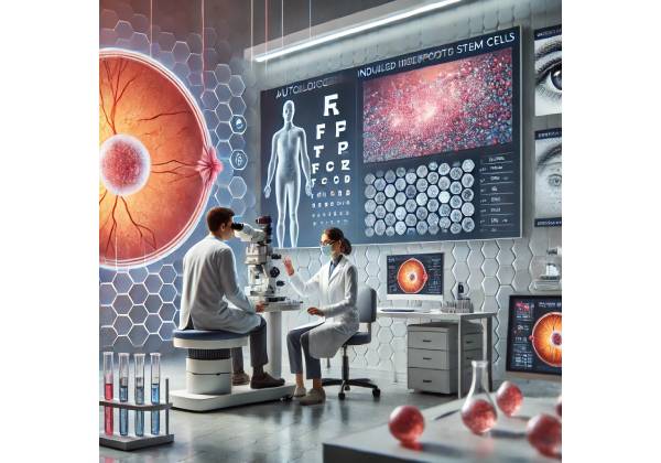
Retinitis Pigmentosa (RP) is a challenging and often debilitating group of inherited retinal disorders that lead to progressive photoreceptor degeneration and eventual vision loss. For decades, ophthalmologists and researchers have sought interventions to slow, halt, or even reverse the damage caused by RP. While existing therapies such as nutritional supplementation, vitamin A, and electronic retinal implants have provided marginal improvements for some patients, the quest continues for a more definitive, regenerative solution. Recent advances in cell-based therapies—especially those involving autologous induced pluripotent stem cells (iPSC)—offer renewed hope of truly preserving or restoring functional vision. This article delves into how autologous iPSC therapy works, why it holds promise for patients with RP, and what the current research reveals about safety, efficacy, accessibility, and costs.
A Game-Changing Approach: Autologous iPSC Therapy for Retinitis Pigmentosa
(Overview of the Therapy)
What Are iPSC?
Induced pluripotent stem cells (iPSC) are laboratory-generated cells that originate from fully differentiated adult somatic cells—often skin cells or blood cells—that are “reprogrammed” back into an embryonic stem cell-like state. This reprogramming is typically accomplished by introducing specific transcription factors (commonly known as the Yamanaka factors: OCT4, SOX2, KLF4, and c-MYC), which reset the epigenetic clock of the adult cell, granting it the ability to self-renew and differentiate into most cell types in the human body. When used in an autologous context, iPSC are derived from a patient’s own tissues, theoretically minimizing immune rejection risks post-transplantation.
Why iPSC Matter for RP
Retinitis Pigmentosa arises from a variety of genetic mutations that ultimately cause retinal photoreceptors (rods and cones) to degrade. Because iPSC can differentiate into many cell types—including retinal pigment epithelium (RPE) cells and photoreceptors—researchers have investigated whether transplanting iPSC-derived cells could replenish or support dying retinal tissue. This approach, if successful, could preserve existing retinal function, restore partial vision, or at least slow the disease’s progression.
Key Advantages of Autologous iPSC
- Reduced Immunogenicity: Cells generated from the patient’s own tissue are less likely to trigger a robust rejection response after transplantation, potentially negating the need for lifelong immunosuppression.
- Tailored Therapy: Because iPSC can theoretically be personalized based on each patient’s genetic makeup, future treatments might incorporate gene editing to fix or bypass the specific mutation causing RP.
- Wide Differentiation Potential: In advanced cases of RP, more than just the photoreceptors may be compromised—RPE cells and other support structures can also degrade. The pluripotent nature of iPSC opens the door for regenerating multiple cell types in the retina.
- Long-Term Sustainability: Once transplanted cells integrate into the retinal environment, they have the potential to continue functioning for extended periods, offering more durable improvements than symptomatic management alone.
Although still in exploratory and early clinical stages, autologous iPSC therapy stands out for its ambitious goal of replacing damaged retinal tissue itself, rather than merely slowing down ongoing degeneration or bypassing lost function.
Retinitis Pigmentosa: The Silent Threat to Retinal Photoreceptors
(Understanding the Condition)
Nature of RP and Genetic Diversity
Retinitis Pigmentosa includes a group of hereditary retinal diseases—over 60 genes have been implicated, and inheritance patterns can be autosomal dominant, autosomal recessive, or X-linked. Regardless of specific genetic pathways, the end result is often similar: gradual loss of rod photoreceptors responsible for peripheral and night vision, followed by cone photoreceptor damage that eventually affects central vision. Patients often notice night blindness (nyctalopia) and peripheral vision constriction early on, with progressive tunnel vision culminating in severe visual impairment.
Given that rods outnumber cones in the human retina, rod dysfunction can remain asymptomatic for years. By the time an individual seeks an ophthalmologic evaluation, significant peripheral damage may have already occurred. Over time, as cones become involved, reading, color discrimination, and central acuity diminish.
Prevalence and Impact
- Incidence: RP affects roughly 1 in 3,500 to 1 in 4,000 individuals globally, making it one of the most common inherited retinal dystrophies.
- Quality of Life: The progressive and irreversible nature of RP can force significant lifestyle adjustments. Many patients are eventually classified as legally blind, though the timeline can vary widely.
- Therapeutic Gap: While interventions like vitamin A or omega-3 fatty acids have shown modest beneficial effects in some subpopulations, they do not effectively reverse damage. For advanced cases, electronic retinal implants (the “bionic eye”) or gene therapy trials have been pursued, but large-scale, definitive cures have remained elusive.
The Role of RPE in RP Pathophysiology
Although rods and cones bear the brunt of the pathology, the retinal pigment epithelium (RPE)—a layer essential for photoreceptor metabolism and waste management—can also deteriorate in many forms of RP. This deterioration exacerbates photoreceptor loss because healthy RPE is crucial for regenerating photopigments, removing debris, and maintaining the delicate retinal environment. iPSC therapy that includes RPE cell replacement or support is thus of keen interest to stabilize or restore normal cellular crosstalk in the retina.
Reprogramming to Rescue Vision: How iPSC Therapy Works
(Mechanism of Action of the Therapy)
iPSC Generation and Differentiation
The process of creating autologous iPSC for RP typically begins with a small biopsy of the patient’s tissue—often a skin punch biopsy or a blood draw. The isolated cells are then cultured in a lab and “reprogrammed” by introducing the reprogramming factors. Once the cells have reverted to a pluripotent state, they can undergo directed differentiation into retinal cells, including:
- Retinal Pigment Epithelial (RPE) Cells: Forming a monolayer that supports photoreceptor health.
- Photoreceptor Precursors: Potentially capable of maturing into rods or cones once transplanted into the host retina.
This differentiation step typically relies on a carefully calibrated cocktail of growth factors and signaling molecules that mimic embryonic retinal development conditions. Successful differentiation is verified through molecular markers (e.g., RPE65 for RPE cells, opsins for photoreceptors) and functional assays.
Implantation and Integration
The next stage involves delivering the iPSC-derived cells into the subretinal space—between the neural retina and the underlying choroid. Surgical approaches can vary, but frequently this is done via a small retinotomy, through which a delicate injection of cell suspension or a pre-fabricated cell sheet is introduced. In theory, the transplanted cells:
- Settle in the subretinal space.
- Integrate with the host tissue, forming or joining a functional RPE monolayer or partially replacing lost photoreceptors.
- Provide Trophic Support, secreting growth factors that help maintain surviving photoreceptors and slow further degeneration.
Potential for Visual Restoration
- Support Mechanisms: Even if photoreceptors are not fully replaced, newly integrated RPE can stabilize existing rods and cones by improving nutrient delivery and waste clearance.
- True Regeneration: If photoreceptor precursor cells mature in situ, they might restore some degree of light sensitivity, improving peripheral or central vision.
- Neuroprotective Benefits: In some experiments, iPSC-derived cells have shown a beneficial paracrine effect—producing substances that reduce retinal inflammation and preserve adjacent neurons.
Still, many scientific challenges remain, including ensuring the grafted cells align properly with the host retina’s architecture, forging synaptic connections with bipolar and ganglion cells, and avoiding immune responses even with autologous cells (particularly if any gene-editing steps introduce subtle antigenic differences).
From Lab to Clinic: Implementing an iPSC Treatment Plan
(Application and Treatment Protocols)
Step-by-Step Treatment Workflow
- Patient Eligibility and Pre-Screening
- Genetic Testing: Identifying the specific mutation, verifying the stage of retinal damage, and confirming that the patient’s retina retains a reasonable potential for functional recovery.
- Ocular Imaging: Optical coherence tomography (OCT) and fundus examinations help assess photoreceptor and RPE integrity.
- Systemic Health Evaluation: Since the iPSC generation process requires a biopsy, patients must be generally healthy enough to undergo the procedure.
- Cell Harvest and Reprogramming
- Tissue Collection: A small tissue sample—often a skin biopsy or a peripheral blood draw—provides the source cells.
- Reprogramming in a GMP-Compliant Lab: Ensuring Good Manufacturing Practice (GMP) standards is crucial for clinical-grade cell therapy.
- Quality Control: The newly generated iPSC are tested for sterility, genetic stability, and pluripotency markers.
- Cell Differentiation and Expansion
- Directed Differentiation Protocols: Lab technicians induce the iPSC to become RPE cells or photoreceptor progenitors using specific media and growth factors.
- Scale-Up: Sufficient cell numbers must be grown to create an implantable population. This can be a time-consuming process, extending for weeks or months.
- Pre-Implantation Characterization: Specialists confirm that the cells display the correct phenotype and gene expression profile, and remain free from contaminants.
- Surgical Delivery
- Procedure Planning: Ophthalmic surgeons determine the ideal injection or transplantation site. Subretinal injection is often performed under local or general anesthesia with micro-scale precision.
- Minimizing Trauma: Techniques such as 25- or 27-gauge vitrectomy instrumentation can limit complications when creating a bleb or retinotomy for cell placement.
- Intra- and Postoperative Monitoring: Surgeons may utilize intraoperative OCT to confirm correct cell placement.
- Follow-Up and Rehabilitation
- Pharmacological Support: Immunosuppressive regimens are typically minimized given the autologous nature of the cells but may still be used short-term to dampen inflammatory responses.
- Visual Function Testing: Regular assessments with electroretinography (ERG), visual field exams, and standard acuity tests track improvements or stability.
- Ongoing Imaging: OCT scans identify changes in retinal thickness, layering, and potential signs of cell graft survival.
Anticipating Potential Hurdles
- Suboptimal Cell Survival: Even carefully transplanted cells can undergo apoptosis if the microenvironment is hostile.
- Surgical Complications: Risks include retinal detachment, hemorrhage, or infection.
- Incomplete Integration: Grafted cells may fail to form functional synapses or orient themselves properly, limiting the degree of vision recovery.
Nevertheless, as research refines these protocols, the potential benefits for patients with few other viable treatment options remain highly compelling.
Measuring Success: Evaluating the Efficacy and Safety of iPSC-Based Therapies
(Effectiveness and Safety)
Early Indicators of Therapeutic Benefit
Because full regeneration of photoreceptor layers is an ambitious goal, success in early iPSC trials often centers on less dramatic but still meaningful milestones. These may include:
- Slowed Progression: A reduction in the rate of visual field narrowing or a slower decline in electroretinogram amplitudes compared to untreated controls.
- Stabilized Central Acuity: Even if only partial photoreceptor function is regained, patients may notice they can read for longer periods or perceive more detail in bright light conditions.
- Quality of Life Measures: Self-reported improvements in daily activities, orientation, and mobility, which can significantly affect mental health and independence.
Safety Considerations
- Tumorigenicity: One of the principal concerns with any pluripotent cell-based therapy is the risk of uncontrolled cell growth or teratoma formation. Rigorous screening and elimination of undifferentiated cells reduce this risk.
- Immune Responses: Even with autologous cells, epigenetic or genetic modifications during reprogramming could elicit immune reactions, though these instances appear rare with thorough characterization.
- Surgical Risks: Introducing cells into the subretinal space can precipitate a detachment or local inflammation, particularly if the retina is already fragile.
Medium- to Long-Term Outcomes
Though many of the current iPSC-based therapies for retinal conditions remain experimental or in early-phase clinical trials, interim data reveal that patients often tolerate these procedures well, with few severe adverse events reported thus far. For those who demonstrate stable or improved retinal function, the effect can last months to years, prompting continued optimism about the long-term trajectory as protocols are refined.
Groundbreaking Trials: What the Data Says on iPSC for Retinitis Pigmentosa
(Current Research Insights)
Pioneering Studies and Proof-of-Concept Research
- First-in-Human RPE Transplants: Around 2014–2015, a landmark trial in Japan led by RIKEN Institute researchers tested autologous iPSC-derived RPE cells in a patient with exudative (wet) age-related macular degeneration. Although not directly targeting RP, this study provided a template for autologous cell transplantation in retinal disease and demonstrated that the procedure was both feasible and safe over an extended follow-up.
- Pilot RP Studies: Several academic centers worldwide have begun smaller pilot or Phase I/II clinical trials investigating iPSC-derived retinal cells in patients with advanced RP. Many of these studies remain ongoing, but preliminary safety data is encouraging, with no major immune rejections or teratoma formation reported in the short term.
Preclinical Models
In rodent and non-human primate models of RP, iPSC-derived photoreceptors have shown the capacity to survive, integrate into the outer nuclear layer, and form synaptic connections with bipolar cells. Visual function improvements, measured through ERG and behavioral tests (e.g., navigation in dim light), support the hypothesis that iPSC can partially restore sight.
Statistical Highlights
- Cell Survival Rates: Depending on the study design and host environment, anywhere from 10% to 50% of transplanted photoreceptor precursors remain viable over several months. Optimization in surgical technique and scaffolding materials (like biodegradable polymers) continue to boost these figures.
- Visual Acuity Changes: Some early-phase human trials report slight improvements—on the order of 5-10 letters on the ETDRS chart—after iPSC-derived RPE transplantation for degenerative diseases. While modest, these results are notable for advanced-stage pathologies traditionally regarded as untreatable.
- Safety Profile: To date, large-scale data on catastrophic complications remain limited, but early-phase trials point to a reassuring safety profile, with minimal incidence of serious adverse events.
Researchers caution that robust, randomized, and controlled trials with longer follow-up durations are still needed to confirm these promising outcomes. Nevertheless, the incremental successes in early-phase investigations have laid a strong foundation for expanding the scope and scale of iPSC-based interventions for RP.
Cost and Coverage: Navigating the Path to iPSC Therapy
(Pricing and Accessibility of the Therapy)
The High Cost of Customization
The promise of autologous iPSC therapy lies in its individualized nature, but that customization can come at a steep financial cost. Production of clinical-grade iPSC lines requires specialized facilities (often with GMP certification) and rigorous quality control. Differentiating these cells into RPE or photoreceptor progenitors further extends lab time and material expenses.
- Cell Harvesting and Reprogramming: Lab fees for reprogramming a single patient’s cells can exceed \$10,000–\$20,000 (USD) under current conditions in high-cost research environments.
- Scaling Up Differentiation: The culture media, growth factors, and advanced quality assays can double or triple overall costs, depending on quantity and complexity.
- Surgical Fees and Hospital Stay: Skilled retina surgeons, operating room time, and post-op care add additional thousands to tens of thousands of dollars to the final bill.
Though widespread insurance coverage for iPSC-based therapies is not yet established, some private insurers in certain countries may consider partial reimbursement if the therapy is part of an approved clinical trial or if conventional treatments have failed. Public healthcare systems rarely cover experimental therapies at full cost, although compassionate use programs occasionally exist for life-altering conditions like advanced RP.
Potential Variants in Pricing
- Government or Institutional Subsidies: Some advanced research institutions offset lab and surgical fees, especially if the patient is enrolled in a clinical study.
- Commercial Partnerships: Biotechnology companies might partner with academic centers to reduce production costs through more standardized iPSC generation processes, though these are still in early stages.
- Gene Editing Add-Ons: In the future, treatments that correct the underlying gene mutation within iPSC lines might raise the price even further but could significantly enhance efficacy.
Toward Greater Accessibility
Biotechnology experts believe that as iPSC reprogramming techniques mature, automation, scaled production, and possibly the development of universal donor cell lines could reduce costs. While universal lines are allogeneic (not autologous), new strategies to “cloak” cells from the immune system (e.g., by knocking out MHC genes) might help them behave similarly to autologous cells. If successful, such off-the-shelf solutions could drastically cut manufacturing costs and broaden patient access.
Nonetheless, in the near term, autologous iPSC therapy remains an advanced, resource-intensive procedure likely to be offered primarily through specialized centers of excellence. For patients determined to pursue it, exploring clinical trials, philanthropic grants, or special early-access programs may be crucial in alleviating financial hurdles.
Disclaimer
This article is intended for educational purposes only and does not replace professional medical advice, diagnosis, or treatment. Always consult a qualified healthcare provider regarding any questions or concerns you may have about a medical condition or treatment options.

