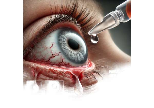
Persistent epithelial defects of the cornea can be profoundly debilitating, undermining not only the clarity of vision but also an individual’s overall comfort and quality of life. In a healthy eye, the cornea’s outermost layer regenerates efficiently thanks to an intricate balance of tears, nutrients, and cellular signals that foster wound repair. However, conditions like severe dry eye, ocular surface disease, autoimmune disorders, or prolonged post-surgical complications can disrupt this process. When the cornea cannot heal on its own, patients may experience chronic pain, light sensitivity, and a heightened risk of infection and scarring.
While artificial tears and conventional ointments offer symptomatic relief, they often do not deliver the bioactive components needed for true tissue regeneration. This shortfall has led to the growing use of autologous serum eye drops, a therapy derived from a patient’s own blood. Loaded with growth factors and other nourishing elements naturally found in human serum, these customized drops have emerged as a powerful intervention for healing persistent epithelial defects. In the following sections, we delve into how autologous serum eye drops work, discuss protocols for their use, explore relevant research data, and examine the associated safety profile and cost considerations.
Understanding the Role of Autologous Serum in Corneal Healing
Autologous serum eye drops harness the concept that the human body produces an array of biological factors essential for cellular repair. When derived from a patient’s own bloodstream, these serum-based solutions can provide a therapeutic milieu that closely mimics the tear film’s natural composition. With an assortment of vitamins, growth factors, and immunoglobulins, autologous serum becomes uniquely suited to repairing damaged corneal epithelium.
The Natural Healing Power of Serum
Human tears do more than keep the eye surface moist; they supply oxygen, nutrients, and defensive molecules to ward off pathogens. Serum from the blood has several of these same qualities in far higher concentrations, especially in terms of growth factors such as:
- Epidermal Growth Factor (EGF): A key element in stimulating epithelial cell proliferation and migration, promoting wound closure.
- Fibronectin: Essential for proper cell adhesion, serving as a scaffold that encourages epithelial cells to spread and cover damaged tissue.
- Vitamin A: Crucial for maintaining epithelial integrity and supporting healthy ocular surface development.
- Transforming Growth Factor-β (TGF-β): Can modulate inflammation and support tissue remodeling.
By integrating these naturally occurring molecules into eye drops, autologous serum solutions support a damaged cornea’s cellular environment more effectively than many synthetic formulations. Clinical experience shows that this synergy of proteins and vitamins can help reestablish the tear film’s normal biochemical composition, making autologous serum eye drops especially beneficial for severe dry eye syndromes or complicated post-surgical healing.
Why Persistent Epithelial Defects Are So Challenging
Persistent epithelial defects—commonly abbreviated as PEDs—occur when the corneal epithelium does not fully close or regenerate within an expected timeframe (often 2 weeks or more). A variety of ocular conditions can lead to this stubborn non-healing state, including:
- Neurotrophic Keratitis: Reduced corneal sensitivity due to nerve damage impedes normal blinking and tear production, slowing wound repair.
- Severe Dry Eye: Inadequate tear film results in recurrent epithelial breakdown.
- Graft-versus-Host Disease (GVHD): Immune dysfunction impairs the eye’s natural healing pathways.
- Post-Keratoplasty: Following corneal transplantation, a compromised ocular surface environment may hinder re-epithelialization.
Because PEDs introduce the risk of infection and corneal perforation, clinicians often pursue aggressive treatments such as bandage contact lenses, punctal occlusion, or amniotic membrane grafting. Yet these approaches do not always deliver the bioactive nutrients necessary for definitive healing. Autologous serum eye drops step in to supply growth factors that accelerate epithelial cell migration and reduce dryness, bridging a vital gap in the typical regimen for corneal disorders.
Tailoring the Therapy to the Patient
One of the most compelling advantages of autologous serum is its individualized formulation. Derived from the patient’s own blood, the therapy bypasses cross-reactivity or rejection issues, making it especially valuable for those with autoimmune conditions. Moreover, the composition of each drop can be fine-tuned by altering dilution ratios. Commonly, clinicians dilute serum with a sterile balanced salt solution (BSS) to levels of 20% to 100%, depending on the severity of the defect and the eye’s tolerance. This customization ensures that each patient receives an optimal concentration of proteins, vitamins, and growth factors without excessive viscosity or potential toxicity.
Despite these promising benefits, autologous serum eye drops demand a precise preparation process, strict handling measures, and careful follow-up. Patients and healthcare providers must be aware of best practices to prevent contamination, maintain sterility, and maximize clinical outcomes. In doing so, this therapy can offer a pathway to relief for individuals who have long struggled with chronic, non-healing corneal wounds.
Guidelines for Proper Use and Administration
Turning a patient’s blood sample into a usable eye drop involves a sequence of meticulous steps, beginning with the blood draw and ending with consistent at-home application. Because the cornea is so vulnerable to microbial infection, each stage mandates careful attention to sterility. Moreover, patients and practitioners must collaborate to ensure consistent usage schedules, correct drop storage, and regular monitoring for improvement or complications.
Step-by-Step Preparation
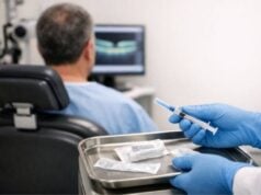
- Blood Collection: Typically, around 10–50 mL of venous blood is drawn from the patient, depending on how many serum vials are required. This procedure usually happens in a certified clinical setting or blood bank.
- Centrifugation: The collected blood is spun in a centrifuge to separate the serum portion from red blood cells and clotting factors. A second spin may be used to clarify the serum further.
- Dilution: Serum is diluted with a sterile solution, often balanced salt solution or 0.9% sodium chloride, to achieve the target concentration. Common dilutions range from 20% to 50% or higher. The ratio chosen depends on the severity of the epithelial defect, plus the patient’s underlying condition.
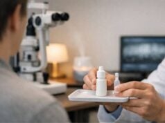
- Aliquoting and Packaging: The finished serum mixture is transferred into multiple dropper bottles or sterile vials, typically with a capacity of 3–5 mL each. These containers are then labeled with the patient’s name, date, and dilution ratio.
- Storage: Serum eye drops must be kept refrigerated or, in some cases, frozen for longer shelf-life. Many protocols advise discarding any opened vial after one week to limit the risk of contamination.
Because the entire supply of autologous serum drops stems from a single venipuncture session, the average yield may last for several weeks. When the supply runs low or therapy must continue, patients schedule another blood draw.
Recommended Application Schedules
The optimal dosing frequency varies based on the severity of the epithelial defect and the patient’s tolerance. In typical use, patients instill autologous serum drops anywhere from 4 to 8 times daily. For more severe, persistent defects, some clinicians recommend as many as 12 or more drops daily, although practical constraints may limit feasibility.
Crucially, the therapy’s success often hinges on adherence. Because corneal healing is an incremental process, skipping doses can delay or undermine progress. Patients should be educated about:
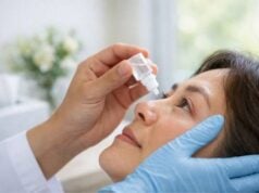
- Hand Hygiene: Washing thoroughly before each application to reduce bacterial contamination risk.
- Dropper Technique: Holding the bottle at a slight angle and avoiding direct contact with eyelashes or eyelid margins that could harbor microbes.
- Single-Use Guidelines: Some clinics opt for single-use droppers or specialized packaging to further diminish contamination risks.
Balancing With Other Therapies
In many cases, autologous serum eye drops complement ongoing treatments like antibiotic prophylaxis, lubricating ointments, or bandage contact lenses. While serum alone can address the biochemical deficits of persistent epithelial defects, other steps—such as controlling underlying inflammation or dryness—can multiply its efficacy. Additionally, some clinicians prefer that patients wait a few minutes between applying different drops to avoid potential dilution or chemical interactions.
When integrating autologous serum into a broader regimen, timing is key. Patients are sometimes advised to administer serum drops last in a series, granting them the maximum contact time with the corneal surface. That said, protocols can differ among practitioners, so it’s essential for each patient to follow individualized directions meticulously.
Patient Education and Follow-Up
Experience shows that patients using autologous serum eye drops can achieve significant epithelial healing within a matter of weeks, depending on the size and chronicity of the defect. But consistent monitoring remains vital. Most clinicians schedule follow-up visits at intervals of 1 to 2 weeks initially, observing corneal epithelial status via slit-lamp examination and assessing tear quality with fluorescein staining.
Additionally, clinicians look for any signs of infection or inflammation. If new infiltration, persistent epithelial defects, or unusual corneal haze arises, therapy may be adjusted—either by increasing concentration or adding supportive measures like punctal occlusion. Conversely, once a defect heals, the dosing frequency can gradually taper, eventually discontinuing if stable re-epithelialization persists.
Insights from Clinical Trials and Ongoing Research
The use of autologous serum eye drops may seem somewhat intuitive—leveraging the body’s own healing powers for corneal repair—but their efficacy has been systematically studied in numerous clinical investigations. These include smaller observational reports and larger prospective trials, each contributing valuable insights into how and why serum-based therapy can outperform standard treatments for chronic ocular surface conditions.
Landmark Studies Supporting Efficacy
Over the past two decades, multiple studies have validated autologous serum’s role in corneal epithelial healing:
- Randomized Controlled Trials: Investigations comparing autologous serum to conventional artificial tears frequently demonstrate faster healing rates, improved tear film stability, and reduced ocular discomfort among serum users. These differences often become evident within the first few weeks of treatment.
- Severe Dry Eye Syndromes: In Sjögren’s syndrome or other immunological conditions that severely compromise tear production, autologous serum repeatedly shows marked improvement in corneal staining scores, tear breakup time, and subjective discomfort levels.
- Neurotrophic Keratopathy: For patients with nerve damage (from conditions like herpes zoster or diabetes), trials indicate that serum-based drops can restore sensation gradually and promote epithelial regrowth more effectively than standard lubricants.
A consistent theme in these studies is the synergy between serum’s growth factors and a chronically inflamed, nutrient-deprived ocular surface. By supplying cellular signals that direct epithelial closure and modulate inflammation, autologous serum sets the stage for a stable corneal environment.
Emerging Therapies and Alternatives
While autologous serum has become a mainstay for many clinicians, researchers continue to explore related biologics to optimize healing further:
- Platelet-Rich Plasma (PRP): Much like serum, PRP is derived from the patient’s blood but with additional concentration of platelets, which release an even richer cocktail of growth factors. Pilot studies show promise in accelerating corneal wound closure and mitigating corneal haze.
- Cord Blood Serum: Collected from umbilical cords donated at childbirth, this resource is rich in growth factors akin to autologous serum. Cord blood-based drops may be suitable for individuals unable to provide adequate serum.
- Recombinant Growth Factor Formulations: Synthetic EGF or nerve growth factor (NGF) drops represent another frontier. Though potential benefits exist, replicating the full biochemical complexity of human serum remains challenging.
In parallel, a wave of cutting-edge research on ocular surface stem cells, 3D bioengineered corneas, and gene editing continues to refine how we approach stubborn corneal defects. Nevertheless, serum-based therapies stand out for their relative simplicity and cost-effectiveness, capitalizing on the body’s innate capacity to regenerate.
Ongoing Questions and Future Directions
Despite demonstrable success, some aspects of autologous serum therapy merit further examination:
- Optimal Dilution Ratios: While most protocols hover between 20% and 50% serum concentration, some specialists favor 100% for severe cases. Additional randomized studies could delineate precise dosing guidelines for each type of corneal defect.
- Long-Term Maintenance: For chronic ocular conditions, is indefinite use of serum drops beneficial, or does repeated short-course therapy suffice once the defect heals? Observational data suggest patient-to-patient variability.
- Storage Stability: Current practice is to refrigerate or freeze serum. Researchers are investigating improved packaging or preservatives that might extend shelf life without reducing efficacy.
- Biochemical Profiling: Systematic analysis of exactly which factors in serum drive corneal healing could spark new generation eye drops that isolate and amplify the most pivotal proteins.
Given the robust data supporting autologous serum, it’s likely that further refinements in technique, storage, and synergy with other biologics will keep shaping how we treat persistent epithelial defects. Clinicians are already adapting the therapy for use in advanced ocular surface disease, pediatric corneal issues, and even mild dryness unresponsive to standard artificial tears.
Safety Profile and Clinical Benefits
Any therapy derived from human blood raises concerns about sterility, contamination, and variable efficacy. Nonetheless, autologous serum eye drops, when prepared under proper protocols, have repeatedly proven safe and well-tolerated for most patient populations. Understanding these safety considerations while highlighting the therapy’s confirmed benefits helps inform discussions between patients and their healthcare providers.
Minimal Risk When Properly Prepared
Safety begins with the handling of the patient’s blood. Modern guidelines stipulate:
- Stringent Aseptic Technique: Blood draws and serum preparation occur under clean, controlled conditions. Reputable clinics or transfusion centers typically have specialized equipment to perform these tasks.
- Testing for Transmissible Diseases: Even though the blood is autologous (used on the same donor-patient), many facilities still screen for infections like HIV or hepatitis for record-keeping and potential contamination concerns.
- Controlled Freezing and Thawing: If serum must be stored for extended periods, flash-freezing in single-use vials helps preserve factor potency while minimizing the bacterial growth that can occur in repeated freeze-thaw cycles.
One of the therapy’s inherent advantages is that it originates from the patient’s own body, eliminating risk of immunologic rejection or cross-contamination from donor material. When standard procedures are followed, the incidence of ocular infections tied to autologous serum use remains extremely low.
Proven Advantages Beyond Wound Healing
While the primary goal is to close persistent epithelial defects, many patients report broader benefits:
- Enhanced Ocular Comfort: Replenishing tear film components often alleviates dryness, burning, and irritation far more effectively than synthetic tears alone.
- Reduced Dependence on Conventional Drops: Patients reliant on frequent artificial tear instillation or antibiotic ointments may find themselves requiring fewer adjunctive treatments once epithelial healing commences.
- Stabilized Visual Acuity: Although resolution of corneal haze or scarring can vary, a healed epithelial surface tends to provide more consistent refraction, leading to crisper vision in some cases.
- Improved Quality of Life: Freed from chronic pain and photophobia, many individuals can resume daily activities with less ocular disruption.
In addition, autologous serum appears to carry minimal risk of steroid-related side effects (such as raised intraocular pressure), since it lacks the potent corticosteroid agents often used in other ocular therapies. That said, some patients may still require mild topical steroids or immunosuppressants in combination with serum therapy if underlying inflammation is significant.
Common Missteps and How to Avoid Them
While the therapy is generally regarded as safe and effective, certain pitfalls can undermine outcomes:
- Poor Compliance: Failing to keep up with recommended dosing schedules and storage protocols can lead to suboptimal results or contamination.
- Inadequate Follow-Up: Without routine eye exams, minor complications like a partial defect or low-grade infection may go unnoticed until they intensify.
- Improper Dilution: Using an overly high concentration in a severely inflamed eye can occasionally cause stinging or mild toxicity. Conversely, an overly low concentration might not deliver enough growth factors to accelerate healing.
- Ignoring Underlying Causes: Serum alone may not suffice if the root problem—like eyelid malposition, severe ocular surface inflammation, or tear film abnormality—remains unaddressed.
In professional practice, success hinges on a tailored approach that respects each patient’s ocular surface dynamics. Through careful patient selection, collaborative planning, and rigorous follow-up, autologous serum eye drops maintain an excellent safety profile and demonstrate remarkable efficacy in closing chronic corneal wounds that once defied standard treatments.
Potential Costs and Access Options
The cost of autologous serum eye drops can vary widely based on where they are produced, how much blood is needed, and the frequency of use. Some eye clinics prepare them onsite, bundling fees for the blood draw, laboratory processing, and packaging into a single charge that may range from a few hundred to over a thousand dollars. Other facilities outsource the work to a specialized compounding pharmacy, which can slightly raise the total expense. Insurance coverage differs by region and provider; certain plans cover autologous serum for medically necessary indications, while others classify it as an experimental therapy. Patients are encouraged to check their policies and discuss payment strategies with the medical staff. Payment plans or financial assistance programs may be available through certain healthcare institutions.
Disclaimer: This article is for educational purposes only and should not be taken as medical advice. Consult with a qualified healthcare provider for personalized information and treatment recommendations.
We invite you to share this article on Facebook, X (formerly Twitter), or any other social media platforms. By doing so, you can help more people learn about the benefits of autologous serum eye drops and potentially find relief from persistent epithelial defects.

