Introduction
Axenfeld-Rieger Syndrome is a rare genetic disorder that primarily affects eye development. It is named after Theodor Axenfeld and Hans Rieger, who were the first to describe the condition. ARS is distinguished by distinctive ocular abnormalities, including defects in the anterior segment of the eye, which can result in serious complications such as glaucoma. Although primarily an ocular condition, ARS can also have systemic effects on the teeth and other parts of the body. Awareness and early detection of ARS are critical for preventing vision loss and effectively managing its associated systemic manifestations.
Detailed overview
Axenfeld-Rieger Syndrome is inherited in an autosomal dominant pattern, which means that the disorder can be caused by a single copy of the mutated gene. Mutations in the PITX2, FOXC1, and occasionally other genes cause ARS. These genes are required for the proper development of the eye and other structures during embryonic development.
Ocular manifestations
- Posterior Embryotoxon: One of the distinguishing features of ARS is posterior embryotoxon, which is defined by a prominent anteriorly displaced Schwalbe’s line. This line appears as a thin, white ridge on the posterior cornea and is visible under a slit lamp.
- Iris Abnormalities: People with ARS frequently have iris hypoplasia, which means the iris is underdeveloped. Other common iris anomalies include polycoria (multiple holes in the iris) and correctopia (pupil displacement). These abnormalities can have an impact on both the appearance and function of the iris.
- Glaucoma: Approximately 50% of ARS patients develop glaucoma, often at a young age. This is due to abnormal development of the trabecular meshwork and Schlemm’s canal, which results in increased intraocular pressure and optic nerve damage. If left untreated, glaucoma can cause significant vision loss.
- Corneal Changes: The cornea may thin and opacify, resulting in visual impairment. Patients may also exhibit corneal endothelial abnormalities, which can impair corneal clarity and health.
Pathophysiology
The genetic mutations associated with ARS disrupt the normal development of the anterior segment of the eye.
- PITX2 Gene: This gene encodes a transcription factor required for the development of the eyes, teeth, and abdominal organs. Mutations in PITX2 disrupt normal developmental pathways, resulting in the anomalies observed in ARS.
- FOXC1 Gene: This gene also encodes a transcription factor involved in eye and brain development. FOXC1 mutations contribute to both the ocular and systemic manifestations of ARS.
Disruption in these genetic pathways affects cell differentiation and migration during embryogenesis, resulting in the ARS-specific characteristics. Abnormalities in the development of the trabecular meshwork and Schlemm’s canal impair aqueous humor drainage, resulting in elevated intraocular pressure and glaucoma.
Clinical Presentation
- Visual Symptoms: Patients with ARS may have blurred vision, photophobia (light sensitivity), and decreased visual acuity as a result of corneal and iris abnormalities. The severity of visual symptoms can vary greatly between individuals.
- Iris Hypoplasia and Corectopia: Underdevelopment of the iris and pupil displacement can have an impact on the eye’s aesthetic appearance as well as vision.
- Secondary Glaucoma: Glaucoma’s increased intraocular pressure can result in progressive vision loss if not treated properly. Patients may experience visual field loss, eye pain, or headaches.
Systemic Features
While ARS primarily affects the eyes, it can also present with the following systemic symptoms:
- Dental Anomalies: Common dental issues in ARS are hypodontia (missing teeth), microdontia (small teeth), and conical teeth. These anomalies can have an impact on both oral health and aesthetics.
- Facial Dysmorphism: ARS has a flattened midface, hypertelorism (wide-set eyes), a broad nasal bridge, and a prominent forehead.
- Periumbilical Skin Redundancy: Extra skin around the belly button is a distinguishing feature of ARS, but it is usually harmless.
- Cardiac and Abdominal Anomalies: Some patients may have congenital heart defects or other abdominal anomalies, but these are uncommon in comparison to ocular and dental symptoms.
Genetic Basis
ARS is caused by mutations in the PITX2 and FOXC1 genes, both of which play critical roles in early development. These genes play a role in the development of the eye’s anterior segment, which includes the cornea, iris, and drainage structures, as well as other body structures.
Epidemiology
ARS is a rare condition with an estimated prevalence of one in every 200,000 individuals. It affects both men and women equally and can occur in any ethnicity. If one parent has the condition, the offspring have a 50% chance of inheriting it.
Effects on Quality of Life
The ocular manifestations of ARS can have a significant impact on a patient’s quality of life. Visual impairment from corneal abnormalities and glaucoma can make daily activities difficult. Additionally, systemic features such as dental and facial anomalies can have an impact on self-esteem and social interactions.
Psychological and Social Concerns
Living with ARS can present a variety of psychological and social challenges. The visible facial and dental anomalies may cause social stigma and psychological distress. Children with ARS may face bullying or social isolation, affecting their mental health and development. Providing psychological support and counseling is critical for assisting affected individuals and their families in dealing with these challenges.
Importance of Multidisciplinary Care
Managing ARS necessitates a collaborative effort involving ophthalmologists, geneticists, dentists, cardiologists, and psychologists. Comprehensive care plans tailored to each individual’s needs can address the syndrome’s diverse manifestations. Early intervention and regular monitoring are critical for effectively managing the condition and improving the quality of life for those affected.
Essential Preventive Tips
- Regular Eye Examinations: Schedule comprehensive eye exams with an ophthalmologist who is familiar with ARS. Early detection and monitoring of glaucoma and other ocular abnormalities is critical to preventing vision loss.
- Genetic Counseling: Families affected by ARS should seek genetic counseling to learn about the inheritance pattern, risks to future offspring, and available genetic testing options. Counseling can also provide useful information for managing the condition.
- Early Dental Care: Consult a specialist who is experienced in treating dental anomalies associated with ARS. Regular dental check-ups and appropriate interventions can help prevent complications and improve oral health.
- Monitor Intraocular Pressure: Regularly monitoring intraocular pressure is critical for detecting early signs of glaucoma. Early intervention can help prevent optic nerve damage and preserve vision.
- Comprehensive Medical Check-Ups: Schedule regular check-ups with a healthcare provider to look for any systemic symptoms, such as cardiac and abdominal abnormalities. Early detection and treatment of these conditions can lead to better overall health outcomes.
- Protective Eyewear: Wear protective eyewear to avoid eye injuries, especially when participating in sports or activities that may cause trauma. This is especially important for people who have corneal abnormalities.
- Educate and Support: Educate people with ARS and their families about the condition, its symptoms, and treatment options. Provide psychological support and resources to help people deal with the social and emotional challenges that come with ARS.
- Healthy Lifestyle: Promote a healthy lifestyle, which includes a balanced diet and regular physical activity, to improve overall health and well-being. Maintaining good overall health can help manage the systemic conditions associated with ARS.
- Regular Genetic Testing Updates: Stay up to date on the latest advances in genetic testing and research. New developments may shed light on the condition and open up new management and treatment options.
- Join Support Groups: Connect with ARS-specific support groups and organizations. These groups can provide valuable resources, emotional support, and a sense of community among people who are facing similar challenges.
Diagnostic methods
Axenfeld-Rieger Syndrome (ARS) is diagnosed using a combination of clinical examination, imaging techniques, and genetic tests. Early and accurate diagnosis is critical for treating the condition and avoiding complications like glaucoma.
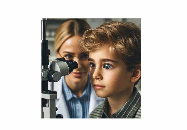
- Slit-Lamp Examination: This is the primary diagnostic tool for identifying the distinctive ocular abnormalities associated with ARS. A slit-lamp microscope allows for a detailed view of the anterior segment of the eye, revealing features such as posterior embryotoxon, iris hypoplasia, and correctopia.
- Gonioscopy: This technique examines the angle of the anterior chamber of the eye. It aids in the evaluation of the trabecular meshwork and Schlemm’s canal, both of which are critical for diagnosing glaucoma in ARS patients.
- Tonometry: Measuring intraocular pressure (IOP) is critical for diagnosing glaucoma, a common complication of ARS. Elevated IOP requires further investigation and management to prevent optic nerve damage.
- Fundus Examination: Ophthalmoscopy is used to examine the back of the eye, which includes the retina and optic nerve. This aids in the detection of any changes or damage caused by elevated IOP or other related conditions.
Innovative Diagnostic Techniques
- Optical Coherence Tomography (OCT): OCT can produce high-resolution cross-sectional images of the retina and optic nerve. It is especially useful for detecting and monitoring glaucoma-related changes in the optic nerve head and retinal nerve fiber layer.
- Anterior Segment OCT (AS-OCT): This specialized type of OCT examines the anterior segment of the eye, which includes the cornea, iris, and anterior chamber angle. AS-OCT can help visualize and assess ARS-specific abnormalities.
- Ultrasound Biomicroscopy (UBM): This technique uses high-frequency ultrasound to produce detailed images of the anterior segment structures, such as the ciliary body and zonules. This technique is useful for detecting anatomical anomalies that are not visible with standard imaging.
- Genetic Testing: Finding mutations in the PITX2, FOXC1, and other ARS-related genes can help confirm the diagnosis and provide valuable information for genetic counseling. Genetic testing is useful in determining the specific mutation and understanding the inheritance pattern.
- Corneal Topography: This imaging technique maps the curvature of the cornea and can detect corneal abnormalities like thinning and opacification, which are common with ARS.
Combining these diagnostic methods ensures a thorough assessment of the ocular and systemic manifestations of ARS, allowing for early intervention and effective management.
Effective Treatments for Axenfeld-Rieger Syndrome
- Management of Glaucoma: Controlling intraocular pressure is critical for preventing optic nerve damage and vision loss in ARS patients with glaucoma. Treatment alternatives include:
- Medications: Topical eye drops containing prostaglandin analogs, beta-blockers, alpha agonists, and carbonic anhydrase inhibitors are frequently used to reduce IOP.
- Laser Therapy: Procedures such as laser trabeculoplasty can increase aqueous outflow and lower IOP.
- Surgical Interventions: Trabeculectomy and drainage implants (e.g., Ahmed or Baerveldt implants) may be required in refractory cases when medications and laser therapy are ineffective.
- Regular Monitoring: Frequent eye exams are critical for early detection and treatment of glaucoma. Monitoring IOP, optic nerve health, and visual fields allows doctors to tailor treatment plans to individual patients’ needs.
- Protective Eyewear: Patients with corneal abnormalities or iris defects should wear protective eyewear to avoid injury and photophobia.
Innovative and Emerging Therapies
- Minimally Invasive Glaucoma Surgery (MIGS): Procedures such as iStent and Xen Gel Stent provide safer and less invasive options for lowering IOP. MIGS can be performed alongside cataract surgery and are associated with shorter recovery times.
- Gene Therapy: Researchers are currently investigating gene therapy approaches for correcting the genetic defects that cause ARS. Gene therapy has the potential to stop or reverse disease progression by addressing the underlying cause.
- Stem Cell Therapy: Advances in stem cell research may lead to new treatments for regenerating damaged ocular tissues. Stem cell therapy may offer new options for treating glaucoma and corneal abnormalities in ARS patients.
- New Medications: Research into new drugs that target specific pathways involved in IOP regulation and neuroprotection is currently underway. These medications are intended to provide more effective and safe options for glaucoma management.
- Custom Ocular Prosthetics: Patients with severe iris abnormalities or significant visual impairment can benefit from custom-designed prosthetic devices that improve both aesthetics and function. These devices are designed to meet individual needs and can improve quality of life.
Managing ARS necessitates a collaborative effort involving ophthalmologists, geneticists, dentists, and other specialists. Coordinated care plans address the syndrome’s various manifestations, ensuring the best outcomes and quality of life for patients.
Trusted Resources
Books
- “Pediatric Ophthalmology and Strabismus” by Kenneth W. Wright and Peter H. Spiegel
- “Genetic Diseases of the Eye” by Elias I. Traboulsi
- “The Glaucoma Book: A Practical, Evidence-Based Approach to Patient Care” by Paul N. Schacknow and John R. Samples


