Introduction
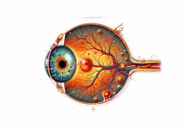
Behçet’s disease is a chronic inflammatory disorder that affects multiple organs, including the eyes. Retinopathy is one of the most serious and vision-threatening complications of the disease. Behçet’s disease-related retinopathy is an inflammation of the retina that can cause significant visual impairment if not treated properly. This condition is distinguished by retinal vasculitis, macular edema, and possible retinal detachment. Early detection and treatment are critical for preserving vision and improving the quality of life for those affected by this debilitating condition.
Behçet’s Disease: Retinopathy Details
The retinopathy associated with Behçet’s disease is caused by the disorder’s systemic vasculitis. Behçet’s disease primarily affects small and large blood vessels, resulting in widespread inflammation of various tissues, including the retina. The retina, a thin layer of tissue at the back of the eye, is essential for vision because it converts light into neural signals that travel to the brain. Inflammation and damage to the retinal blood vessels can significantly impair this function.
Pathophysiology
The exact pathogenesis of Behçet’s disease-related retinopathy is a complex interaction of genetic, immunological, and environmental factors. The HLA-B51 allele is strongly associated with an increased risk of developing Behçet’s disease. The root cause is thought to be an abnormal immune response to an environmental trigger, such as an infection, in genetically predisposed people. This immune response causes widespread inflammation and vasculitis, which can affect retinal vessels.
In Behçet’s disease-related retinopathy, the inflammatory process targets small blood vessels in the retina, resulting in retinal vasculitis. This condition is characterized by inflammation of the retinal blood vessels, which causes them to leak and result in exudates and hemorrhages. Inflammation can also occlude retinal blood vessels, reducing blood flow to the retina and resulting in ischemia and neovascularization (the formation of new, abnormal blood vessels). These new blood vessels are fragile and prone to bleeding, which further impairs vision.
Clinical Manifestations
Behçet’s disease-related retinopathy can cause a variety of symptoms, depending on the severity and extent of the inflammation. Common symptoms include:
- Blurry Vision: Inflammation and fluid accumulation in the retina can cause vision to blur.
- Floaters: Small spots or threads that float across the field of vision may be caused by inflammation and debris in the vitreous humor.
- Photophobia: One of the most common symptoms of retinal inflammation is light sensitivity.
- Visual Field Defects: Patients may experience blind spots or reduced vision in their visual field.
- Sudden Vision Loss: Severe inflammation can cause sudden and significant vision loss, particularly when combined with retinal detachment or extensive hemorrhage.
Retinal Vasculitis
Retinal vasculitis is characteristic of Behçet’s disease-related retinopathy. It is characterized by retinal blood vessel inflammation, which causes vascular leakage, hemorrhages, and the formation of cotton-wool spots (localized microinfarctions of the retinal nerve fiber layer). Retinal vasculitis can cause extensive damage to the retinal tissue, resulting in vision loss.
Macular Edema
Macular edema, or the buildup of fluid in the macula (the central part of the retina responsible for sharp vision), is a common side effect of retinal vasculitis. Macular edema causes the macula to swell, resulting in blurred or distorted vision. Persistent macular edema can permanently damage the macular tissue and cause irreversible vision loss.
Retinal Ischemia and Neovascularization
Retinal ischemia, or decreased blood flow to the retina, can result from the occlusion of inflamed retinal blood vessels. Ischemia causes the release of vascular endothelial growth factor (VEGF), which promotes the development of new, abnormal blood vessels (neovascularization). These new vessels are fragile and prone to bleeding, resulting in vitreous hemorrhage and further vision loss. Neovascularization can also cause tractional retinal detachment, in which the abnormal blood vessels pull on the retina, causing it to separate from the underlying tissue.
Complications
If not treated properly, Behçet’s disease-related retinopathy can cause a variety of complications. This includes:
- Permanent Vision Loss: Persistent inflammation and retinal damage can result in irreversible vision loss.
- Vitreous Hemorrhage: Bleeding into the vitreous humor can impair vision and may necessitate surgical treatment.
- Retinal Detachment: Tractional retinal detachment can occur as a result of neovascularization and requires immediate surgical repair to avoid permanent vision loss.
- Glaucoma: Elevated intraocular pressure caused by inflammation or neovascularization can damage the optic nerve, resulting in glaucoma and further vision loss.
- Secondary Cataracts: Long-term use of corticosteroids, which are frequently required to control inflammation, can cause cataracts.
Effects on Quality of Life
The visual impairment associated with Behçet’s disease-related retinopathy can have a significant impact on a patient’s quality of life. Vision loss can have an impact on daily activities, work, and independence, causing psychological distress and a reduction in overall well-being. The chronic nature of the disease, with the possibility of recurrent flares and ongoing treatment, increases the burden on patients and their families.
Prognosis
The prognosis for Behçet’s disease-related retinopathy is dependent on the severity of the condition and the timing of treatment. With prompt and aggressive treatment, inflammation can be controlled and vision preserved or restored. However, recurrent episodes of retinopathy can result in cumulative damage and progressive vision loss, emphasizing the importance of ongoing monitoring and treatment.
Essential Preventive Tips
- Regular Eye Examinations: Schedule regular eye exams with an ophthalmologist to detect early signs of retinopathy and other ocular complications. Early detection is critical to preventing vision loss.
- Protective Eyewear: Wear protective eyewear to reduce the possibility of eye injuries that cause inflammation. This is especially important during activities that carry a risk of trauma.
- Manage Systemic Inflammation: Collaborate with your healthcare provider to manage systemic inflammation with appropriate medications and lifestyle changes. Reducing overall inflammation can help to avoid ocular flares.
- Adherence to Treatment: Stick to your prescribed treatment plan, including the use of topical and systemic medications. Consistent treatment helps to control inflammation and avoid complications.
- Healthy Lifestyle: Eat a well-balanced diet high in anti-inflammatory foods, exercise frequently, and avoid smoking. These lifestyle choices can help improve overall health and reduce inflammation.
- Stress Management: Try stress-reduction techniques like mindfulness, meditation, and yoga. Stress can worsen inflammation and cause disease flares.
- Avoid Triggers: Determine and avoid potential triggers that can cause retinopathy flares, such as certain medications, environmental factors, and infections. Discuss any new symptoms or concerns with your doctor.
- Be Informed: Learn more about Behçet’s disease and its ocular manifestations. Join support groups and connect with other people who have the same condition to share your experiences and strategies for managing symptoms.
- Vaccinations: Stay up to date on recommended vaccinations to avoid infections that may cause or worsen inflammation.
- Hydration and Eye Care: Stay hydrated and practice good eye hygiene to keep your eyes healthy and reduce the risk of infection.
Diagnostic methods
Diagnosing Behçet’s disease-related retinopathy necessitates a multifaceted approach that includes clinical evaluation, advanced imaging techniques, and laboratory tests to accurately identify and assess retinal involvement.
Clinical Evaluation
Diagnosing Behçet’s disease-related retinopathy begins with a thorough clinical evaluation by an ophthalmologist. This includes conducting a thorough patient history to identify symptoms and systemic manifestations of Behçet’s disease, such as oral and genital ulcers, skin lesions, and joint pain. The ophthalmologist will conduct a thorough eye examination, including a slit-lamp examination to assess the anterior segment and a fundoscopic examination to evaluate the retina and optic nerve.
Fundus Photography
Fundus photography is a common diagnostic tool that takes detailed images of the retina, allowing for the tracking of retinal changes over time. It is especially useful for diagnosing retinal vasculitis, hemorrhages, and cotton-wool spots.
Fluorescein Angiography(FA)
Fluorescein angiography (FA) is a specialized imaging technique that involves injecting a fluorescent dye into the bloodstream and taking multiple photographs of the retina. FA aids in the visualization of retinal and choroidal circulation, revealing areas of vascular leakage, occlusion, and neovascularization. This technique is critical for detecting and monitoring retinal vasculitis and macular edema.
Optical Coherence Tomography(OCT)
Optical Coherence Tomography (OCT) is a non-invasive imaging technique that produces high-resolution cross-sections of the retina. OCT is an invaluable tool for detecting macular edema, retinal thickness, and structural changes in the retinal layers. It enables a thorough examination of the macula and aids in monitoring the efficacy of treatment.
Indocyanine green angiography (ICGA)
Indocyanine Green Angiography (ICGA) is similar to FA, but it employs indocyanine green dye, which is more readily absorbed by the choroidal circulation. ICGA is especially useful for visualizing choroidal vasculature and detecting choroidal inflammation, providing valuable diagnostic information in cases of posterior uveitis and choroiditis.
Visual Field Testing
Visual field testing evaluates the patient’s peripheral vision and identifies visual field defects. Automated perimetry can identify areas of vision loss, which is important for assessing the functional impact of retinal vasculitis and other complications.
Lab Tests
Laboratory tests help diagnose Behçet’s disease-related retinopathy. Blood tests that measure inflammatory markers such as erythrocyte sedimentation rate (ESR) and C-reactive protein (CRP) can reveal more about systemic inflammation. Genetic testing for the HLA-B51 allele can also be used to determine genetic susceptibility.
Behçet’s Disease Retinopathy Treatments
The treatment for Behçet’s disease-related retinopathy aims to reduce inflammation, prevent relapses, and preserve vision. A combination of medications and, in some cases, surgical interventions are used depending on the severity of the condition.
Corticosteroids
Corticosteroids are the main treatment for acute retinal inflammation in Behçet’s disease. They can be given orally, intravenously, or as periocular injections to reduce inflammation quickly. High-dose intravenous corticosteroids are frequently used first, followed by a tapering course of oral steroids to prevent relapse. Long-term corticosteroid use is associated with side effects such as increased intraocular pressure, cataracts, and systemic complications, which necessitate close monitoring.
Immunosuppressive Agents
Azathioprine, cyclosporine, and methotrexate are common immunosuppressive agents used to treat chronic or refractory retinopathy without the use of steroids. These medications help to regulate the immune response and reduce inflammation, lowering the likelihood of disease flares. Regular monitoring for possible side effects, such as liver toxicity and bone marrow suppression, is required.
Biological Therapies
Biologic therapies have transformed the management of Behçet’s disease-related retinopathy. TNF inhibitors, such as infliximab and adalimumab, have demonstrated excellent efficacy in treating severe and refractory retinal inflammation. These agents target specific components of the inflammatory process, providing better results with potentially fewer side effects than traditional immunosuppressive drugs.
Anti-VEGF Therapies
Anti-VEGF (vascular endothelial growth factor) therapy is used to treat the neovascularization and macular edema caused by Behçet’s disease-related retinopathy. Medications like bevacizumab, ranibizumab, and aflibercept are injected into the vitreous humor to stop the growth of abnormal blood vessels and reduce retina swelling.
Surgery
Surgical interventions are typically used to treat complications like vitreous hemorrhage or retinal detachment. Vitrectomy, a surgical procedure that removes the vitreous humor, may be used to clear blood and debris from the vitreous cavity and treat retinal detachment. Laser photocoagulation can also be used to repair leaking blood vessels and prevent further bleeding.
Innovative and Emerging Therapies
Interleukin Inhibitors
Interleukin inhibitors, including tocilizumab (an IL-6 inhibitor) and secukinumab (an IL-17 inhibitor), are being studied for their ability to control inflammation in Behçet’s disease. These biologic agents target specific cytokines involved in the inflammatory process, providing a novel way to treat retinopathy.
Janus Kinase (JAK) inhibitors
Janus Kinase (JAK) inhibitors, like tofacitinib, are a promising class of drugs for treating Behçet’s disease-related retinopathy. JAK inhibitors disrupt intracellular signaling pathways that contribute to inflammation, offering a more targeted treatment option with the potential for fewer adverse effects.
Stem Cell Therapy
Stem cell therapy is a developing field that has the potential to regenerate damaged retinal tissues and restore vision. Early-stage clinical trials are looking into using mesenchymal stem cells to treat severe retinopathy and other complications of Behçet’s disease.
Genetic Therapy
Gene therapy seeks to correct the underlying genetic flaws that contribute to the abnormal immune response in Behçet’s disease. Researchers are working to develop safe and effective gene therapy techniques that could provide a long-term solution for patients with this condition.
Trusted Resources
Books
- “Behçet’s Disease: From Genetics to Therapeutics” by Yusuf Yazici
- “Behçet’s Disease: A Guide to Its Clinical Understanding and Management” by Joanne Zeis
- “Behçet’s Syndrome” by Christos Zouboulis
Online Resources
- American Behçet’s Disease Association: https://www.behcets.com
- Behçet’s Syndrome Society: https://www.behcetssyndrome.org.uk
- National Institute of Arthritis and Musculoskeletal and Skin Diseases: https://www.niams.nih.gov
- National Organization for Rare Disorders (NORD): https://rarediseases.org


