What is Blepharochalasis?
Blepharochalasis is a rare and chronic eyelid condition characterized by recurring episodes of eyelid edema (swelling), which over time causes the eyelid skin to stretch, thin, and wrinkle. This condition frequently results in an excess of eyelid skin that hangs over the eyelashes, potentially impairing vision. Blepharochalasis usually develops in adolescence and can affect one or both eyes. The exact cause of blepharochalasis is unknown, but it is thought to be linked to an abnormal immune response or an underlying inflammatory process. Understanding and treating blepharochalasis is critical for maintaining both ocular health and the appearance of the eyelids.
Blepharochalasis Insights
Blepharochalasis is caused by a complex interplay of factors that results in the characteristic changes in the eyelids. The condition is distinguished by recurring episodes of painless eyelid swelling that can last several days before resolving spontaneously. These episodes of edema cause progressive stretching and thinning of the eyelid tissues, resulting in the typical blepharochalasis signs.
Pathophysiology
The pathophysiology of blepharochalasis is unclear, but several theories have been proposed. According to one theory, the condition could be caused by an abnormal immune response, in which the body’s immune system attacks the tissues of the eyelids, causing inflammation and swelling. Another theory suggests that blepharochalasis is caused by recurring episodes of angioedema, a condition characterized by sudden and severe swelling of the deeper layers of the skin that is frequently triggered by allergic reactions, infections, or stress.
The recurrent swelling and inflammation cause the eyelid skin to lose elasticity and structural integrity. The collagen and elastin fibers that give the skin its strength and flexibility are damaged and fragmented. Blepharochalasis is characterized by thin, wrinkled, and redundant eyelid skin.
Clinical Features
Blepharochalasis manifests as a variety of clinical features that differ depending on the stage and severity of the condition. Common symptoms and signs are:
- Recurrent Eyelid Edema: Blepharochalasis is characterized by painless swelling of the eyelids. These episodes can last anywhere from a few hours to a few days and usually end spontaneously.
- Eyelid Skin Changes: Repeated episodes of swelling cause the eyelid skin to thin, wrinkle, and relax. During the acute phase of swelling, the skin may turn red or purple.
- Redundant Eyelid Skin: Over time, stretched and thinned eyelid skin causes an excess of skin to hang over the eyelashes. This can result in a hooded appearance and, in severe cases, impair vision by blocking the visual field.
- Ptosis: Ptosis, or drooping of the upper eyelid, can coexist with blepharochalasis. This is caused by the weakening of the muscles and tissues that support the eyelid.
- Eyelid Malposition: Blepharochalasis can occasionally cause eyelid malposition, such as ectropion (outward turning of the eyelid) or entropion (inward turning of the eyelid). These conditions can lead to additional irritation and discomfort.
- Asymmetry: Blepharochalasis can affect one or both eyelids, and the severity varies between the eyes, resulting in an asymmetrical appearance.
Risk Factors
Several risk factors have been identified as potentially increasing the likelihood of developing blepharochalasis. This includes:
- Genetic Predisposition: A family history of blepharochalasis or similar conditions may increase the likelihood of developing the disorder.
- Allergic Reactions: People who have a history of allergies or angioedema are more likely to experience recurrent eyelid swelling, which can contribute to the development of blepharochalasis.
- Chronic Inflammation: Conditions that cause chronic inflammation, such as autoimmune diseases or chronic blepharitis, can cause long-term damage to eyelid tissues.
- Trauma: Repeated trauma or irritation to the eyelids, whether caused by rubbing, cosmetic procedures, or environmental factors, can lead to skin weakening and thinning.
Complications
Blepharochalasis, if left untreated, can cause a number of complications that impact both ocular health and overall well-being. The complications include:
- Impaired Vision: Excess and redundant eyelid skin can hang over the eyelashes, blocking the visual field and impairing vision. This can have an impact on daily activities like reading, driving, and working.
- Cosmetic Concerns: The appearance of thin, wrinkled, and drooping eyelid skin can raise cosmetic concerns and lower self-esteem. Patients may seek medical attention primarily for aesthetic purposes.
- Functional Issues: Ptosis and eyelid malposition can result in discomfort, irritation, and difficulty blinking. This can cause dry eyes, tearing, and a higher risk of eye infections.
- Emotional Impact: Blepharochalasis’ chronic and visible nature can have an emotional and psychological impact on patients, causing anxiety, depression, and social isolation.
Essential Preventive Tips
- Maintain Good Eyelid Hygiene: Use a gentle cleanser or prescribed eyelid scrub to remove debris and reduce the risk of inflammation and infection.
- Avoid Rubbing Your Eyes: Avoid rubbing or touching your eyes frequently, as this can aggravate swelling and irritation, causing additional damage to the eyelid tissues.
- Manage Allergies: If you have known allergies, consult with your doctor about how to effectively manage and treat them. Use antihistamines and other medications as directed to reduce the risk of allergic reactions that can cause eyelid swelling.
- Protect Your Eyes: Use UV-protective sunglasses to shield your eyes from harmful ultraviolet rays and environmental irritants like dust and wind.
- Avoid Cosmetic Procedures: Avoid using cosmetic procedures or products around the eyes that may irritate or traumatize the delicate eyelid skin.
- Stay Hydrated: Drink plenty of water to stay hydrated and promote skin health. Dehydration can make the skin more susceptible to damage and irritation.
- Healthy Diet: Eat a well-balanced diet high in vitamins and antioxidants to promote skin health and reduce inflammation. Omega-3-rich foods, such as fish and flaxseeds, can be especially beneficial.
- Avoid Stress: Relaxation techniques such as yoga, meditation, and deep breathing exercises can help you manage your stress. Stress can cause inflammatory responses in the body, exacerbating symptoms.
- Regular Check-Ups: Make regular appointments with your ophthalmologist or dermatologist to monitor the condition of your eyelids and receive appropriate treatment if necessary.
- Use Prescribed Medications: Follow your doctor’s instructions for using any prescribed medications or treatments to manage inflammation and prevent recurring episodes of swelling.
Blepharochalasis Diagnosis
Blepharochalasis is diagnosed using a combination of clinical evaluation, patient history, and, in some cases, advanced imaging techniques to accurately assess the condition and rule out other related disorders.
Clinical Evaluation
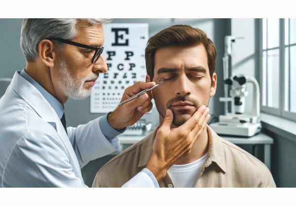
The initial step in diagnosing blepharochalasis is a thorough clinical examination by an ophthalmologist or dermatologist. The doctor will examine the eyelids thoroughly, looking for characteristic signs such as redundant, thin, and wrinkled eyelid skin. The examination also evaluates eyelid function, ptosis, and any associated asymmetry.
Patient History
Obtaining a thorough patient history is critical for diagnosis. The clinician will ask about the frequency, duration, and causes of eyelid swelling episodes, as well as any associated symptoms like itching, redness, or pain. Information about known allergies, autoimmune conditions, or a family history of similar symptoms can help identify potential underlying causes.
Slit Lamp Examination
A slit lamp examination provides a magnified view of the eyelids and anterior segment of the eye. This aids in detecting specific changes in the eyelid skin and meibomian gland function. The slit-lamp can also reveal signs of other conditions, such as blepharitis, which can coexist with blepharochalasis.
Photography
Photographic documentation of the eyelids is commonly used to track the progression of blepharochalasis over time. Comparing images taken at various stages of the condition can aid in determining treatment efficacy and disease progression.
Allergy Testing
If an allergic component is suspected, allergy testing may be used to identify specific allergens that are causing the eyelid swelling. Skin prick tests or blood tests can help identify allergens and guide management strategies.
Biopsy
In rare cases where the diagnosis is uncertain, a biopsy of the eyelid tissue may be required. Histopathological examination of the tissue can reveal blepharochalasis-related changes such as dermal thinning and collagen fiber fragmentation. This can help distinguish blepharochalasis from other conditions that share clinical characteristics.
Advanced Imaging Techniques
Innovative imaging techniques, such as high-resolution optical coherence tomography (OCT) and ultrasound biomicroscopy (UBM), can produce detailed images of the eyelid structures. These noninvasive methods can determine the thickness and integrity of the eyelid tissues, as well as the function of the meibomian glands. These techniques are especially useful in complex cases and for monitoring treatment response rates.
Management of Blepharochalasis
Blepharochalasis treatment aims to alleviate symptoms, improve eyelid appearance, and address underlying causes. A combination of medical and surgical approaches is frequently needed.
Medical Management
Topical corticosteroids
Topical corticosteroids can be used to reduce inflammation during acute episodes of eyelid swelling. These medications help to reduce the immune response and relieve symptoms. Corticosteroids, on the other hand, can cause skin thinning and other side effects if used for an extended period of time, so they are usually prescribed in short courses.
Antihistamines
For patients with an allergic component, oral or topical antihistamines can help control symptoms and prevent recurrent swelling. Antihistamines reduce the release of histamines and other inflammatory mediators, relieving itching and swelling.
Immunosuppressive Agents
If blepharochalasis is associated with an underlying autoimmune condition, immunosuppressive medications such as cyclosporine or methotrexate may be prescribed. These medications help to regulate the immune system and reduce inflammation.
Surgical Management
Blepharoplasty
Blepharoplasty is a surgical procedure that removes excess and redundant eyelid skin, improves their appearance, and restores normal function. During the procedure, the surgeon removes excess skin and tightens the underlying tissue. This can have significant cosmetic and functional benefits, particularly in cases where redundant skin impairs vision.
Ptosis Repair
If ptosis is present, the upper eyelid drooping may require surgical repair. Ptosis is repaired by tightening or repositioning the muscles and tendons that elevate the eyelid. This improves the visual field and enhances the appearance of the eyelids.
Innovative and Emerging Therapies
Laser Skin Resurfacing
Laser skin resurfacing is a new treatment that uses laser technology to improve the texture and tone of eyelid skin. This procedure can help to reduce wrinkles and tighten the skin, resulting in a more youthful appearance. Laser skin resurfacing is a minimally invasive procedure that can be combined with other surgical treatments.
Radiofrequency Therapy
Radiofrequency (RF) therapy is another novel approach that uses RF energy to stimulate collagen production and tighten skin. This non-invasive treatment can help to improve the elasticity and firmness of the eyelid skin, resulting in fewer wrinkles and excess skin.
Platelet Rich Plasma (PRP) Therapy
Platelet-rich plasma (PRP) therapy involves injecting a high concentration of the patient’s platelets into the affected area. PRP contains growth factors, which promote healing and tissue regeneration. This treatment can help to improve the quality of eyelid skin while also reducing inflammation, providing both cosmetic and therapeutic benefits.
Trusted Resources
Books
- “Eyelid and Periorbital Surgery” by Mark A. Codner and Clinton D. McCord
- “Oculoplastic Surgery: The Essentials” by William P. Chen
- “Surgery of the Eyelid, Orbit, and Lacrimal System” by Bradley N. Lemke and Michael T. Yen
Online Resources
- American Academy of Ophthalmology: https://www.aao.org
- National Eye Institute: https://www.nei.nih.gov
- Ophthalmology Times: https://www.ophthalmologytimes.com
- American Society of Ophthalmic Plastic and Reconstructive Surgery: https://www.asoprs.org


