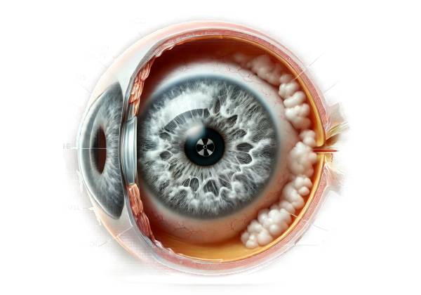
Radiation cataract is a type of cataract caused by exposure to ionizing radiation. Unlike the more common age-related cataracts, which develop gradually as a result of the natural aging process, radiation cataracts are caused by external factors—specifically, exposure to high-energy radiation, which damages the eye’s lens. Understanding radiation cataracts necessitates a thorough examination of the causes of cataracts, the effects of radiation on biological tissues, and the specific vulnerabilities of the eye’s lens.
Radiation’s Effects on the Eye
Radiation can cause significant damage to the human eye, especially the lens. The lens is a transparent, flexible structure that sits behind the iris and in front of the vitreous body. Its primary function is to focus light on the retina, which transmits visual information to the brain. The clarity of the lens is critical to maintaining sharp vision. The lens is made of water and proteins, which are arranged in such a way that they remain transparent. However, the lens is avascular, which means it does not have its own blood supply and does not regenerate cells like other tissues. These characteristics make the lens especially susceptible to cumulative damage, such as that caused by ionizing radiation.
Ionizing Radiation’s Effects
Ionizing radiation refers to types of radiation that have enough energy to remove tightly bound electrons from atoms, resulting in ions. This category of radiation includes X-rays, gamma rays, and particles such as protons and neutrons. Ionizing radiation can cause significant damage to biological tissues by directly breaking chemical bonds in DNA and other critical molecules or by producing reactive oxygen species (ROS). ROS, or free radicals, are highly reactive molecules that can cause oxidative damage to cells and tissues.
When ionizing radiation enters the eye’s lens, it can disrupt the structure and function of lens epithelial cells. These cells are in charge of keeping the lens transparent and regulating the slow turnover of lens fibers. Radiation can cause DNA damage in these cells, resulting in cell death or dysfunction. Over time, this damage can accumulate, causing protein clumping and the formation of opacities in the lens, known as cataracts.
Pathology of Radiation Cataract
Radiation cataracts typically form in a dose-dependent manner, which means that the risk of developing cataracts increases with radiation exposure. However, even low doses of radiation, if administered repeatedly or over an extended period of time, can eventually result in cataract formation. The rate of exposure, the type of radiation, and individual susceptibility factors, such as age and genetic predisposition, all have an impact on radiation cataract development.
One of the distinguishing characteristics of radiation cataracts is their location within the lens. Radiation-induced cataracts are commonly characterized as posterior subcapsular cataracts (PSC). These cataracts develop at the back of the lens, just below the lens capsule. The posterior subcapsular region is especially vulnerable to radiation because it contains a population of lens epithelial cells that are essential for maintaining lens transparency. When radiation damages these cells, they can form clumps of abnormal proteins that scatter light, resulting in the characteristic visual disturbances associated with cataracts.
The time it takes for cataracts to develop after radiation exposure can range from several months to several decades. Factors influencing this variability include the total dose of radiation, the fractionation (or splitting) of the dose over time, and the individual’s age at the time of exposure. Younger people and those who have received higher doses of radiation are more likely to develop cataracts. The progression of radiation cataracts can also vary; in some cases, the cataracts may remain stable for years, while in others, they may progress quickly, resulting in significant visual impairment.
Sources of Radiation Exposure
Radiation exposure that can cause cataract formation can occur in a variety of settings. These include therapeutic radiation, occupational exposure, and unintentional exposure.
Therapeutic Radiation
Medical treatment, particularly radiation therapy for cancer, is one of the most common sources of ionizing radiation exposure. Radiation therapy is the use of high-energy radiation to eliminate cancer cells. When this radiation is directed at or near the head, neck, or chest, the lens of the eye may receive a significant dose, even if it is not the intended treatment target. Despite advances in radiation therapy techniques that aim to reduce exposure to surrounding healthy tissues, the lens can still be damaged, particularly if shielding is insufficient or not possible. Patients who receive radiation therapy for brain tumors, head and neck cancers, or ocular malignancies are at an increased risk of developing radiation cataracts.
Occupational Exposure
Certain professions expose workers to higher levels of radiation for extended periods of time, increasing their risk of developing radiation cataracts. Healthcare workers, particularly those in radiology, interventional cardiology, and radiation oncology, may be exposed to ionizing radiation on the job. Although protective measures like leaded glasses and shielding devices are widely used, long-term exposure can increase the risk of cataract formation. Employees in the nuclear industry, such as those working at nuclear power plants, are also at risk. Furthermore, due to the altitudes at which they work, airline pilots and cabin crew members are exposed to higher levels of cosmic radiation, putting them at a greater risk.
Accidental Exposure
Accidental radiation exposure, while less common, can result in significant doses that cause cataracts. Such exposure could occur during nuclear accidents like the Chernobyl disaster or the Fukushima Daiichi nuclear disaster. Individuals exposed to high levels of radiation during such events are significantly more likely to develop radiation cataracts, often within a few years. The rapid onset and progression of cataracts in these cases is usually due to the high radiation doses received.
Clinical Features of Radiation Cataracts
Radiation cataracts typically have a distinct set of clinical features that set them apart from other types of cataracts, such as those caused by aging or metabolic conditions like diabetes. As previously stated, radiation cataracts commonly manifest as posterior subcapsular cataracts (PSC), which form at the back of the lens. This location is especially problematic because it is along the visual axis, which can significantly impair vision.
Patients with radiation cataracts may initially experience subtle visual symptoms such as difficulty seeing in low light, increased sensitivity to glare, and a gradual decrease in overall visual acuity. As the cataract progresses, these symptoms usually worsen, resulting in significant visual impairment. Patients may also notice their vision becoming blurred or hazy, and colors appearing less vibrant. Unlike other types of cataracts, which typically develop symmetrically in both eyes, radiation cataracts may initially affect only one eye, especially if the radiation exposure was localized.
Radiation cataracts can have a significant impact on an individual’s quality of life, especially if the condition progresses to severe visual impairment. Individuals who rely heavily on their vision for work or daily activities, such as healthcare workers, pilots, or drivers, may find radiation cataracts particularly debilitating. Radiation cataracts may necessitate early retirement or a change in career due to loss of visual function.
The Importance Of Early Detection
Given the potentially rapid progression and severe consequences of radiation cataracts, early detection is critical. Individuals who have been exposed to ionizing radiation, particularly those in high-risk occupations or who have received radiation therapy, require regular eye examinations. Early detection enables monitoring of the cataract’s progression and timely intervention to avoid severe visual impairment.
Diagnostic methods
Radiation cataracts require a comprehensive approach that includes a detailed patient history, a thorough clinical examination, and specialized imaging techniques. Accurate diagnosis is critical for timely intervention and management, especially in individuals with a history of radiation exposure.
Patient History
The first step in diagnosing radiation cataracts is to gather a detailed patient history. This includes inquiring about the patient’s exposure to radiation, whether it was occupational, therapeutic, or accidental. Significant aspects of history include:
- Type and Duration of Exposure: Identifying the source of radiation, the dose received, and the duration of exposure is critical. Patients should be asked about any radiation therapy they have received, especially if it involved the head, neck, or chest. Occupational history should include jobs that require exposure to ionizing radiation, such as healthcare, nuclear industry work, or aviation.
- Onset of Symptoms: Knowing when the patient first noticed symptoms, such as blurred vision or increased sensitivity to light, can aid in correlating these symptoms to known radiation exposure.
- Previous Medical Conditions: Examine the patient’s complete medical history, including any other conditions that may contribute to cataract formation, such as diabetes or long-term corticosteroid use.
Clinical Examination
A comprehensive eye examination is required to accurately diagnose radiation cataracts. This usually includes:
- Visual Acuity Test: This standard test assesses the clarity of the patient’s vision and helps determine how the cataract affects their ability to see fine details.
- Slit-Lamp Examination: A slit lamp is a microscope that allows for a highly magnified view of the eye’s structures. During this examination, the ophthalmologist can assess the lens’s transparency and detect the presence of cataracts. Because of their location at the back of the lens, posterior subcapsular cataracts are more visible under a slit-lamp examination. The slit-lamp examination also allows the ophthalmologist to assess the cataract’s size, shape, and density, all of which are important in determining the severity of the condition.
Imaging Techniques
In addition to the standard clinical examination, specialized imaging techniques can be used to confirm the diagnosis of radiation cataracts and track their progress. These imaging modalities provide detailed views of the lens and other structures within the eye, allowing for more accurate assessment.
- Optical Coherence Tomography (OCT): OCT is a non-invasive imaging test that produces detailed cross-sectional images of the eye. It is especially useful for seeing the posterior subcapsular region of the lens, which is where radiation cataracts usually form. OCT can detect early changes in the lens that would not be visible during a slit-lamp examination, making it an important tool for early diagnosis and monitoring.
- Scheimpflug Imaging: Scheimpflug imaging is a specialized technique that produces detailed, three-dimensional images of the eye’s anterior segment, including the lens. This imaging method is especially useful for determining the density and location of cataracts. It provides a comprehensive view of the lens’s structure, allowing for the differentiation of radiation cataracts from other types of cataracts as well as the quantification of lens opacities.
Additional Tests
In some cases, additional tests may be required to assess the impact of radiation cataracts on vision and rule out other possible causes of visual impairment.
- Glare Sensitivity Testing: This test determines the patient’s ability to see in the presence of bright lights or glare, which is a common problem with posterior subcapsular cataracts. Patients with radiation cataracts frequently report increased sensitivity to glare, particularly when driving at night or in bright sunlight.
- Contrast Sensitivity Testing: This test evaluates the patient’s ability to distinguish between various shades of gray, which can be impacted by cataracts. Decreased contrast sensitivity is a common early sign of cataract development, and it can have a significant impact on the patient’s quality of life.
Radiation Cataract Management
Radiation cataract management entails a variety of approaches, including preventive measures, medical management, and surgical intervention. The severity of the cataract, the degree of visual impairment, and the individual patient’s needs and preferences all influence the management strategy chosen.
Preventive Measures
To prevent the development or progression of radiation cataracts, limit your exposure to ionizing radiation. Individuals undergoing radiation therapy require careful planning and shielding techniques to protect their eyes. This could include wearing leaded glasses or other protective barriers to limit the amount of radiation that reaches the lens. To reduce risk in occupational settings, it is critical to follow safety protocols, such as wearing personal protective equipment (PPE) and monitoring radiation levels on a regular basis.
Regular eye examinations are recommended for healthcare workers and others who may be exposed to radiation on the job in order to detect early signs of cataract formation. Early detection allows for timely interventions, which may slow the progression of the cataract or reduce its impact on vision.
Medical Management
When the symptoms of radiation cataract development are mild, medical treatment may be sufficient to improve or maintain vision. This may include using prescription eyeglasses or contact lenses to correct refractive errors caused by the cataract. Anti-glare lens coatings can help reduce sensitivity to bright lights, a common symptom of posterior subcapsular cataracts.
While no medications are currently available to reverse cataracts, ongoing research is looking into potential treatments to slow their progression. For example, some studies have looked into the use of antioxidants and other compounds to reduce oxidative stress in the lens, potentially delaying cataract formation. However, these treatments remain experimental and not widely available.
Surgical Intervention
Surgical removal of the cataract is the most definitive treatment for radiation cataracts, especially if the cataract has progressed to the point where it significantly limits vision. Cataract surgery removes the cloudy lens and replaces it with an artificial intraocular lens (IOL). This procedure is extremely effective at restoring vision and is one of the most popular and successful surgeries performed worldwide.
The decision to proceed with cataract surgery is based on several factors, including the severity of the cataract, the patient’s overall health, and their visual requirements. Patients who have difficulty performing daily activities like reading, driving, or working due to cataract-related vision loss are usually considered good candidates for surgery.
Cataract surgery for radiation cataracts is similar to that for other types of cataracts. The surgeon makes a small incision in the eye, removes the clouded lens with techniques like phacoemulsification (in which ultrasound waves break up the lens), and inserts an IOL to restore vision. The type of IOL used can be tailored to the patient’s specific requirements, including options for correcting astigmatism, presbyopia, and other refractive errors.
Post-surgical Care
Following cataract surgery, patients are typically given anti-inflammatory and antibiotic eye drops to prevent infection and inflammation. Most patients notice significant improvements in their vision within a few days to weeks of surgery. To ensure a smooth recovery, patients should carefully follow their surgeon’s post-operative care instructions.
Regular follow-up appointments are required to monitor the healing process and rule out any potential complications, such as increased intraocular pressure, infection, or IOL dislocation. While cataract surgery is generally safe, some patients may experience complications, especially if they have other underlying health conditions or if the procedure is performed on an eye that has previously been exposed to high doses of radiation.
Trusted Resources and Support
Books
- “Cataract Surgery: Techniques, Complications, and Management” by Roger F. Steinert, MD – A comprehensive guide covering all aspects of cataract surgery, including radiation cataracts.
- “Ocular Radiation Therapy: Eye Preservation and Cancer Control” by Simone Graue – Provides detailed information on managing ocular conditions, including radiation cataracts, after radiation therapy.
Organizations
- American Academy of Ophthalmology (AAO) – Offers resources and guidelines for the diagnosis and management of cataracts, including those induced by radiation.
- Radiation Oncology Society (ASTRO) – Provides information on radiation safety, therapy, and potential side effects like cataract formation.


