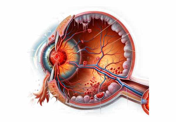
Introduction to Central Retinal Artery Occlusion
Central Retinal Artery Occlusion (CRAO) is a serious and vision-threatening condition marked by a sudden loss of blood flow to the retina, the light-sensitive layer at the back of the eye. This blockage causes sudden, painless vision loss in one eye, which is often described as a curtain falling over the visual field. CRAO is similar to a stroke in the eye and requires immediate medical attention to avoid permanent visual impairment. Although the condition is uncommon, it can result in significant disability, emphasizing the importance of early detection and intervention.
Understanding The Condition
Central Retinal Artery Occlusion (CRAO) occurs when the central retinal artery that supplies blood to the retina becomes clogged. The retina requires a constant blood supply to receive oxygen and nutrients needed for visual function. When this blood flow is disrupted, the retinal tissues begin to suffer ischemic damage, which can quickly result in irreversible cell death and permanent vision loss.
Pathophysiology
The central retinal artery branches from the ophthalmic artery, which is a branch of the internal carotid artery. The central retinal artery enters the eye via the optic nerve and then divides into several smaller branches that supply the retina’s inner layers. A blockage in the central retinal artery reduces blood flow to these critical areas.
The blockage is typically caused by an embolus, which is a small particle or clot that travels through the bloodstream and becomes lodged in the narrow retinal arteries. Emboli can come from a variety of sources, including:
- Atherosclerotic Plaques: These are fatty deposits that accumulate in the walls of arteries, usually the carotid arteries, and can break off and travel to the retinal artery.
- Cardiac Emboli: Blood clots in the heart, particularly in people with atrial fibrillation or valvular heart disease, can become dislodged and travel to the retinal artery.
- Giant Cell Arteritis: This inflammatory condition in the blood vessels can cause arterial occlusion, including the central retinal artery.
In addition to emboli, CRAO can be caused by thrombosis, which is the formation of a blood clot within an artery. Thrombosis can be linked to systemic vascular diseases like hypertension, diabetes, and hyperlipidemia.
Clinical Presentation.
CRAO is typically characterized by sudden, painless vision loss in one eye. Patients frequently report a dramatic and abrupt decrease in visual acuity, which can lead to complete blindness in the affected eye if left untreated. The onset of symptoms can occur in seconds to minutes, making medical intervention timely.
The severity of vision loss is determined by the extent of the blockage and the duration of ischemia. Patients may be able to maintain some peripheral vision if the blockage is partial or if there is collateral blood flow from the ciliary arteries, which can sometimes provide limited blood supply to the retina.
Epidemics and Risk Factors
CRAO is relatively rare, with an estimated 1-2 cases per 100,000 people per year. It primarily affects older adults, with the majority of cases involving people over the age of sixty. The condition is more common in males than females and is associated with a number of systemic risk factors, including:
- Hypertension: High blood pressure can promote atherosclerosis and the formation of emboli.
- Diabetes Mellitus: This metabolic disorder raises the possibility of vascular complications, including CRAO.
- Hyperlipidemia: High levels of cholesterol and triglycerides can cause the formation of atherosclerotic plaques.
- Cardiovascular Disease: Conditions like coronary artery disease and atrial fibrillation raise the risk of embolic events.
- Smoking: Tobacco use is a major risk factor for atherosclerosis and vascular occlusion.
- Giant Cell Arteritis: This condition, which primarily affects older adults, can result in inflammation and occlusion of the central retinal artery.
Pathological consequences
The retina is one of the body’s most metabolically active tissues, requiring a high amount of oxygen. When blood flow is obstructed, retinal tissue quickly becomes ischemic. Within 90 minutes of occlusion, irreversible damage to the retinal ganglion cells and inner retinal layers may occur. Prolonged ischemia causes cell death, which results in permanent vision loss.
Complications
CRAO can cause a variety of complications, including neovascularization, in which new, abnormal blood vessels form on the retina or optic nerve head. These vessels are fragile and prone to bleeding, which can exacerbate vision issues. Another possible complication is neovascular glaucoma, a severe type of glaucoma characterized by high intraocular pressure caused by the formation of new blood vessels on the iris and the eye’s drainage angle.
Prognosis
The prognosis for visual recovery in CRAO is generally poor, especially if treatment is not initiated immediately. Despite medical interventions, the majority of patients develop significant, permanent vision loss. However, some vision recovery is possible if blood flow is restored quickly, highlighting the importance of early detection and treatment.
Psychosocial impact
Patients who experience sudden vision loss as a result of CRAO may suffer significant psychosocial consequences. It can cause a loss of independence, difficulties with daily tasks, and emotional distress. Patients may experience anxiety, depression, and a lower quality of life. Support from healthcare providers, family, and support groups is critical in helping patients cope with the difficulties associated with this condition.
Essential Preventive Measures
- Regular Medical Checkups
- Schedule regular health screenings to monitor and manage systemic conditions like hypertension, diabetes, and hyperlipidemia, which are all major risk factors for CRAO.
- Control your blood pressure
- Maintain optimal blood pressure levels by making lifestyle changes and taking medications as prescribed by a healthcare provider to reduce the risk of vascular occlusion.
- Manage Diabetes
- To avoid vascular complications, maintain blood sugar control through appropriate dietary measures, medications, and regular monitoring.
- Monitor and manage cholesterol levels
- To reduce the risk of atherosclerosis, eat a well-balanced diet, exercise regularly, and take medications as needed.
- Quit smoking
- Avoid smoking to reduce your risk of atherosclerosis and other vascular diseases. Seek help and resources for quitting smoking if needed.
- Exercise regularly
- Exercise regularly to improve cardiovascular health and lower the risk of CRAO-related conditions.
- Healthy diet
- Eat a heart-healthy diet high in fruits, vegetables, whole grains, and lean proteins to improve overall vascular health and lower your risk of occlusive events.
- Regular Eye Examination
- Get regular comprehensive eye exams, especially if you have risk factors for CRAO, to detect any early signs of vascular problems and receive timely treatment.
- Monitor Heart Health
- Regularly monitor and manage heart conditions such as atrial fibrillation and valvular heart disease, which can contribute to embolic events that result in CRAO.
- Be aware of symptoms
- Recognize the signs of CRAO, such as sudden, painless vision loss, and seek medical attention right away if these symptoms appear to improve your chances of preserving your sight.
Diagnostic methods
To begin treatment on time, Central Retinal Artery Occlusion (CRAO) must be diagnosed quickly and accurately. The standard diagnostic approach begins with a thorough patient history and an in-depth eye examination. The key diagnostic techniques are:
Fundoscopy
The retina and optic disc are visualized using either direct or indirect ophthalmoscopy. In cases of CRAO, the retina is often pale and swollen, with a distinctive “cherry-red spot” at the macula, while the central fovea is relatively unaffected by the ischemia. This distinctive sign is an important indicator of CRAO.
Fluorescein Angiogram
In fluorescein angiography, a fluorescent dye is injected into the bloodstream and photographed as it travels through the retinal vessels. This technique aids in determining the location and severity of the arterial blockage, as well as assessing retinal blood flow. A delayed or absent filling of the central retinal artery can confirm the diagnosis of CRAO.
Optical coherence tomography (OCT)
OCT is a non-invasive imaging technique for obtaining high-resolution cross-sectional images of the retina. OCT in CRAO may reveal thickening of the inner retinal layers due to edema, followed by thinning due to atrophy. OCT can also be useful for tracking retinal changes over time.
Electroretinography (ERG).
ERG measures the retina’s electrical response to light stimulation. In CRAO, the b-wave amplitude is typically reduced, indicating impaired function of the inner retinal layers. This test can help you determine the extent of retinal damage.
Color Doppler Imaging (CDI)
CDI is an ultrasound technique that measures blood flow in the retina and orbital vessels. It can detect abnormalities in the central retinal artery’s blood flow and assess the presence of collateral circulation, both of which can influence the prognosis.
Optical Coherence Tomography Angiography (OCTA)
OCTA is a new non-invasive imaging modality that provides detailed visualization of the retinal and choroidal vasculature without the need for dye injection. It can detect microvascular changes and flow deficits in CRAO, providing an accurate assessment of the affected retinal circulation.
These diagnostic methods, which range from traditional fundoscopy to advanced imaging techniques such as OCT and OCTA, provide detailed insights into retinal structure and blood flow, allowing for accurate diagnosis and effective monitoring of CRAO.
Central Retinal Artery Occlusion Treatment Methods
The goal of treating Central Retinal Artery Occlusion (CRAO) is to restore retinal blood flow while preventing further ischemia. Immediate intervention is critical, as delayed treatment significantly reduces the chances of visual recovery. Standard and emerging treatments include the following:
Immediate measures
- Ocular massage
- Applying gentle pressure to the closed eyelid may help dislodge the embolus by increasing and then releasing intraocular pressure, potentially moving the blockage downstream.
- Lowering intraocular pressure
- Medications like acetazolamide, mannitol, or topical glaucoma medications can lower intraocular pressure, potentially helping to dislodge the embolus.
- Breathing into a paper bag
- This technique raises carbon dioxide levels in the blood, causing vasodilation and possibly improving retinal blood flow.
Hyperbaric oxygen therapy (HBOT)
HBOT involves breathing pure oxygen in a pressurized chamber, which improves oxygen delivery to the retina via the choroidal circulation. This treatment can help improve retinal oxygenation and reduce ischemic damage, particularly if started within a few hours of symptom onset.
Thrombolysis Therapy
In some cases, thrombolytic agents like tissue plasminogen activator (tPA) are used to dissolve the clot blocking the central retinal artery. This treatment is typically given intra-arterially or intravenously and is most effective when started within the first few hours of CRAO onset. However, it carries a high risk of serious complications and necessitates careful patient selection.
Antiplatelet and Anticoagulant Therapy
Patients with CRAO frequently require long-term treatment with antiplatelet or anticoagulant medications to avoid recurrent vascular events. Aspirin, clopidogrel, or anticoagulants such as warfarin may be prescribed depending on the underlying cause of the occlusion and the patient’s overall health.
Emerging Therapies
Research is being conducted to identify new treatments for CRAO. Potential treatments include:
- Neuroprotective agents
- These agents are intended to protect retinal neurons from ischemic damage. Apoptosis (programmed cell death) inhibitors and oxidative stress reducers are among the experimental treatments.
- stem cell therapy
- Stem cell transplantation shows promise in repairing ischemic retinal tissue and restoring function. This innovative approach is still in the experimental stage, but it has the potential to provide long-term benefits.
- Genetic Therapy
- Gene therapy techniques are being investigated to improve retinal cell survival and function after ischemic injury. This groundbreaking study aims to provide long-term solutions for retinal ischemia.
- Advanced Laser Therapy
- Laser treatments, such as photocoagulation and laser-induced choroidal neovascularization, are being studied for their ability to improve retinal perfusion and repair.
- VEGF (vascular endothelial growth factor) inhibitors
- These agents, which are commonly used to treat retinal vascular diseases, are being investigated for their ability to control abnormal blood vessel growth and improve retinal oxygenation in CRAO.
In summary, the treatment of CRAO consists of immediate measures to restore blood flow, long-term management to prevent recurrence, and ongoing research into novel therapies. Early intervention remains the cornerstone of effective treatment, emphasizing the importance of receiving prompt medical care.
Trusted Resources
Books
- “Retinal Vascular Disease” by A.M. Joussen, T.W. Gardner, B. Kirchhof, S.J. Ryan
- “Clinical Ophthalmology: A Systematic Approach” by Jack J. Kanski
- “Retina” by Stephen J. Ryan, SriniVas R. Sadda


