What is Chalazion?
A chalazion is a common and usually harmless condition affecting the eyelids. It appears as a small, painless lump or swelling that gradually develops on the upper or lower eyelid. This condition develops when a meibomian gland, which secretes the oily layer of the tear film, becomes blocked and inflamed. While chalazia can be unpleasant and cosmetically unappealing, they are usually not contagious and resolve on their own over time. Understanding chalazion is critical for those affected because it aids in identifying appropriate preventive measures and determining when medical intervention is required. This article will go over the nature, causes, and preventive measures for chalazion in detail.
Chalazion: Comprehensive Insight
A chalazion is a slow-growing, painless nodule or cyst that develops on the eyelid as a result of a blocked meibomian gland. The meibomian glands are sebaceous glands found in the tarsal plates of the eyelids. These glands secrete meibum, an oily substance that helps to form the tear film and prevents tears from evaporating. When a meibomian gland becomes blocked, the trapped oil can trigger a localized inflammatory response, resulting in the formation of a chalazion.
Anatomy and Physiology
Understanding chalazion requires knowledge of the anatomy and function of the meibomian glands. These glands are located within the tarsal plate of the eyelids and open onto the eyelid margin, producing meibum. Meibum is required to maintain a stable tear film, which is critical for ocular surface health and vision. The tear film is composed of three layers: the outer lipid layer (meibum), the middle aqueous layer (produced by the lacrimal glands), and the inner mucin layer (produced by conjunctival goblet cells). The lipid layer retards evaporation of the aqueous layer, resulting in a smooth optical surface.
Pathophysiology
A meibomian gland duct becomes obstructed, which causes chalazion formation. Several factors can contribute to this obstruction, including inflammation (meibomianitis), poor eyelid hygiene, and underlying skin conditions like rosacea or seborrheic dermatitis. When the duct is blocked, meibum accumulates within the gland, causing distention and rupture. The leaked meibum causes a granulomatous inflammatory response in the surrounding eyelid tissue, as evidenced by the infiltration of macrophages and other immune cells. This inflammatory response eventually forms a palpable nodule or cyst.
Clinical Presentation
Chalazia are typically seen as non-tender, firm nodules on the eyelid. They can develop on either the upper or lower eyelid and range in size from a few millimeters to more than a centimeter in diameter. Unlike styes (hordeola), which are acute and painful infections of the eyelid glands, chalazia are typically painless and chronic. However, larger chalazia can cause mechanical ptosis (eyelid drooping), astigmatism (pressure on the cornea), and cosmetic issues.
Patients with chalazia may have a history of similar episodes or underlying conditions that make them vulnerable to meibomian gland dysfunction. These conditions include chronic blepharitis, rosacea, and seborrheic dermatitis. Furthermore, certain environmental factors, such as wind, dust, or smoke, can aggravate meibomian gland dysfunction and increase the likelihood of chalazion formation.
Differential Diagnosis
When evaluating a patient with a chalazion, it is critical to consider alternative diagnoses. The differential diagnosis for an eyelid lump includes the following:
- Hordeolum (Stye) is an acute, painful infection of the eyelid glands (meibomian or Zeis glands). Hordeola are typically red, tender, and may include a pustule.
- Sebaceous Cyst: A benign cyst that forms on the eyelid and is usually filled with sebaceous material. These cysts are typically non-inflammatory and lack the firm consistency of a chalazion.
- Basal Cell Carcinoma: A malignant tumor that may appear as a nodular lesion on the eyelid. These lesions can have a pearly appearance, telangiectasias (small blood vessels), and central ulcers.
- Sebaceous Gland Carcinoma is a rare but aggressive cancer of the sebaceous glands. It can look like a chalazion, but it usually causes recurrent or persistent lesions, as well as eyelash loss (madarosis).
- Blepharitis: Chronic inflammation of the eyelid margins that can result in the formation of several small chalazia. Blepharitis is characterized by redness, swelling, and crusting at the eyelid margins.
Complications
While chalazia are usually harmless and self-limiting, they can cause complications if left untreated or become recurrent. Possible complications include:
- Infection: Although chalazia are not infectious, they can cause secondary bacterial infection, resulting in acute hordeolum or preseptal cellulitis.
- Visual Disturbance: Large chalazia can put pressure on the cornea, resulting in astigmatism and blurred vision. This is more common when chalazia appears on the upper eyelid.
- Eyelid Deformity: Recurrent or untreated chalazia can cause scarring and deformity of the eyelid margin. This can lead to chronic irritation and cosmetic issues.
- Chronic Inflammation: Recurrent chalazia can cause chronic meibomian gland dysfunction and blepharitis, resulting in a vicious cycle of eyelid problems.
Avoiding Chalazion Formation
- Maintain Eyelid Hygiene: Clean the eyelid margins on a regular basis with a mild cleanser or commercial lid scrubs to avoid the accumulation of oil and debris that can clog the meibomian glands.
- Use Warm Compresses: Apply warm compresses daily to keep meibomian gland secretions flowing and prevent blockages.
- Manage Underlying Conditions: Treat and manage underlying conditions like blepharitis, rosacea, and seborrheic dermatitis to reduce the risk of chalazion formation.
- Avoid Eye Makeup Contamination: Apply eye makeup, such as mascara and eyeliner, cleanly, and avoid sharing these products with others to reduce the risk of gland blockage.
- Protect Your Eyes: Keep your eyes away from environmental irritants like dust, wind, and smoke, which can aggravate meibomian gland dysfunction.
- Regular Eye Exams: Schedule regular eye exams with an ophthalmologist or optometrist to monitor eyelid health and detect early signs of meibomian gland dysfunction.
- Healthy Diet: Eat a well-balanced diet high in omega-3 fatty acids, which can help improve the quality of meibomian gland secretions and overall vision health.
Diagnostic methods
A chalazion is typically diagnosed through a straightforward clinical examination by an eye care professional. A detailed patient history is frequently used to begin the process, including any previous episodes of similar lesions, underlying skin conditions, and associated symptoms such as pain or visual disturbance.
Clinical Examination
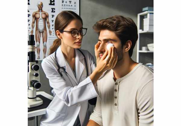
During the clinical examination, the eye care professional will look for chalazion-specific signs, such as a firm, non-tender nodule. The inner surface of the eyelid may be everted in order to examine the meibomian gland openings for inflammation or blockage. The clinician may also look for signs of blepharitis, which can be linked to chalazion formation.
Differential Diagnosis
It is critical to distinguish chalazion from other eyelid lesions, such as hordeolum (stye), sebaceous cysts, or more serious conditions like basal cell carcinoma or sebaceous gland carcinoma. A thorough examination helps to ensure an accurate diagnosis and appropriate treatment.
Innovative Diagnostic Techniques
While chalazia are typically diagnosed based on clinical appearance, novel diagnostic techniques can improve the diagnostic process:
- Meibography: This imaging technique shows the structure and function of the meibomian glands. Meibography can help identify gland dysfunction and blockages that cause chalazion formation.
- Ocular Surface Analysis: Advanced devices can evaluate the tear film’s lipid layer, providing information about the overall health of the meibomian glands and the ocular surface.
- High-Resolution Ultrasonography: This non-invasive imaging technique can be used to determine the size and extent of a chalazion, especially in complex or recurring cases.
- Histopathological Examination: If there is a suspicion of cancer, a biopsy of the lesion may be performed to rule out sebaceous gland carcinoma or other malignant conditions.
Combining traditional clinical examination with these innovative diagnostic techniques allows eye care professionals to gain a more complete understanding of chalazion and tailor management strategies accordingly.
Chalazion Treatment Options
The treatment for chalazion is determined by its size, duration, and associated symptoms. The primary goal is to reduce inflammation, promote drainage, and address any discomfort or cosmetic concerns.
- Warm Compresses: The first step in treatment is to apply warm compresses to the affected eyelid. This softens the meibum and promotes drainage. Compresses should be used for 10-15 minutes, three to four times per day.
- Eyelid Hygiene: Maintaining good eyelid hygiene by gently cleaning with a dilute baby shampoo solution or commercial lid scrubs can help prevent further meibomian gland blockages.
- Massage: Gently massaging the affected eyelid can aid in the release of the blocked gland’s contents, promoting chalazion resolution.
- Topical Antibiotics: If a secondary bacterial infection is suspected, topical antibiotic ointments containing erythromycin or bacitracin may be prescribed.
- Steroid Injections: Intralesional corticosteroid injections can be used to reduce inflammation and speed up resolution, particularly in larger or persistent chalazia.
- Surgical Intervention: If the chalazia does not respond to conservative treatment or is particularly large, surgical drainage may be required. This minor procedure is usually performed under local anesthesia and involves making a small incision on the inner aspect of the eyelid to drain the contents of the chalazion.
Innovative and Emerging Therapies
Emerging therapies for chalazion are constantly being investigated to improve patient outcomes and reduce recurrence rates.
- Thermal Pulsation Devices: These devices use heat and gentle pressure on the eyelids to stimulate the production of meibomian gland secretions. They can help with meibomian gland dysfunction and prevent chalazion formation.
- Intense Pulsed Light (IPL) Therapy: IPL therapy has shown promise in treating meibomian gland dysfunction by reducing inflammation and increasing gland function. It may help to prevent chalazia from recurring in patients with chronic eyelid inflammation.
- Laser Therapy: Laser treatments, such as CO2 lasers, can be used to precisely target and excise chalazia, lowering the risk of scarring and recurrence when compared to traditional surgical procedures.
- Botulinum Toxin Injections: Research is being conducted into the use of botulinum toxin injections to reduce meibomian gland secretion and inflammation, which could provide another treatment option in refractory cases.
Trusted Resources
Books
- “Clinical Ophthalmology: A Systematic Approach” by Jack J. Kanski and Brad Bowling
- “Ocular Pathology” by Myron Yanoff and Joseph W. Sassani


