Introduction to CHARGE Syndrome
CHARGE syndrome is a multifaceted genetic disorder that stands for coloboma, heart defects, atresia choanae, growth retardation, genital abnormalities, and ear abnormalities. Among its many symptoms, ocular manifestations are prominent and must be addressed because they have the potential to impair vision and quality of life. Children with CHARGE syndrome frequently exhibit eye abnormalities, which can range from minor visual impairment to complete blindness. Understanding the ocular manifestations of CHARGE syndrome is critical for early detection, appropriate management, and preventive measures to reduce complications and improve visual outcomes.
– Detailed CHARGE Syndrome Eye Manifestations
CHARGE syndrome is primarily caused by mutations in the CHD7 gene, which is essential for the early development of many tissues, including the eyes. The ocular manifestations of CHARGE syndrome are diverse and can affect a variety of eye structures. These manifestations are often present at birth and can have a significant impact on visual acuity and ocular health.
Cloboma
One of the most distinguishing ocular features of CHARGE syndrome is coloboma, a structural defect in the eye caused by incomplete closure of the optic fissure during embryonic development. Colobomas can affect the iris, retina, choroid, or optic disc, with varying degrees of severity. An iris coloboma can appear as a keyhole-shaped pupil, whereas retinal or optic disc colobomas can cause significant visual impairment or blindness, depending on their size and location.
Microphthalmia and anophthalmia
Children with CHARGE syndrome may have microphthalmia, in which one or both eyes are abnormally small, or anophthalmia, in which one or both eyes are missing. These conditions are caused by disruptions in ocular development during early embryogenesis. Microphthalmia and anophthalmia can cause severe visual impairment and are frequently accompanied by other structural eye abnormalities.
Strabismus
Strabismus, also known as eye misalignment, is another common ocular manifestation of CHARGE syndrome. It can manifest as esotropia (inward turning of the eye), exotropia (outward turning of the eye), or hypertropia. Strabismus can cause amblyopia, also known as “lazy eye,” where the brain favors one eye over the other, resulting in decreased vision in the affected eye.
Nystagmus
Individuals with CHARGE syndrome frequently exhibit nystagmus, a condition characterized by involuntary, repetitive eye movements. These movements can be horizontal, vertical, or rotary, and they frequently interfere with the ability to focus on objects, resulting in poor visual acuity. Nystagmus can be congenital or develop later in childhood as a result of other ocular or neurological issues related to CHARGE syndrome.
Refractive Errors
Refractive errors, such as myopia (nearsightedness), hyperopia (farsightedness), and astigmatism, are common among children with CHARGE syndrome. These errors occur due to abnormalities in the shape of the eye, which impair the eye’s ability to focus light accurately on the retina. Uncorrected refractive errors can cause visual discomfort, eye strain, and poor visual development.
Retinal and Optic Nerve Abnormalities
Aside from coloboma, CHARGE syndrome is characterized by other retinal and optic nerve abnormalities. These may include retinal dysplasia, which occurs when the retina fails to develop properly, and optic nerve hypoplasia, which is defined by an underdeveloped optic nerve. These conditions can cause significant visual impairment and are frequently accompanied by other ocular and systemic abnormalities in CHARGE syndrome.
Ocular Surface Disorders
Children with CHARGE syndrome may also have ocular surface disorders, such as keratoconjunctivitis sicca (dry eye syndrome) or exposure keratopathy. Inadequate tear production or incomplete eyelid closure causes dryness, irritation, and potential corneal surface damage. Proper treatment of these conditions is critical to avoiding complications like corneal ulcers and infections.
Cataracts
Although cataracts are uncommon, they can occur in people with CHARGE syndrome. Cataracts are caused by the natural lens of the eye becoming clouded, resulting in decreased vision. They can be present from birth (congenital cataracts) or develop later in life. Surgical intervention may be required to restore vision, depending on the severity of the cataract and its effect on visual acuity.
Lacrimal System Abnormalities
CHARGE syndrome is also characterized by abnormalities in the lacrimal system, which is responsible for tear production and drainage. These may include nasolacrimal duct obstruction, which results in chronic tearing and recurring eye infections. Proper diagnosis and treatment are required to alleviate symptoms and avoid further complications.
Impact on Development and Learning
The ocular manifestations of CHARGE syndrome can have a significant impact on a child’s development and learning. Visual impairment can disrupt cognitive development, motor skills, and social interactions. Early intervention with appropriate visual aids, therapies, and educational support is critical for improving the developmental outcomes of children with CHARGE syndrome.
– Eye Health Tips for CHARGE Syndrome
Preventive measures and risk-reduction strategies are critical for managing CHARGE syndrome’s ocular manifestations. Here are some key steps to consider:
- Schedule regular ophthalmic examinations with a pediatric ophthalmologist to detect ocular abnormalities early.
- Early Intervention for Refractive Errors: – Correct refractive errors with glasses or contact lenses to promote optimal visual development and prevent amblyopia.
- Strabismus Management: – Treat strabismus early and consistently, using patching, exercises, or surgical correction to prevent amblyopia and improve alignment.
- Managing Nystagmus: – Use therapies and low vision aids to improve visual function.
- Protective Eyewear: – Use protective eyewear during high-risk activities to avoid further eye trauma.
- Environmental Adaptations: – Modify the child’s environment to accommodate visual impairments, such as proper lighting, high contrast materials, and tactile cues.
- Addressing Ocular Surface Disorders: – Maintain proper hydration and use lubricating eye drops to prevent dry eye syndrome and corneal damage.
- Regular Monitoring and Support: – Follow-up with healthcare providers to monitor eye health and address any emerging issues promptly.
- Genetic Counseling: – Provide genetic counseling to families to understand the hereditary nature of CHARGE syndrome and its impact on ocular health.
- Education and Support Services: – Provide specialized educational and support services for children with visual impairments to promote their learning and development.
Diagnostic methods
The ocular manifestations of CHARGE syndrome are diagnosed using both standard and advanced techniques. Comprehensive eye examinations are essential for detecting and treating the syndrome’s various ocular abnormalities. These examinations typically include visual acuity tests, refraction assessments, and detailed ophthalmoscopic examinations of the eye’s internal structures, such as the retina and optic nerve.
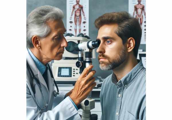
Slit Lamp Examination: A slit-lamp examination allows for a thorough examination of the anterior segment of the eye, which includes the cornea, iris, and lens. This technique is critical for detecting colobomas, cataracts, and other anterior segment abnormalities.
Fundus photography and optical coherence tomography (OCT): Fundus photography produces detailed images of the retina, allowing for the detection of retinal colobomas, retinal detachment, and other retinal abnormalities. OCT is a non-invasive imaging technique that provides high-resolution cross-sectional images of the retina, allowing for accurate assessment of retinal structure and pathological changes.
Electrophysiological Tests: Electroretinography (ERG) and visual evoked potentials (VEP) are used to evaluate the function of the retina and visual pathways. ERG measures the retina’s electrical response to light stimulation and can help detect retinal dysfunction. VEP tests measure electrical activity in the visual cortex in response to visual stimuli, revealing information about the visual pathway from the eyes to the brain.
Genetic Testing: Testing for mutations in the CHD7 gene can confirm the diagnosis of CHARGE syndrome and help predict the likelihood of associated ocular symptoms. Identifying CHD7 mutations using next-generation sequencing (NGS) can provide critical information for genetic counseling and family planning.
Ultrasound Biomicroscopy (UBM): UBM is a cutting-edge imaging technique that uses high-frequency ultrasound to examine the anterior segment of the eye. It is especially useful for assessing microphthalmia and other structural abnormalities that are difficult to detect using standard examination methods.
Magnetic Resonance Imaging (MRI) and Computed Tomography (CT): MRI and CT scans are commonly used to evaluate the overall craniofacial structure and associated anomalies in people with CHARGE syndrome. These imaging techniques can provide detailed views of the ocular and orbital anatomy, assisting with the diagnosis and treatment of complex cases.
– Managing CHARGE Syndrome Eye Conditions
The treatment of ocular manifestations in CHARGE syndrome is multifaceted and tailored to each individual’s unique needs. Standard treatments include both medical and surgical interventions, and novel and emerging therapies are constantly being investigated to improve patient outcomes.
Medical Management: To improve visual acuity in people with refractive errors, corrective lenses such as glasses or contact lenses are prescribed. Photophobia, which is commonly associated with iris colobomas, can be treated with tinted lenses or sunglasses that reduce light sensitivity. Medication may also be prescribed to treat associated conditions such as glaucoma, which uses intraocular pressure-lowering drugs to prevent optic nerve damage.
Surgical Interventions: Surgery is frequently required for severe ocular anomalies. Cataract surgery, for example, consists of removing the cloudy lens and replacing it with an artificial intraocular lens to restore vision. In cases of retinal detachment, surgical reattachment is required to prevent permanent vision loss. Vitrectomy and scleral buckling are common procedures used for this purpose.
Strabismus Surgery: Individuals with strabismus may require surgical correction to properly align their eyes. This surgery involves repositioning the eye muscles to achieve proper alignment, which can significantly improve the appearance and function of the eyes.
Prosthetic Eyes: In severe cases of microphthalmia or anophthalmia, prosthetic eyes may be required. These prostheses serve both cosmetic and functional purposes, supporting the surrounding facial structures. They can be tailored to match the appearance of the other eye, improving the person’s overall aesthetic and psychological well-being.
Innovative Therapies: Emerging therapies show promise for treating CHARGE syndrome’s ocular manifestations. Gene therapy, for example, is being studied as a potential treatment for retinal diseases caused by genetic mutations. This method attempts to correct the underlying genetic defect, potentially restoring normal function to the affected cells. Stem cell therapy is another novel approach being studied, with the potential to regenerate damaged ocular tissues and restore vision.
Vision Rehabilitation: Vision rehabilitation is an essential part of treating visual impairment in CHARGE syndrome. This may include using low vision aids, orientation and mobility training, and adaptive technologies to improve functional vision. Vision therapists collaborate with patients to develop personalized strategies that maximize their remaining vision and improve their overall quality of life.
Trusted Resources
Books
- “CHARGE Syndrome” by Timothy S. Hartshorne, Margaret W. Hefner, Sandra L. Davenport, and James C. M. Chaney
- “Pediatric Ophthalmology: Current Thought and A Practical Guide” by Edward M. Wilson, Richard A. Saunders, Trivedi Rupal
- “Genetic Disorders and the Eye” by Elias I. Traboulsi


