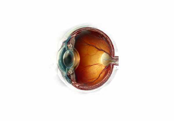
What is Chorioretinitis Sclopetaria?
Chorioretinitis sclopetaria, also known as sclopetaria, is a traumatic ocular condition caused by high-velocity projectile injuries to the eye or orbit. This uncommon condition is distinguished by severe damage to the choroid and retina, resulting in inflammation, scarring, and potential vision loss. The term “sclopetaria” is derived from the Latin word “sclopetum,” which means firearm, indicating its association with ballistic trauma. Sclopetaria is more common in military or conflict zones, but it can also occur in civilian settings as a result of gunshot injuries or high-velocity foreign bodies. Understanding the etiology, pathophysiology, and clinical manifestations of sclopetaria is critical for successful management and prevention of this vision-threatening condition.
Understanding Chorioretinitis Sclopetaria
Etiology
Chorioretinitis sclopetaria is most commonly caused by high-velocity trauma from projectiles such as bullets, shrapnel, or other fast-moving foreign bodies. The energy transferred by the projectile as it passes through the orbit generates a shock wave and rapid deformation of the ocular tissues, resulting in extensive damage. This condition can occur even if the projectile does not directly strike the eye but passes close enough to cause significant concussion forces.
Pathophysiology
The pathophysiological mechanisms underlying chorioretinitis sclopetaria include mechanical and inflammatory processes. The initial trauma disrupts the structural integrity of the choroid and retina. The shock wave produced by the projectile causes immediate and severe damage, including:
- Bruch’s Membrane Rupture: The high-energy impact may cause the rupture of Bruch’s membrane, a critical structure that supports the retinal pigment epithelium (RPE) and the retina.
- Choroidal Rupture: Damage to the choroid, a vascular layer that provides oxygen and nutrients to the outer retina, can cause significant hemorrhage and subsequent fibrosis.
- Retinal Detachment: The force of the trauma can cause retinal detachment, which occurs when the neurosensory retina separates from the underlying RPE, potentially resulting in vision loss.
- Vitreous Hemorrhage: Blood can enter the vitreous humor, a gel-like substance that fills the eye, as a result of ruptured blood vessels, complicating the condition.
After the initial mechanical injury, an inflammatory response develops. This involves the activation of immune cells and the release of inflammatory cytokines, which results in chorioretinitis, or inflammation of the choroid and retina. The inflammatory process can aggravate tissue damage and promote the formation of scar tissue, further impairing vision.
Clinical Manifestations
The clinical presentation of chorioretinitis sclopetaria differs according to the severity of the trauma and the extent of tissue damage. Common symptoms include:
- Vision Loss: Patients frequently report sudden and severe vision loss in the affected eye. The severity of vision impairment can range from partial to complete loss, depending on the extent of retinal and choroidal damage.
- Floaters: The presence of blood or inflammatory cells in the vitreous humor can result in floaters, which appear as dark spots or strands floating in the visual field.
- Photopsia: Due to retinal traction or detachment, patients may experience flashes of light, also known as photopsia.
- Pain and Discomfort: While not always present, some patients may experience pain and discomfort, especially if the injury affects the orbital bones or surrounding tissues.
- Visual Field Defects: Patients with retinal damage may have scotomas (blind spots) or other visual field defects.
Complications
If not treated promptly and adequately, chorioretinitis sclopetaria can cause a number of serious complications, including
- Chronic Inflammation: Persistent inflammation can lead to chronic chorioretinitis, which causes ongoing tissue damage and vision loss.
- Proliferative Vitreoretinopathy (PVR): This condition is characterized by the formation of membranes on the retina’s surface, which can contract and cause recurring retinal detachments.
- Neovascularization: The injury and subsequent healing process can promote the formation of abnormal blood vessels (neovascularization), which can cause bleeding or additional retinal damage.
- Macular Scarring: Damage to the macula, the central part of the retina responsible for fine detail vision, can cause macular scarring and permanent central vision loss.
- Sympathetic Ophthalmia: Although uncommon, sympathetic ophthalmia is a bilateral inflammatory condition that can develop following ocular trauma and may affect the uninjured eye.
Risk Factors
Several factors can increase the risk of developing chorioretinitis sclopetaria after an ocular trauma.
- High-Velocity Injuries: Sclopetaria is most commonly caused by injuries involving high-velocity projectiles, such as bullets or shrapnel.
- Orbital Fractures: Trauma that causes fractures in the orbital bones can increase the risk of severe ocular damage and sclopetaria.
- Close-Range Injuries: Injuries that occur at close range, where the energy transfer from the projectile is greatest, are more likely to result in significant eye damage.
- Penetrating Injuries: While sclopetaria can occur without direct eye penetration, penetrating injuries involving high-velocity projectiles carry a higher risk.
Essential Preventive Measures
The primary goal of preventing chorioretinitis sclopetaria is to reduce the risk of ocular trauma, especially from high-velocity projectiles. Here are some important preventative measures:
- Use Protective Eyewear: Wearing protective eyewear, such as safety goggles or ballistic-rated glasses, can significantly reduce the risk of ocular injuries in high-risk environments such as the military, law enforcement, and certain industries.
- Safe Handling of Firearms: Proper firearm handling, storage, and use can help prevent accidental shootings and ocular injuries. This includes adhering to safety protocols, using gun safes, and receiving appropriate training.
- Awareness and Education: Educating people about the dangers of high-velocity projectiles, as well as the importance of wearing eye protection, can help reduce the number of traumatic eye injuries.
- Workplace Safety Measures: Implementing and enforcing safety measures in workplaces that use high-velocity projectiles or machinery can help prevent ocular trauma. This includes using protective barriers and ensuring that employees are wearing proper safety gear.
- Child Safety: Keeping firearms and other potential sources of high-velocity projectiles out of children’s reach can help prevent accidental injuries. Educating children about the dangers of handling such objects is also essential.
- Military and Law Enforcement Training: Providing comprehensive training on the use of protective gear and situational awareness to military and law enforcement personnel can reduce the risk of ocular injuries on the job.
- Emergency Preparedness: Having protocols in place for the immediate response and treatment of ocular injuries can help to reduce the severity of damage and improve outcomes. This includes having access to first-aid supplies and emergency medical assistance.
- Regular Eye Examinations: For people working in high-risk jobs, regular eye exams can help detect early signs of trauma-related complications and allow for timely intervention.
Diagnostic methods
Chorioretinitis sclopetaria is diagnosed using a combination of clinical evaluation and advanced imaging techniques to determine the extent and nature of the damage. The following are the standard and innovative diagnostic methods used in clinical practice:
- Clinical Examination: The first step is a thorough clinical examination, which includes:
- Visual Acuity Testing: To assess the degree of vision impairment.
- Slit-Lamp Examination: Examine the anterior segment of the eye for signs of inflammation or injury.
- Fundoscopy: Examine the retina and choroid for signs of rupture, hemorrhage, and inflammation.
- Optical Coherence Tomography (OCT): OCT is a non-invasive imaging technique for obtaining high-resolution cross-sectional images of the retina and choroid. It aids in detecting structural damage like retinal detachment, choroidal rupture, and macular involvement.
- Fluorescein Angiography: This imaging technique involves injecting a fluorescent dye into the bloodstream to obtain detailed images of the retinal and choroidal blood vessels. It aids in detecting vascular abnormalities, neovascularization, and areas of leakage or ischemia.
- B-Scan Ultrasonography: This modality is useful when the retina is obscured by media opacities, such as vitreous hemorrhage. B-scan ultrasonography can reveal important information about retinal detachment, vitreous hemorrhage, and posterior segment integrity.
Innovative Diagnostic Techniques
- Indocyanine Green Angiogram (ICGA): ICGA is similar to fluorescein angiography, but it uses indocyanine green dye, which is more readily absorbed by the choroidal circulation. This technique produces detailed images of the choroidal vasculature, which aids in the diagnosis of choroidal ruptures and neovascularization.
- Fundus Autofluorescence (FAF): This imaging technique detects the retinal pigment epithelium’s natural fluorescence. It is useful for assessing RPE health and detecting areas of atrophy or hyperactivity, which can indicate the presence of ongoing diseases.
- Adaptive Optics Imaging: This cutting-edge technology enables cellular-level imaging of the retina, resulting in unprecedented detail. Adaptive optics can help detect microscopic changes in retinal cells after trauma.
- Electroretinography (ERG): ERG assesses the electrical responses of different cell types in the retina. This functional test can help determine the extent of retinal damage as well as the trauma’s functional impact.
Treatment
The treatment of chorioretinitis sclopetaria consists of dealing with the immediate trauma as well as any secondary complications that may arise. Treatment strategies are broadly classified as standard treatments or emerging therapies.
- Medications: – Anti-inflammatory Drugs: Corticosteroids are often used to reduce inflammation and prevent further damage. They can be given topically, orally, or as periocular injections.
- Antibiotics: If there is a risk of infection from an open wound or foreign body, prophylactic antibiotics may be prescribed to prevent bacterial infections.
- Surgery: – Vitrectomy: This surgical procedure removes the vitreous humor, especially in cases of significant vitreous hemorrhage. It clears the visual axis, allowing for better visualization and treatment of the retina.
- Retinal Repair: Laser photocoagulation, cryopexy, and scleral buckling are surgical techniques used to repair retinal detachments and ruptures.
- Choroidal Repair: In cases of severe choroidal damage, surgery may be required to stabilize the structure and prevent further hemorrhage or fibrosis.
Innovative and Emerging Therapies
- Anti-VEGF Therapy: Vascular endothelial growth factor (VEGF) inhibitors, including ranibizumab and bevacizumab, are used to treat neovascularization and macular edema. These medications are administered intravitreally and can help reduce abnormal blood vessel growth and leakage.
- Gene Therapy: Researchers are investigating gene therapy approaches that could potentially repair damaged retinal cells or prevent further degeneration. While still experimental, these therapies show promise in treating severe cases of sclopetaria.
- Stem Cell Therapy: Stem cell therapy attempts to regenerate damaged retinal tissue by introducing stem cells capable of differentiating into retinal cells. This innovative treatment is still in the experimental stages, but it holds promise for future applications in ocular trauma.
- Retinal Prostheses: In cases of severe vision loss, retinal prostheses or “bionic eyes” may restore some visual function. These devices convert light into electrical signals that stimulate the remaining retinal cells, allowing for visual perception.
Trusted Resources
Books.
References: Ferenc Kuhn’s “Ocular Trauma: Principles and Practice” and J. Fernando Arévalo’s “Retinal and Choroidal Manifestations of Selected Systemic Diseases”.
- “Intraocular Inflammation: Uveitis and Ocular Immunology” by Manfred Zierhut, Hans-Georg Rammensee, and Nicholas A. Jones


