
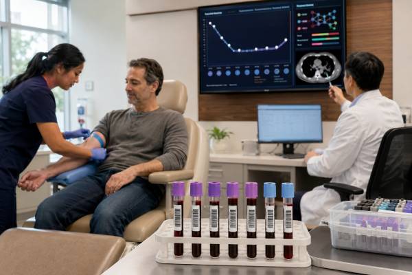
A circulating tumor DNA test analyzes small fragments of cancer-derived DNA released into the bloodstream. It can identify tumor mutations without a new tissue biopsy, track molecular changes during treatment, and detect molecular residual disease after apparently curative therapy. The same term covers several different tests, however, and their meaning depends on why the test was ordered.
In advanced cancer, plasma ctDNA profiling may find an actionable mutation or a resistance mechanism. After surgery, a highly sensitive assay may look for tiny amounts of residual tumor DNA that predict recurrence risk before a scan becomes abnormal. During systemic therapy, serial levels can provide an early signal of response or progression. A positive result can be informative, but a negative result does not prove that cancer is absent because some tumors shed little DNA or are below the assay’s detection limit. Results must be interpreted with tissue pathology, imaging, treatment history, timing of collection, tumor type, and assay design. Clonal hematopoiesis from blood cells can also produce misleading mutations unless the laboratory controls for it.
- ctDNA is the tumor-derived portion of cell-free DNA; most cell-free DNA in blood comes from normal cells.
- A plasma-positive actionable mutation can guide treatment in validated settings, but a plasma-negative result may require tissue testing.
- Post-treatment ctDNA positivity often signals high recurrence risk, yet treatment changes are not standardized for every cancer.
- Tumor-informed and tumor-naive assays use different strategies and have different strengths.
- Serial trends are usually more informative than a single low-level measurement.
Table of Contents
- What ctDNA Is and What the Test Measures
- When ctDNA Testing Is Used
- How Blood Is Collected and Analyzed
- Tumor-Informed and Tumor-Naive Assays
- How to Read Positive, Negative, and Quantitative Results
- ctDNA for Treatment Selection and Resistance
- ctDNA for Molecular Residual Disease and Recurrence
- Limitations, False Results, and Questions to Ask
What ctDNA Is and What the Test Measures
Cell-free DNA, or cfDNA, consists of short DNA fragments released into body fluids as cells die or actively secrete material. In a person with cancer, a fraction of plasma cfDNA may come from tumor cells. That fraction is called circulating tumor DNA. It can carry substitutions, small insertions and deletions, copy-number changes, rearrangements, methylation patterns, or other features found in the cancer.
The tumor fraction can range from undetectable to a substantial share of total cfDNA. It tends to be higher with greater tumor burden, active metastatic disease, liver involvement, and rapidly proliferating tumors. It may be very low in small localized cancers, indolent tumors, central nervous system tumors, or disease confined to certain body compartments.
A ctDNA test is often called a liquid biopsy, but liquid biopsy is a broader term that can include circulating tumor cells, extracellular vesicles, RNA, proteins, or DNA from fluids other than blood. Most routine oncology ctDNA testing uses plasma obtained from peripheral blood.
The assay may report individual mutations and their variant allele fractions, or VAFs. VAF is the proportion of sequencing reads containing a variant. It is influenced by tumor fraction, copy-number changes, normal cfDNA, treatment, and technical factors. A 2% VAF does not mean that 2% of the body is cancer, and VAF should not be converted directly into tumor size.
For molecular residual disease, the test often gives a qualitative “detected” or “not detected” result and sometimes a concentration such as mean tumor molecules per milliliter. These values are assay-specific. Results from different laboratories are not necessarily interchangeable.
ctDNA is biologically dynamic. Its half-life in blood is generally measured in minutes to hours, so levels can change quickly after surgery or treatment. This makes ctDNA responsive but also sensitive to collection timing. Tissue injury can temporarily increase background cfDNA and dilute the tumor fraction.
A cell-free DNA test can serve very different purposes in prenatal care, transplantation, and oncology. The ordering indication and laboratory method must be clear before a result is interpreted.
When ctDNA Testing Is Used
There are four major clinical uses: genomic profiling in advanced cancer, detection of acquired resistance, response monitoring, and molecular residual disease assessment. The evidence and regulatory status differ among these uses.
In advanced solid tumors, plasma next-generation sequencing may be ordered when tissue is unavailable, insufficient, unsafe to obtain, or too old to reflect current disease. It can identify mutations linked to approved targeted therapies, resistance pathways, or clinical trials. It may also produce results faster than arranging a new biopsy.
At progression, ctDNA can reveal new clones that emerged under treatment pressure. Examples include secondary EGFR mutations in lung cancer, ESR1 mutations in hormone-receptor-positive breast cancer, RAS pathway alterations after anti-EGFR therapy in colorectal cancer, and resistance changes in other targeted pathways. Because blood samples DNA from multiple tumor sites, ctDNA may capture heterogeneity missed by a single biopsy.
Serial testing during therapy can show falling ctDNA with response or rising ctDNA before radiographic progression. It may help clarify whether a treatment is suppressing the molecular clone, but routine scan schedules and clinical assessment usually remain necessary. A transient early rise can occur when treatment kills tumor cells, and not every fluctuation represents progression.
After surgery, radiation, or definitive chemoradiation, highly sensitive testing may look for molecular residual disease, also called minimal residual disease. A positive result often identifies patients at much higher recurrence risk. This use is most mature in colorectal cancer and is expanding in lung, breast, bladder, and other cancers.
ctDNA testing may also be used after recurrence to select another therapy, monitor a molecularly targeted drug, or determine whether a previously resistant clone has declined. The clinical value depends on whether an action has been validated for that tumor and biomarker.
Routine ctDNA testing is not a substitute for diagnosing a suspicious mass. Tissue biopsy establishes histology, grade, receptor status, and the tumor microenvironment. Nor is standard tumor ctDNA testing recommended as a general screening test in asymptomatic people. Multi-cancer early detection assays are a separate, evolving category with unresolved questions about false positives, overdiagnosis, diagnostic workup, and mortality benefit.
Testing should answer a defined clinical question. Repeating a broad panel without a plan for acting on the result can add cost and uncertainty without improving care.
How Blood Is Collected and Analyzed
A ctDNA test usually requires one or more tubes of blood. No fasting is needed. Specialized stabilizing tubes preserve white blood cells during shipping; if cells break down, they release large amounts of normal genomic DNA that dilute ctDNA and reduce sensitivity. Processing time, temperature, tube type, centrifugation, and storage all affect quality.
Plasma is separated from blood cells and cfDNA is extracted. The laboratory then uses one of several methods:
- Digital PCR or droplet digital PCR for one or a few known variants
- Targeted next-generation sequencing for dozens to hundreds of genes
- Error-corrected sequencing with molecular barcodes for very low variant fractions
- Methylation or fragment-pattern analysis for tumor detection or tissue-of-origin signals
- Whole-genome or broad sequencing approaches in selected platforms or research settings
Digital PCR is highly sensitive and useful when the exact mutation is known, but it cannot discover unexpected resistance mechanisms. Broad NGS can examine many genes at once, although sensitivity varies by variant class and depth. Fusions, copy-number losses, and complex rearrangements may be harder to detect in plasma than hotspot substitutions.
For advanced-cancer profiling, the report may include actionable variants, potential therapies, clinical trials, and variants of uncertain significance. For MRD, the report may provide a binary result, quantitative level, and comparison with prior samples. Laboratories should state the limit of detection, sample quality, and whether matched white-blood-cell sequencing was used.
Timing depends on the purpose. A pretreatment sample establishes baseline shedding. During therapy, samples are collected at protocol-defined intervals. After surgery, testing too soon can be less sensitive because trauma releases abundant normal cfDNA; many studies collect the first postoperative sample after approximately two to four weeks, with timing adjusted for tumor type and treatment plan. Serial postoperative samples increase the opportunity to detect residual disease.
A positive unexpected mutation in TP53, DNMT3A, TET2, ASXL1, JAK2, or another blood-associated gene may come from clonal hematopoiesis. Sequencing paired white blood cells helps distinguish blood-cell variants from tumor variants. Without that control, clinical context and tissue comparison are essential.
Turnaround ranges from days to several weeks. A delayed result should not postpone urgent treatment when standard clinical evidence already supports action.
Tumor-Informed and Tumor-Naive Assays
A tumor-informed assay begins by sequencing the person’s tumor, sometimes with matched normal DNA. The laboratory selects a set of patient-specific tumor variants and then builds a blood assay to track them. Because the variants are known to come from the tumor, this approach can achieve high specificity and reduce confusion from clonal hematopoiesis.
Tumor-informed MRD testing is particularly useful after curative-intent treatment. Tracking multiple private variants increases the chance of detecting a small residual clone. The disadvantages are the need for adequate tumor tissue, additional setup time, and inability to detect a completely new cancer or a clone that no longer carries the selected markers.
A tumor-naive, or plasma-only, assay does not require prior tumor sequencing. It searches a fixed panel of recurrent cancer variants and may incorporate methylation, fragmentomics, or machine-learning signals. It can be ordered when tissue is unavailable and may return results more quickly. It can also discover variants not represented in an old tissue specimen.
The tradeoff is specificity. A plasma-only assay must distinguish tumor DNA from clonal hematopoiesis and technical noise without knowing the original tumor profile. Some platforms sequence white blood cells or use bioinformatic filters. Others may report a low-level variant that requires tissue correlation.
For treatment selection in advanced cancer, many commercial tests are tumor-naive broad panels. For postoperative MRD, both tumor-informed and tumor-naive platforms are used. Their sensitivity, reporting units, sampling schedules, and positive thresholds differ, so results should not be compared as if they were the same laboratory measurement.
A negative tumor-informed result may occur if residual cells do not shed enough DNA or if the tracked variants are lost through clonal evolution. A negative tumor-naive result may reflect low shedding, incomplete panel coverage, or filtering. Neither method can guarantee absence of microscopic disease.
The distinction also affects turnaround. Tumor-informed testing requires retrieval and sequencing of a tissue block, which may be delayed by transfers between institutions. A plasma-only test can begin immediately after blood collection. When an adjuvant decision has a narrow time window, the team should order early and know whether the assay can report in time.
Ask whether the test is intended for broad mutation profiling or ultra-sensitive MRD. A broad 300-gene panel designed for metastatic disease usually cannot detect the same extremely low concentrations as a personalized MRD assay, even though both analyze ctDNA.
How to Read Positive, Negative, and Quantitative Results
A positive genomic profiling result means the assay detected one or more alterations above its reporting threshold. The finding may be an approved companion-diagnostic biomarker, a potentially actionable alteration supported by another assay or cancer type, a resistance marker, or a VUS. The report’s evidence tier matters more than the presence of a long mutation list.
A plasma-positive result for a well-validated activating mutation is often reliable, especially when the variant fits the cancer type and has adequate allele fraction. Some companion diagnostic indications permit treatment selection directly from an approved plasma assay. Other findings require confirmation with an FDA-approved or locally validated test.
A negative plasma profile does not mean the tumor is genetically normal. It can mean there was too little ctDNA, the variant lies outside the panel, the alteration type is poorly detected, or treatment suppressed shedding. When a negative result would deny an effective targeted therapy, tissue testing is recommended if feasible.
A positive MRD result after curative-intent treatment means tumor-associated DNA remains detectable in blood. Across many studies, this strongly predicts recurrence, often months before imaging. It does not identify where recurrence is located, and it does not guarantee that clinical relapse will occur on a specific date.
A negative MRD result lowers estimated recurrence risk but does not reduce it to zero. Sensitivity improves with serial testing. A single negative sample collected too early, during low-shedding disease, or after recent chemotherapy may be less reassuring than repeated negative results over time.
Quantitative changes must be interpreted consistently. A decline from 10 to 1 tumor molecule per milliliter may indicate response, but confidence depends on assay precision and sampling conditions. Small changes around the detection threshold can reflect statistical variation. Laboratories may use different normalization methods, so cross-platform numerical comparisons are unreliable.
“Not detected” differs from “test failed.” A failed sample has inadequate cfDNA, poor quality, or a technical problem and provides no biological reassurance. “No actionable variants” may still include nonactionable tumor variants and does not equal “no ctDNA.”
A VUS should not guide treatment. A possible germline alteration—especially in BRCA1, BRCA2, PALB2, ATM, mismatch-repair genes, or another predisposition gene—requires confirmatory germline testing because plasma profiling cannot reliably distinguish inherited from somatic origin.
ctDNA for Treatment Selection and Resistance
Plasma genomic profiling is established in several advanced cancers when performed with a validated assay. It may identify EGFR, ALK, ROS1, RET, MET, BRAF, KRAS, ERBB2, PIK3CA, ESR1, BRCA1/2, and other alterations, depending on the tumor type and panel. The drug indication may require a specific variant, cancer type, disease stage, and prior treatment.
A molecular match does not automatically mean treatment will work. The alteration must be an oncogenic driver, present in a meaningful clone, and linked to evidence in that disease. Co-mutations and resistance pathways can reduce response. The report should distinguish an on-label companion-diagnostic indication from off-label evidence or trial eligibility.
ctDNA has an advantage when disease is heterogeneous. A biopsy samples one site at one time, whereas plasma may contain DNA from several metastases. This can expose a resistant subclone that is absent from the biopsied lesion. The same feature can make interpretation complex because multiple competing resistance mechanisms may appear simultaneously.
In colorectal cancer, RAS or EGFR-pathway alterations can emerge during anti-EGFR therapy and later decline after the drug is stopped. Serial ctDNA can support rechallenge strategies in selected settings, though protocols and access vary. In breast cancer, ESR1 mutations detected in plasma may influence endocrine therapy selection where an approved indication applies. In lung cancer, plasma can identify targetable drivers at diagnosis and resistance changes at progression.
Tissue remains essential when histologic transformation is possible, such as small-cell transformation of EGFR-mutant lung cancer or neuroendocrine transformation of prostate cancer. ctDNA may suggest a genomic change but cannot show cell morphology. A biopsy also permits immunohistochemistry and assessment of the tumor environment.
Treatment-response monitoring is promising but not uniformly standardized. A rapid molecular decline can precede radiographic response, while persistent ctDNA may identify poor prognosis. Changing therapy solely because ctDNA rises, before clinical or radiographic progression, can expose a patient to an unproven switch. The action should be supported by a trial, guideline, or validated pathway.
A tumor genomic test and plasma profiling are complementary. Tissue often has higher sensitivity when available; plasma is faster, less invasive, and can be repeated.
ctDNA for Molecular Residual Disease and Recurrence
After a tumor has been removed or eradicated on imaging, microscopic cancer cells may remain. MRD assays try to detect their DNA before conventional recurrence becomes visible. Post-treatment ctDNA positivity is one of the strongest prognostic markers studied in localized solid tumors.
In colorectal cancer, randomized and prospective studies have shown that ctDNA-guided strategies can reduce chemotherapy use in selected stage II disease without clearly worsening recurrence outcomes, while ongoing trials are testing escalation for ctDNA-positive patients. Evidence is developing in stage III colon cancer, rectal cancer, lung cancer, breast cancer, bladder cancer, and other tumors.
Prognostic value does not always equal predictive value. A test is prognostic when it identifies high recurrence risk. It is predictive when it shows that a particular treatment changes that risk. Many MRD assays are clearly prognostic, but evidence that every ctDNA-positive patient benefits from more treatment—or that every ctDNA-negative patient can safely omit treatment—is incomplete.
The clinical response to a positive result varies. Options may include confirmatory repeat testing, earlier imaging, enrollment in an MRD-directed trial, or standard adjuvant therapy when the result reinforces an existing indication. Starting an unproven drug solely because of molecular positivity can cause toxicity without established benefit.
Serial testing can be more informative than one sample. Persistently positive or rising ctDNA after therapy suggests ongoing disease. Conversion from positive to negative may indicate molecular clearance, although relapse can still occur. Conversion from negative to positive may precede imaging by months and should prompt coordinated evaluation rather than panic.
Lead time is not automatically beneficial. Detecting recurrence earlier improves outcome only if an effective intervention works better at the earlier molecular stage. More frequent scans can also produce incidental findings, radiation exposure, cost, and anxiety. Surveillance plans should be evidence-based and tailored to the cancer.
Insurance coverage for MRD testing varies by cancer, stage, assay, and jurisdiction. Patients should ask whether the result will change a covered treatment, whether serial samples are included, and whether participation in a clinical trial is available.
The term minimal residual disease is also used in blood cancers, where methods and evidence differ substantially from solid-tumor ctDNA testing.
Limitations, False Results, and Questions to Ask
Low tumor shedding is the central limitation. Brain tumors are partly separated from blood by the blood-brain barrier, and small lung, peritoneal, pleural, or low-volume lesions may release little detectable DNA. Some histologies shed less than others. A negative plasma result therefore cannot be interpreted without knowing tumor burden and site.
False-positive or misleading findings can arise from clonal hematopoiesis, sequencing artifacts, germline variants, or an unrelated second cancer. Clonal hematopoiesis becomes more common with age, smoking, prior chemotherapy, and radiation. Matched white-cell sequencing, tissue comparison, and variant-pattern review reduce this problem.
Preanalytic errors include delayed processing, incorrect tubes, hemolysis, inadequate blood volume, and recent major tissue injury. Analytical limitations vary for substitutions, indels, fusions, copy-number changes, methylation, and low-complexity regions. The report’s limit of detection applies only under validated conditions.
Useful questions include:
- Is this test for treatment selection, response monitoring, or MRD?
- Is it tumor-informed or tumor-naive, and was white-cell DNA analyzed?
- What is the validated limit of detection for the relevant mutation type?
- Could a negative result reflect low shedding, and should tissue be tested?
- Is the reported alteration an approved companion-diagnostic biomarker in this exact cancer?
- Could the mutation come from clonal hematopoiesis or the germline?
- What does the quantitative unit mean, and can results be compared over time?
- Will a positive MRD result change treatment outside a clinical trial?
- How often should testing be repeated, and what imaging remains necessary?
- What is the plan for an unexpected positive or a test failure?
ctDNA should be ordered within a care pathway that defines what happens after each possible result. A molecular tumor board can help when findings are complex, discordant with tissue, or linked only to early evidence.
Contact the oncology team promptly for new severe pain, neurologic symptoms, breathing difficulty, jaundice, bleeding, rapidly worsening weakness, or other concerning changes. A ctDNA result should never delay evaluation of symptoms or replace urgent imaging and examination.
References
- ESMO recommendations on the use of circulating tumour DNA assays for patients with cancer: a report from the ESMO Precision Medicine Working Group 2022 (Recommendation)
- Circulating tumor DNA to monitor treatment response in solid tumors 2025 (Review)
- Utility of ctDNA-molecular residual disease in predicting recurrence in solid tumors 2025 (Review)
- Minimal residual disease in solid tumors 2026 (Review)
- Summary of Safety and Effectiveness Data: Guardant360 Liquid CDx 2026 (FDA)
Disclaimer
This information is educational and does not replace advice from an oncologist, pathologist, genetic counselor, or laboratory professional familiar with the specific assay. ctDNA performance and recommended actions differ by cancer, stage, treatment setting, and regulatory jurisdiction. Do not start, stop, or change cancer treatment based only on a ctDNA result without clinical review.





