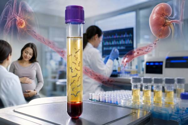
A cell-free DNA test analyzes short DNA fragments that circulate outside cells, most often in blood plasma. The same basic specimen can answer very different clinical questions. During pregnancy, placental DNA is measured to estimate the chance of selected fetal chromosome conditions. In cancer care, circulating tumor DNA may reveal tumor mutations or help track disease. After an organ transplant, donor-derived cell-free DNA can signal injury to the transplanted organ. These uses share laboratory technology, but their reports are not interchangeable. A high-risk prenatal screen does not diagnose a fetal condition, an undetected tumor variant does not rule out cancer, and an elevated transplant result does not identify the cause of graft injury by itself. The result only becomes meaningful when the laboratory method, tested targets, timing, clinical setting, and follow-up plan are considered together.
- Prenatal cfDNA is a screening test: A high-risk result usually needs confirmation with chorionic villus sampling or amniocentesis before irreversible decisions are made.
- Cancer cfDNA looks for tumor-related material in blood: A detected actionable variant may guide therapy, but a negative result can occur when a tumor sheds little DNA.
- Transplant cfDNA measures graft-derived DNA: A rise may reflect rejection or other organ injury and is interpreted with graft function, antibodies, imaging, and sometimes biopsy.
- Most prenatal tests can start around 10 weeks: Early testing may fail when the placental DNA fraction is too low.
- Collection is usually a standard blood draw: Special tubes and prompt plasma processing help prevent white blood cell DNA from diluting the signal.
- Trends may be more informative than one value: This is especially true for cancer monitoring and post-transplant surveillance.
Table of Contents
- What Cell-Free DNA Is and How Testing Works
- cfDNA in Prenatal Screening
- cfDNA and Circulating Tumor DNA in Cancer
- Donor-Derived cfDNA After Transplantation
- Sample Collection and Laboratory Methods
- How to Understand cfDNA Results
- Limitations, False Results, and Incidental Findings
- Questions and Follow-Up After Testing
What Cell-Free DNA Is and How Testing Works
Cell-free DNA consists of short DNA pieces released into body fluids as cells die, turn over, or become injured. In blood, most cfDNA comes from blood-forming cells. A smaller fraction may come from the placenta during pregnancy, a tumor in a person with cancer, or a transplanted organ. Laboratories distinguish these sources by looking for chromosome patterns, sequence variants, differences between donor and recipient DNA, methylation marks, fragment size, or combinations of these features.
Most plasma cfDNA fragments are roughly the length of DNA wrapped around a nucleosome, often near 166 base pairs. They remain in circulation for a short time, generally minutes to hours rather than days. This rapid turnover makes cfDNA responsive to changing biology. It also means that collection timing, recent treatment, inflammation, surgery, exercise, and tissue injury can affect the amount present.
A cfDNA test may measure one of several things:
- The proportion of DNA with a selected chromosome pattern, as in prenatal aneuploidy screening.
- A specific sequence change, such as an EGFR or KRAS variant in cancer.
- Many genes at once through a targeted next-generation sequencing panel.
- The fraction or absolute quantity of donor-derived DNA after transplantation.
- Epigenetic or fragment-based patterns that suggest tissue of origin.
The phrase “cell-free DNA test” therefore describes a specimen and analyte, not one universal assay. The report should identify what was tested, the technology used, the reportable range, and the clinical purpose. A person considering broader genetic testing may also benefit from understanding the difference between genetic screening and diagnostic testing, because cfDNA can serve either a screening or a disease-monitoring role depending on context.
cfDNA in Prenatal Screening
Prenatal cfDNA screening analyzes DNA released mainly by placental cells into the pregnant patient’s blood. Although it is often called “fetal DNA,” the measurable material usually comes from the placenta, which generally shares the fetus’s chromosome makeup but can occasionally differ. The test is also called noninvasive prenatal testing or noninvasive prenatal screening.
Screening is commonly offered from about 10 weeks of pregnancy. It is the most sensitive screening method for common fetal trisomies, especially trisomy 21, and it can also screen for trisomy 18 and trisomy 13. Many laboratories offer sex chromosome analysis. Some add microdeletions, rare autosomal trisomies, or selected single-gene conditions, but performance and professional recommendations are not equally strong for every added target.
What a prenatal report may show
Reports usually use terms such as low risk, high risk, screen negative, screen positive, no result, or uninterpretable. They may include a risk estimate and fetal fraction—the percentage of total cfDNA attributed to the placenta.
A high-risk result means the tested chromosome pattern was detected at a level associated with increased probability. It does not prove that the fetus has the condition. Positive predictive value changes with the condition, maternal age, gestational age, and pretest probability. A result for trisomy 21 generally has a higher positive predictive value than a result for a rare microdeletion because the underlying condition is more common and test performance is better established.
A low-risk result substantially lowers the chance of the screened conditions but does not guarantee an unaffected pregnancy. The test does not evaluate every chromosome problem, birth defect, developmental condition, or single-gene disorder. A second-trimester anatomy ultrasound remains important even after a low-risk screen.
Why confirmation matters
A high-risk screen should be discussed with a clinician experienced in prenatal genetics. Diagnostic testing using chorionic villus sampling or amniocentesis can directly analyze placental or fetal cells. The choice depends on gestational age, ultrasound findings, the suspected condition, and patient preferences. More information about the distinction is available in a broader discussion of prenatal genetic screening and diagnosis.
Discordant results can arise from confined placental mosaicism, a vanished twin, maternal chromosome variation, maternal transplant history, or rarely maternal cancer. A no-call result also deserves follow-up. Low fetal fraction is more common early in pregnancy and with higher maternal body weight, but it can also be associated with placental or chromosome-related factors. Repeating the blood draw may succeed, yet ultrasound and diagnostic testing may be more appropriate in some situations.
cfDNA and Circulating Tumor DNA in Cancer
Circulating tumor DNA, or ctDNA, is the tumor-derived portion of cfDNA. It may carry point mutations, insertions or deletions, copy-number changes, rearrangements, methylation patterns, or other features found in cancer cells. A blood-based assay that evaluates ctDNA is one type of liquid biopsy.
Cancer cfDNA testing is most established when a known cancer is already present. It may help identify a treatment target when tissue is unavailable, insufficient, unsafe to obtain, or too slow to acquire. It may also capture DNA released from several tumor sites, providing a broader view than one small tissue sample. In selected cancers, plasma testing can detect variants that affect eligibility for targeted drugs, such as alterations in EGFR, ESR1, PIK3CA, KRAS, BRAF, or gene fusions.
A detected tumor-associated variant should be interpreted through the laboratory’s evidence tier and the cancer type. The same variant can have different treatment implications in different tumors. Some reports separate variants with approved therapy, variants supported by clinical evidence, resistance markers, and variants of uncertain significance. The principles of somatic genetic testing apply because the result usually describes changes acquired by the tumor rather than inherited changes present in every cell.
Monitoring treatment and residual disease
Repeated ctDNA measurements can sometimes show whether molecular disease burden is falling or rising. A decline after effective therapy may precede changes on imaging. A new resistance mutation may explain progression and suggest another treatment. Tumor-informed minimal residual disease assays use variants identified in a patient’s tumor to search for tiny amounts of matching ctDNA after surgery or treatment. Tumor-naive assays use a fixed panel or broader molecular signature without first sequencing the tumor.
The meaning of a positive post-treatment result depends on the cancer, test, timing, and evidence supporting that use. In some settings, detectable ctDNA is strongly associated with recurrence risk. That does not always mean that changing therapy based only on the result has been proven to improve survival. Clinical trials and disease-specific guidelines determine whether the result should alter care.
Why a negative plasma result may need tissue testing
Tumors differ greatly in how much DNA they release. Small, slow-growing, early-stage, brain-confined, or anatomically isolated tumors may shed little ctDNA into blood. Treatment can also reduce the signal. A negative plasma result may therefore be “uninformative” rather than truly negative. When a treatment decision depends on finding a mutation, tissue testing is often recommended if plasma testing finds nothing and a suitable biopsy is available. A broader tumor genomic test may also examine alterations that the plasma assay does not cover.
Donor-Derived cfDNA After Transplantation
After a solid organ transplant, dying or injured cells from the donated organ release donor-derived cell-free DNA, abbreviated dd-cfDNA. Laboratories distinguish donor DNA from recipient DNA by measuring genetic differences between them. The result may be reported as a percentage of total cfDNA, an absolute concentration, or both.
Dd-cfDNA is most widely used in kidney, heart, lung, and liver transplant monitoring, although evidence and thresholds differ by organ and assay. Levels are often high immediately after surgery because the graft has experienced procurement, storage, implantation, and reperfusion injury. They usually fall as the organ recovers. A later rise can indicate new graft injury.
Rejection is one possible cause, not the only cause. Infection, ischemia, vascular problems, biliary complications, drug toxicity, trauma, or other inflammatory injury may also increase graft-derived DNA. For this reason, a transplant cfDNA test is usually combined with organ-function measurements, symptoms, imaging, donor-specific antibody testing, medication levels, and sometimes tissue biopsy.
Fractions, absolute values, and trends
A percentage result can change because the numerator or denominator changes. For example, the donor fraction may appear lower when the recipient’s total cfDNA rises during infection or systemic inflammation. An absolute dd-cfDNA concentration may add context, but assay units and thresholds are not interchangeable across laboratories.
Serial values are often more useful than a single isolated measurement. A stable low baseline can support a low probability of active injury, while a clear increase from that individual baseline may prompt closer evaluation. Transplant type, time since surgery, recent procedures, rejection history, and concurrent infection affect interpretation. A result should be compared with the reference framework validated for that specific assay and organ.
Dd-cfDNA does not replace all biopsies. A biopsy can show the pattern and severity of tissue injury and may distinguish rejection subtypes. CfDNA may help decide when a biopsy is more or less urgent, but clinical practice varies by transplant center and organ.
Sample Collection and Laboratory Methods
Most cfDNA testing begins with a peripheral blood draw. Plasma is preferred because serum preparation can release additional DNA from white blood cells. Many collection kits use specialized tubes that stabilize nucleated cells during transport. Standard EDTA tubes may also be suitable when plasma is separated promptly according to the laboratory’s instructions.
Before analysis, the laboratory separates plasma, often with two centrifugation steps, extracts the short DNA fragments, checks quantity and quality, and applies the selected assay. Common methods include:
| Method | Typical strength | Common use | Main limitation |
|---|---|---|---|
| Quantitative PCR | Fast targeted measurement | Known sequences or total DNA estimates | Limited number of targets |
| Digital PCR | Highly sensitive absolute counting | Low-level known variants or donor markers | Requires predefined targets |
| Targeted NGS | Many genes with deep coverage | Cancer mutation panels | Only covered regions are assessed |
| Genome-wide counting | Chromosome representation | Prenatal aneuploidy screening | Screening resolution and placental biology |
| Methylation or fragmentomics | Tissue-of-origin information | Emerging cancer and transplant applications | Many uses remain under validation |
Preparation is usually minimal. Fasting is rarely required unless another blood test being drawn at the same time requires it. The ordering team should tell the laboratory about pregnancy details, multiple gestation, recent transfusion, organ or bone marrow transplant, active cancer, and prior treatment because these facts can influence interpretation.
Preanalytical errors can be decisive. If white blood cells rupture before plasma separation, their abundant genomic DNA dilutes the low-level signal. Delayed processing, wrong tubes, inadequate plasma volume, hemolysis, repeated freeze-thaw cycles, and low DNA input may lead to failure or reduced sensitivity. The laboratory report may state that quality thresholds were not met rather than issuing a clinical interpretation.
How to Understand cfDNA Results
Start by identifying the test’s intended use. The words positive and negative mean different things in screening, tumor genotyping, and graft surveillance.
| Setting | Positive or elevated result | Negative or low result | Usual next step |
|---|---|---|---|
| Prenatal screening | Higher probability of a screened chromosome condition | Lower probability, not exclusion | Genetic counseling; diagnostic testing after a positive screen |
| Cancer genotyping | A tumor-associated alteration was detected | No covered alteration detected; shedding may be low | Match to cancer type and therapy evidence; consider tissue after an uninformative negative result |
| Cancer monitoring | Molecular disease is detectable or increasing | Below assay detection; microscopic disease can still exist | Compare with prior values, imaging, and disease-specific guidance |
| Transplant surveillance | Increased graft-derived DNA suggests graft injury | Lower probability of active injury within assay limits | Review organ function, antibodies, infection, and need for biopsy |
Read the technical details as well as the headline. Important fields include specimen date, gestational age or treatment timing, fetal or tumor fraction, variant allele fraction, assay coverage, limit of detection, quality-control status, reference interval, and comments about confirmatory testing.
Variant allele fraction is the proportion of DNA molecules carrying a specific variant. In cancer, a 1% allele fraction does not mean that 1% of the tumor cells carry the variant. It is influenced by tumor DNA shedding, tumor purity, copy number, normal DNA dilution, and biological subclones. In transplant testing, a percentage is likewise not a direct measure of the percentage of damaged graft cells.
A result may also be indeterminate. This can reflect insufficient cfDNA, low fetal fraction, low total DNA, contamination, poor sequencing quality, or a signal near the assay’s decision threshold. Recollection is sometimes appropriate, but repeating the same test without addressing the reason for failure may produce the same outcome.
Limitations, False Results, and Incidental Findings
CfDNA testing samples a mixture of DNA from many tissues. That feature makes blood testing possible, but it also creates ambiguity.
In prenatal screening, placental mosaicism can produce a positive result when fetal diagnostic testing is normal. Maternal mosaicism or a maternal chromosome variant may also affect sex chromosome or copy-number findings. A vanished twin can contribute DNA for weeks. Rarely, an unusual pattern across several chromosomes may lead to evaluation for maternal disease, including cancer, but the prenatal test is not designed to diagnose cancer.
In oncology, age-related clonal hematopoiesis can produce blood-cell variants that resemble tumor variants. Genes such as DNMT3A, TET2, ASXL1, JAK2, and TP53 may be altered in blood-cell clones without representing the solid tumor. Some assays compare plasma with white blood cell DNA to reduce this problem. Others use bioinformatic filtering and clinical interpretation.
Technical false negatives occur when the target is outside the panel, the molecular change is a type the assay detects poorly, the DNA input is inadequate, or the biological fraction falls below the limit of detection. False positives can arise from sequencing artifacts, contamination, index misassignment, low-complexity regions, or statistical noise near the cutoff. Well-validated laboratories use duplicate molecules, unique molecular identifiers, controls, and confirmatory rules to limit these errors.
A cfDNA finding can also raise a germline question. A cancer-associated variant detected near a 50% allele fraction across time may represent an inherited change rather than a tumor-only alteration, although fraction alone cannot prove this. Confirmation requires a suitable non-tumor specimen and a dedicated germline genetic test. This distinction affects relatives, future cancer screening, and reproductive choices.
No cfDNA test should be interpreted as a complete survey of health. Each assay has a defined menu, threshold, and intended population. Screening healthy people for many cancers with broad cfDNA signatures is developing rapidly, but positive results may require extensive localization work, and negative results cannot guarantee that cancer is absent.
Questions and Follow-Up After Testing
A useful follow-up discussion should connect the laboratory result to a specific clinical decision. Consider asking:
- What exact DNA source and condition was this assay designed to assess?
- Was the sample technically adequate, and what fraction or concentration was measured?
- Is this a screening, diagnostic, predictive, prognostic, or monitoring result?
- What is the assay’s limit of detection and what alterations are outside its scope?
- Does a positive result require confirmation in another specimen or by another method?
- Could pregnancy, transplant history, clonal hematopoiesis, infection, treatment, or recent surgery affect the result?
- Would a repeat value, tissue biopsy, imaging study, ultrasound, or diagnostic procedure change management?
- Is genetic counseling appropriate for possible inherited or reproductive implications?
During pregnancy, urgent emergency care is not usually required simply because a cfDNA screen is high risk, but timely genetic counseling and diagnostic planning are important because options depend on gestational age. In cancer care, new severe symptoms should be assessed based on the symptoms themselves rather than waiting for a molecular result. After transplantation, a rising dd-cfDNA result combined with reduced graft function, fever, pain over the graft, shortness of breath, low urine output, jaundice, or other organ-specific symptoms warrants prompt contact with the transplant team.
Keep copies of the full report, not only the patient portal summary. Future clinicians need the laboratory name, test version, specimen, date, methodology, genes or chromosomes assessed, and exact variant notation. Because assays and evidence change, an older result may later be reinterpreted or repeated with a more capable method. The most reliable interpretation combines the molecular result with the patient’s clinical history, imaging, pathology, family history, and the reason testing was ordered.
References
- A Brief Guide to SMFM’s Updated Prenatal Genetic Screening Recommendations 2025 (Guideline Summary)
- Technical Advances in Circulating Cell-Free DNA Detection and Analysis for Personalized Medicine in Patients’ Care 2024 (Review)
- A review of cell-free DNA and epigenetics for non-invasive diagnosis in solid organ transplantation 2024 (Review)
- A Review of Circulating Tumor DNA (ctDNA) and the Liquid Biopsy in Precision Oncology 2025 (Review)
- Donor-derived cell-free DNA monitoring for early diagnosis of antibody-mediated rejection after kidney transplantation: a randomized trial 2025 (RCT)
Disclaimer
Cell-free DNA results require interpretation in the exact clinical setting and with the specific assay used. This information is educational and does not replace prenatal counseling, oncology care, transplant evaluation, diagnostic testing, or advice from a qualified clinician. Seek prompt medical guidance for concerning pregnancy findings, cancer symptoms, or signs of transplant dysfunction.