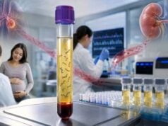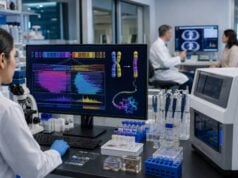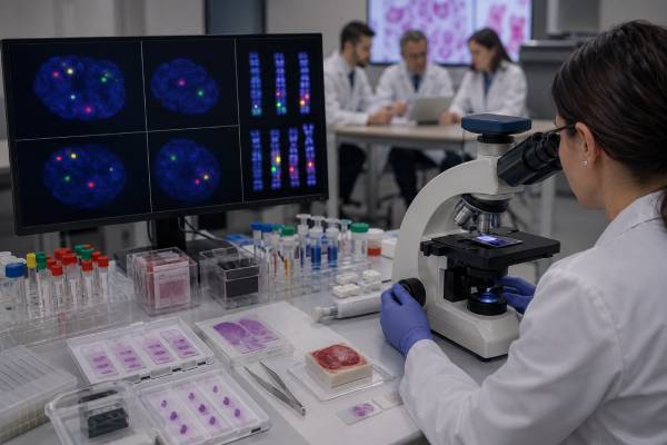
Fluorescence in situ hybridization, or FISH, uses colored DNA probes to locate selected chromosome or gene regions inside cells. A pathologist or cytogenetic technologist examines the fluorescent signals under a microscope and compares their number, position, and spacing with the expected pattern. FISH can quickly detect targeted chromosome gains or losses, gene deletions, amplifications, and rearrangements such as fusions. It is widely used in leukemia, lymphoma, solid tumors, prenatal diagnosis, and confirmation of inherited chromosome findings. Its main advantage is that it shows molecular information in individual cells and often works without growing cells in culture. Its main limitation is narrow scope: a FISH test sees only the regions covered by the ordered probes. A normal result does not exclude changes elsewhere in the genome, small sequence variants, or rearrangements that the probe design cannot reveal. Accurate interpretation therefore begins with knowing exactly which probe set was used.
- FISH is a targeted chromosome test: It detects only the genes, chromosome regions, or rearrangements covered by the selected fluorescent probes.
- Signal pattern determines the result: Extra, missing, separated, or fused colored signals can indicate amplification, deletion, or rearrangement.
- Break-apart and dual-fusion probes answer different questions: One detects disruption of a gene region; the other looks for a specific partner fusion.
- FISH can analyze nondividing cells: It often produces results faster than a full karyotype because cell culture may not be required.
- A negative FISH result is not a genome-wide negative result: Microarray, sequencing, or karyotyping may still be needed.
- Cutoffs are assay- and specimen-specific: The percentage of abnormal cells must be compared with the laboratory’s validated threshold.
Table of Contents
- How FISH Probes Produce Colored Signals
- Main FISH Probe Designs
- Where FISH Is Used Clinically
- How FISH Signal Patterns Are Read
- Percent Positive, Cutoffs, and Result Language
- FISH Compared With Karyotype, Microarray, and Sequencing
- Limitations and Interpretation Pitfalls
- Specimens, Preparation, and Follow-Up
How FISH Probes Produce Colored Signals
FISH begins with a short or long laboratory-made DNA sequence called a probe. The probe is designed to match a specific location in the human genome and is labeled with a fluorescent dye. When the probe is placed on prepared cells or tissue, it binds—hybridizes—to its complementary DNA sequence. Unbound probe is washed away, and the slide is viewed with a fluorescence microscope.
A normal cell usually shows a predictable number and arrangement of signals. For an autosomal locus, two signals are commonly expected in a diploid cell, one from each chromosome copy. A deletion may produce one signal. A duplication or amplification may produce extra signals. A rearrangement may separate two colors that normally overlap or bring two colors together that are normally apart.
FISH can be performed on interphase nuclei or metaphase chromosomes. Interphase FISH evaluates cells that are not actively dividing. It is fast and useful when a laboratory needs to count many nuclei or when tumor cells do not grow well in culture. Metaphase FISH places signals on condensed chromosomes, which can show where a segment is located and help characterize a rearrangement.
The cells are fixed on a slide, DNA is denatured so the two strands separate, and the probe mixture is applied. Hybridization may take several hours or overnight. A counterstain such as DAPI colors the nuclei and chromosomes, allowing the technologist to see cell boundaries and assess signal quality.
The laboratory does not interpret every colored dot as a true copy. Signals may overlap, split, fade, or lie outside the focal plane. Tissue sections can cut through nuclei and remove part of a signal. Technologists follow probe-specific scoring rules, count a defined number of cells, and use controls to distinguish biological abnormalities from preparation artifacts.
FISH is an example of targeted testing. If an ALK break-apart probe is ordered, it can assess the ALK region but will not evaluate every other fusion gene. This differs from a broad tumor genomic test, which may survey many genes through sequencing.
Main FISH Probe Designs
Probe design determines what an abnormal pattern means. The most common clinical designs include enumeration, locus-specific, deletion, break-apart, dual-fusion, and chromosome-painting probes.
Enumeration probes
Enumeration probes bind repetitive DNA near a chromosome centromere or another stable region. They count copies of a chromosome or chromosome arm. Three signals for chromosome 8 in a nucleus may support trisomy 8, while one X-chromosome signal in an appropriate prenatal or constitutional context may support monosomy X.
In cancer, extra centromere signals may reflect aneuploidy or polyploidy. Enumeration probes are also used as internal references for gene-amplification ratios, such as comparing HER2 signals with chromosome 17 enumeration signals in breast cancer testing.
Locus-specific and deletion probes
A locus-specific probe binds a gene or critical region. It is often paired with a control probe on the same chromosome. If the control appears twice but the target appears once, the pattern supports a heterozygous deletion. This approach is used for selected microdeletion syndromes and cancer-associated losses.
A normal two-signal pattern does not exclude a smaller deletion inside the gene if the probe still binds outside the missing segment. It also does not evaluate sequence variants.
Break-apart probes
Break-apart probes use different colors on opposite sides of a gene’s usual breakpoint region. In a normal chromosome, the colors are close enough to appear fused or adjacent. When the gene is rearranged, the colors separate beyond a defined distance.
A break-apart assay shows that the gene region is disrupted, but it generally does not identify the fusion partner. This is useful for genes that can fuse with many partners, including ALK, ROS1, RET, BCL6, MYC, EWSR1, and NTRK genes. Some rearrangements generate atypical patterns, such as loss of one colored signal, so probe-specific rules are essential.
Dual-fusion probes
Dual-fusion probes target two known partner genes in different colors. A translocation brings the colors together, creating one or more fusion signals. This design is used for specific rearrangements such as BCR::ABL1, PML::RARA, IGH::CCND1, and selected lymphoma fusions.
Dual-fusion assays are highly specific for the named partners but can miss rearrangements involving a different partner. They may also detect complex or variant signal patterns when extra derivative chromosomes are present.
Gene-amplification probes
Amplification assays compare the number of target-gene signals with a reference or with the number of nuclei. A tight cluster of many signals can indicate high-level amplification. HER2 FISH is a familiar example, but amplification testing is also used for genes such as MYC, MDM2, and others in selected tumors.
Whole-chromosome painting
Chromosome-painting probes label much of an entire chromosome. They can clarify large translocations and marker chromosomes in metaphase cells. Their resolution is lower than locus-specific FISH, but they show chromosome origin and structural relationships.
| Probe type | Normal pattern | Typical abnormal pattern | Main question answered |
|---|---|---|---|
| Enumeration | Expected number of same-color signals | Extra or missing signals | Chromosome or region copy number |
| Deletion | Two target and two control signals | One target with retained controls | Is a selected locus missing? |
| Break-apart | Paired or fused colors | Separated colors or validated atypical pattern | Is this gene rearranged? |
| Dual-fusion | Separate partner colors | New fusion signals | Are these two specific genes fused? |
| Amplification | Balanced target and reference counts | Excess target signals or clusters | Is the gene present in high copy number? |
Where FISH Is Used Clinically
FISH is used most often when a specific chromosome abnormality is strongly suspected or when a rapid, cell-based answer will affect diagnosis, prognosis, or treatment.
Leukemia and myeloid disorders
Blood and bone marrow FISH panels can detect recurrent abnormalities in acute and chronic leukemias, myelodysplastic syndromes, and myeloproliferative neoplasms. Examples include BCR::ABL1, PML::RARA, KMT2A rearrangements, CBFB rearrangements, RUNX1::RUNX1T1, and deletions involving 5q, 7q, or 17p.
The test may support diagnosis, define a risk category, identify a treatment target, or monitor a known abnormal clone. However, molecular PCR or sequencing may be more sensitive for measurable residual disease once the exact fusion transcript is known.
Lymphoma and plasma-cell neoplasms
FISH is central to evaluating many lymphomas and multiple myeloma. Break-apart or dual-fusion probes can assess MYC, BCL2, BCL6, CCND1::IGH, and other rearrangements. Plasma-cell FISH often requires enrichment because abnormal plasma cells may represent a small fraction of marrow cells. Prognostic interpretation depends on the exact disease and combination of findings.
Solid tumors
FISH can evaluate HER2 amplification in breast and gastric cancers, ALK or ROS1 rearrangements in lung cancer, MDM2 amplification in selected sarcomas, and EWSR1 rearrangements in tumors with compatible morphology. It is often used when immunohistochemistry is equivocal or when a rearrangement defines the tumor type.
A positive break-apart result does not always prove a specific histologic diagnosis because the same gene can be rearranged in more than one tumor. Morphology, immunostains, clinical site, and fusion partner information may still be needed.
Constitutional and pediatric genetics
Targeted FISH can confirm a suspected microdeletion, detect a familial rearrangement, identify a marker chromosome, or test relatives. Chromosomal microarray has replaced FISH as the broad first-line test for many developmental disorders because it searches the genome rather than one locus. FISH remains useful for rapid confirmation, mosaic analysis, and structural localization.
Prenatal testing
Interphase FISH can rapidly assess selected chromosomes—commonly 13, 18, 21, X, and Y—in chorionic villus or amniotic fluid cells. It can provide an initial answer before the full karyotype or microarray is complete. Because it targets only a few chromosomes, a normal rapid FISH result cannot exclude other chromosome abnormalities. Prenatal decisions should use the complete diagnostic workup and counseling, not a limited rapid panel alone.
Urine and other cytology specimens
Some assays use urine cells to detect chromosome abnormalities associated with urothelial cancer. FISH can also be applied to effusions, sputum, cerebrospinal fluid, and other cytology samples when validated. Sensitivity depends on the number and quality of abnormal cells shed into the specimen.
How FISH Signal Patterns Are Read
A FISH result is based on a population of scored cells, not one striking nucleus. The technologist first confirms that the slide has adequate nuclei, strong control signals, limited overlap, and acceptable background. Cells that are truncated, damaged, clumped, or poorly focused may be excluded.
Missing signals
One target signal instead of two can indicate a heterozygous deletion. In paraffin tissue, nuclear truncation can also remove a signal from the section. Laboratories compensate by using control probes, counting many nuclei, and setting cutoffs from normal tissue.
Loss of one side of a break-apart probe can represent an unbalanced rearrangement, a deletion, or a technical artifact. The clinical meaning depends on the gene and validated pattern. It should not automatically be called a standard rearrangement.
Extra signals
Three signals may indicate duplication, trisomy, or polysomy. Numerous dispersed signals or a tight cluster may indicate amplification. Whole-genome duplication and tumor aneuploidy can raise both target and reference counts, so ratios may be more informative than raw counts.
Split signals
In a break-apart assay, colors must be separated by a probe-specific distance to qualify as split. Two colors that touch or are one signal diameter apart may still count as fused, depending on the validated scoring rule. Nuclear geometry can make signals appear farther apart in a two-dimensional image than they are in three dimensions.
Fusion signals
A dual-fusion assay expects a characteristic combination of separate and fused colors. Classic BCR::ABL1 patterns, for example, include signals from normal chromosomes plus fusion signals from derivative chromosomes. Variant translocations, extra copies, or deletions can alter the pattern.
Mosaic and subclonal patterns
If only a portion of cells show the abnormal pattern, the result may reflect mosaicism, a tumor subclone, sample mixture, or background artifact. The observed percentage must exceed the assay’s cutoff. In cancer, a low percentage can still be important when the abnormality is disease-defining, but tumor content and enrichment must be considered.
Images in a patient portal are usually representative rather than exhaustive. The final interpretation should rely on the laboratory’s full cell count, scoring rules, and pathologist review.
Percent Positive, Cutoffs, and Result Language
A FISH report usually identifies the probe set, specimen, number of cells scored, observed pattern, percentage of abnormal nuclei, laboratory cutoff, and interpretation. It may also include standardized cytogenetic nomenclature.
A result can be reported as positive, negative, abnormal, normal, equivocal, borderline, or insufficient. These words are assay-specific.
Positive or abnormal
The validated abnormal pattern exceeds the laboratory threshold. In cancer, the report may state that a rearrangement, deletion, or amplification was detected. In constitutional testing, it may describe a deletion or chromosome count. Positive does not automatically mean inherited; tumor FISH findings are usually somatic unless separate germline testing shows otherwise.
Negative or normal
The targeted abnormality was not detected above the validated cutoff in the scored cells. This does not exclude another rearrangement, a cryptic breakpoint outside the probe span, a low-level clone below sensitivity, or abnormalities elsewhere.
Equivocal or borderline
The abnormal-cell percentage lies near the cutoff, signals are technically difficult, or results differ among readers. Repeat scoring, another block, an enriched specimen, or an orthogonal test may be recommended.
Insufficient or failed
There may be too few cells, degraded tissue, weak hybridization, high autofluorescence, decalcification damage, or failed controls. A new specimen or a different method may be needed.
Cutoffs are developed from normal or negative specimens and validated for each probe, tissue type, and preparation. A 5% abnormal-cell threshold for one assay cannot be applied to another. Paraffin sections often have higher normal-background signal loss than intact blood or marrow nuclei.
HER2 testing illustrates the importance of detailed criteria. Breast cancer interpretation uses target and chromosome 17 signals, ratios, average copy numbers, and integration with immunohistochemistry under current pathology guidelines. A raw HER2 signal count without the scoring group and IHC context is incomplete.
FISH Compared With Karyotype, Microarray, and Sequencing
Each method sees a different layer of genome structure.
| Method | Scope | Main advantage | Main limitation |
|---|---|---|---|
| FISH | Selected loci | Fast, cell-based, works in interphase, shows location and heterogeneity | Cannot discover unprobed abnormalities |
| Karyotype | All chromosomes | Shows large balanced and unbalanced structure | Lower resolution and often needs dividing cells |
| Chromosomal microarray | Genome-wide copy number | Detects small deletions and duplications | Usually misses balanced rearrangements and cell morphology |
| PCR | Known sequence or fusion | Highly sensitive and fast | Requires specific primer-compatible target |
| NGS | Many genes | Broad variant and fusion discovery | Longer workflow and variable structural-variant performance |
FISH is often chosen because it preserves cell context. In a tumor, the pathologist can score cells within a selected area and see whether all or only some cells carry an amplification. Sequencing may report an averaged signal from a mixture of tumor and normal cells.
Karyotyping shows the whole chromosome architecture and can identify an unexpected balanced translocation. FISH can then confirm which genes lie at the breakpoints. A karyotype test and FISH therefore often complement each other.
Microarray provides broader and usually finer copy-number detection in constitutional genetics, but it cannot show where an extra copy is inserted. FISH can localize that material on metaphase chromosomes. RNA sequencing can identify the exact expressed fusion partner and breakpoint, while break-apart FISH may only show that one gene is rearranged.
The best method depends on the decision. A rapid PML::RARA result in suspected acute promyelocytic leukemia may require a targeted assay immediately. An undiagnosed developmental disorder needs broad discovery. A morphologically defined sarcoma may need FISH for one characteristic rearrangement, followed by RNA sequencing if the pattern is atypical.
Limitations and Interpretation Pitfalls
The most common misunderstanding is treating FISH as comprehensive. A normal probe set answers only its stated targets. A five-probe leukemia panel does not exclude other leukemia-associated changes. A negative ALK break-apart test does not assess EGFR, KRAS, ROS1, RET, MET, or every possible ALK alteration.
Probe location creates another limitation. A small intragenic deletion can leave both flanking break-apart probes intact. A rearrangement breakpoint outside the usual probe span may appear normal. Conversely, a deletion of one probe region can create an atypical split-like pattern.
Tissue preparation affects results. Acid decalcification can damage marrow or bone-tumor DNA. Formalin fixation, old blocks, crush artifact, necrosis, and autofluorescence reduce signal quality. Thick sections cause overlapping nuclei; thin sections increase truncation.
Biological heterogeneity can produce discordant results between blocks, metastatic sites, or time points. A small amplified clone may be missed if the tested area lacks it. Treatment can select a resistant subclone with a new rearrangement or copy-number pattern.
Observer interpretation matters. Borderline split distances, signal clusters, and overlapping nuclei require experience. Laboratories use blinded second review or adjudication for equivocal cases. Automated image analysis can assist but does not remove the need for validated human oversight.
A positive break-apart result can be biologically nonspecific. It confirms disruption near the gene but not necessarily a functional transcript. Some rearrangements are nonproductive, and some genes have multiple partners with different clinical effects. RNA or DNA sequencing may be needed to define the fusion.
FISH can also reveal a tumor-associated abnormality that resembles a constitutional change. If a deletion is present in nearly all cells from a non-tumor specimen or family history suggests inheritance, dedicated germline testing may be appropriate. Tumor FISH alone should not be used to assign inherited risk.
Specimens, Preparation, and Follow-Up
FISH can use blood, bone marrow, fresh or frozen tissue, formalin-fixed paraffin-embedded tissue, cytology slides, urine, amniotic fluid, chorionic villi, and cultured cells. Preparation depends on the specimen.
Patients usually do not need to fast for a blood or marrow FISH test. The main preparation is procedural: a marrow biopsy, amniocentesis, or tissue biopsy has its own instructions and risks. The ordering team should tell the laboratory the suspected diagnosis so the correct probes and tissue area are selected.
Before testing, useful questions include:
- Which exact genes or chromosome regions are covered?
- Is the probe an enumeration, deletion, break-apart, dual-fusion, or amplification design?
- Will interphase cells, metaphase cells, or both be examined?
- How many cells are scored and what cutoff defines positivity?
- Is tumor-cell enrichment or microdissection needed?
- What abnormal patterns are recognized by this assay?
- What follow-up method will resolve an equivocal or atypical pattern?
- Does a negative result leave an important treatment or diagnostic gap?
After a positive result, the next step may be immediate treatment, risk classification, confirmatory sequencing, family testing, or broader chromosome analysis. After a negative result, another method should be chosen according to the remaining clinical suspicion rather than simply repeating the same probe.
Keep the full report, including the probe names and percentage of abnormal cells. “FISH positive” is not a complete medical result. A future clinician needs to know whether the finding was a deletion, amplification, break-apart pattern, specific fusion, or chromosome count and which specimen contained it.
Urgent communication is important when FISH identifies a result that changes immediate care, such as PML::RARA in suspected acute promyelocytic leukemia. In other settings, the result can wait for integrated pathology review. The laboratory pattern should always be interpreted with morphology, immunophenotype, clinical history, and other molecular findings.
References
- Fluorescence in Situ Hybridization (FISH Test) 2024 (Official Clinical Resource)
- HER2 Testing in Breast Cancer – 2023 Guideline Update 2023 (Guideline)
- Challenges and prospects in utilizing technologies for gene fusion detection 2024 (Review)
- The implications of abnormal signal patterns of break-apart fluorescence in situ hybridization probes used in the diagnosis of bone and soft tissue tumors 2025 (Study)
- ISCN 2024: Summary of Revisions and New Nomenclature 2025 (Review)
Disclaimer
FISH interpretation depends on the exact probe, specimen, cell count, cutoff, and clinical context. This article is educational and does not replace pathology review, cytogenetic consultation, prenatal counseling, or treatment decisions based on an integrated diagnostic report.