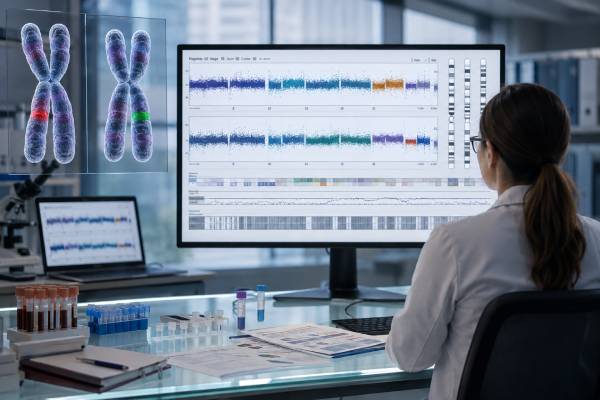
A copy number variant test looks for sections of DNA that are present in fewer or more copies than expected. These changes range from a missing exon within one gene to a gain or loss spanning millions of DNA bases and many genes. CNV testing is not one single laboratory technique. Chromosomal microarray, deletion/duplication analysis, MLPA, quantitative PCR, digital PCR, next-generation sequencing, and genome sequencing can all detect copy-number changes, but they differ in scope and resolution. A test chosen for one gene may miss a clinically important deletion elsewhere, while a genome-wide method may detect uncertain findings unrelated to the original question. Results also require biological interpretation: a deletion can disrupt a dosage-sensitive gene, expose a recessive variant on the remaining copy, or have no known effect. The best report explains what region changed, how many copies were measured, how confidently the change was detected, and whether current evidence supports a diagnosis.
- CNVs are DNA gains or losses: Deletions reduce copy number, while duplications or amplifications increase it.
- Test coverage determines what can be found: A single-gene assay, panel, microarray, exome, and genome test do not examine CNVs in the same way.
- A pathogenic CNV may involve one exon or many genes: Clinical impact depends on dosage sensitivity, breakpoints, inheritance, and the person’s features.
- A negative result is method-specific: It rules out only the CNV types and regions the laboratory validated.
- Parental or family testing often improves interpretation: It can show whether a constitutional CNV is inherited or de novo.
- Tumor copy-number results use different language: Amplification, homozygous deletion, tumor purity, and treatment relevance are interpreted as somatic findings.
Table of Contents
- Copy Number Variants and Gene Dosage
- Methods Used to Detect CNVs
- Choosing the Right CNV Test
- Reading a CNV Laboratory Report
- Pathogenic, Uncertain, and Benign Results
- Mosaic, Recessive, Prenatal, and Tumor CNVs
- False Results, Technical Gaps, and Confirmation
- Follow-Up for Patients and Families
Copy Number Variants and Gene Dosage
Most autosomal DNA regions are present in two copies. A deletion may leave one copy, called a heterozygous deletion, or remove both copies, called a homozygous deletion. A duplication often produces three copies, although triplications and larger gains can create four or more. X- and Y-chromosome copy number depends on chromosomal sex and the region being tested.
CNVs vary enormously in size. A change can remove one coding exon, several exons, an entire gene, a cluster of genes, or part of a chromosome. The effect is not predicted by size alone. A small deletion that removes one critical exon of a dosage-sensitive gene can cause disease, while a larger CNV in a gene-poor or variation-tolerant region may be benign.
Gene dosage describes how the number of functional copies affects biology. Some genes tolerate one missing copy. Others are haploinsufficient, meaning one functional copy does not produce enough gene product for normal function. A duplication can cause disease when a gene is triplosensitive and extra dosage disrupts development or cell regulation. Many genes have no proven dosage relationship, so a CNV involving them may remain uncertain.
Breakpoints also matter. Two people may both be described as having a deletion in the same chromosome band, but one deletion may interrupt a critical gene while the other stops outside it. A duplication may be inserted next to the original region, inverted, or moved to another chromosome. Routine copy-number testing often establishes the amount of DNA gained but not its orientation or exact location.
CNVs can be constitutional or somatic. Constitutional variants are present from conception in most or all cells and may be inherited or de novo. Somatic CNVs arise later in a subset of cells, commonly in tumors or clonal blood-cell populations. The laboratory must know which question is being asked because the specimen, detection threshold, classification system, and clinical meaning differ.
A person unfamiliar with broader testing terminology may find it helpful to review how a genetic variant result is classified. CNVs use the same broad five-category language in constitutional testing, but evidence is evaluated across a genomic interval rather than at one DNA letter.
Methods Used to Detect CNVs
No method detects every copy-number change equally well. The laboratory chooses a platform based on the expected size, target region, specimen, turnaround needs, and whether genome-wide discovery or focused confirmation is required.
| Method | Typical scope | Strength | Important limitation |
|---|---|---|---|
| Chromosomal microarray | Genome-wide | Strong detection of submicroscopic deletions and duplications | Usually does not define balanced structure or exact breakpoints |
| MLPA | Selected exons or regions | Efficient exon-level dosage testing | Only probe-covered targets are assessed |
| qPCR or digital PCR | One or a few targets | Fast, quantitative confirmation or familial testing | Not suitable for broad discovery |
| Targeted NGS panel | Panel genes | Sequence and CNV analysis in one test | Coverage variability can reduce sensitivity |
| Exome sequencing | Protein-coding exons | Can add CNV calls to sequence analysis | Poorer resolution in sparsely captured or noncoding regions |
| Genome sequencing | Genome-wide | Can combine read depth, split reads, and breakpoint evidence | Complex regions and interpretation remain challenging |
| FISH or karyotype | Cell-based chromosome analysis | Shows location, cell-to-cell pattern, or large structure | Lower resolution or targeted scope |
Microarray infers dosage from probe intensity. SNP arrays add allele-pattern information and can reveal regions of homozygosity or some forms of uniparental disomy. A detailed explanation of this platform is available in the chromosomal microarray test article.
MLPA uses pairs of probes that bind next to each other at selected sites. After ligation and amplification, peak heights are compared with controls. It is widely used for genes such as DMD, BRCA1, BRCA2, SMN1, and others in which exon-level deletions or duplications are clinically important.
NGS CNV calling usually relies on read depth: regions with fewer sequence reads than expected may be deleted, while regions with more reads may be duplicated. Genome sequencing can add paired-end and split-read clues that reveal breakpoint structure. Exome and panel assays compare each exon with reference samples, which makes batch quality, GC content, capture efficiency, and coverage normalization critical.
Orthogonal confirmation means verifying a finding with a different method. A panel CNV might be confirmed by MLPA, qPCR, array, or another validated assay. Confirmation is most important when the signal is borderline, the change would drive major care, or the original method has known weaknesses in that region.
Choosing the Right CNV Test
The clinical question should determine test scope. A family with a known deletion needs targeted testing for that exact variant, not a broad assay that increases cost and the chance of unrelated findings. A child with unexplained developmental delay and multiple congenital anomalies may benefit from genome-wide analysis because no single gene or region is obvious. A person with classic Duchenne muscular dystrophy features needs a DMD method with strong exon-level deletion and duplication coverage.
Before ordering, confirm four points:
- What regions are assessed? “CNV analysis included” may mean all genes, selected genes, or only larger events.
- What is the validated resolution? Ask about single-exon sensitivity, minimum event size, and poorly covered regions.
- What specimen and mosaic level are accepted? Blood may not represent a tissue-limited variant.
- What alteration types are outside scope? Balanced rearrangements, repeat expansions, pseudogene regions, and triplet-repeat changes often need separate assays.
For hereditary disease panels, sequence analysis and deletion/duplication analysis are complementary. Some reports use “full-gene analysis” to mean both, while others describe them separately. A negative sequence result without dosage testing may leave a meaningful gap, especially in genes where exon-level CNVs account for a notable share of pathogenic variants.
A broad exome or genome test can consolidate several analyses, but the report should explicitly state whether CNVs were called and validated. Not every exome pipeline includes clinically reportable CNV analysis. Even when it does, a dedicated assay may be required for genes with homologous sequences, pseudogenes, repetitive exons, or complex rearrangements.
In prenatal testing, the choice may be between karyotype, microarray, targeted familial testing, or sequencing. In cancer, the choice may involve tumor tissue, plasma, bone marrow, or blood and must account for tumor fraction. An assay designed for constitutional two-versus-one copy discrimination may not be validated for highly aneuploid tumor genomes.
Cost and turnaround time matter, but they should not override analytical fit. A cheaper test that cannot detect the suspected event may delay diagnosis. Conversely, genome-wide testing is not automatically better when a highly sensitive targeted method answers the exact question.
Reading a CNV Laboratory Report
A complete report contains more than the words deletion or duplication. The following fields help define the finding.
Genomic location and reference build
Coordinates are tied to a reference genome such as GRCh37 or GRCh38. A region described as chr17:41,000,000–41,200,000 on one build may have different numbers on another. Coordinate conversion is necessary before comparing reports.
Copy number and zygosity
Reports may state x1 for a one-copy deletion, x3 for a duplication, or x0 for a homozygous deletion. Gene-level reports may use heterozygous deletion, biallelic deletion, exon 3–5 duplication, or whole-gene gain. In tumors, terms such as focal amplification or deep deletion may be used rather than a simple integer.
Size and breakpoints
Microarray boundaries are usually approximate because the true breakpoint lies between the last normal and first abnormal probes. Sequencing may define breakpoints more precisely, but repetitive DNA can still prevent exact mapping. A report may list a minimum and maximum interval.
Genes and critical regions
The laboratory identifies genes fully or partially included. Partial-gene deletions can disrupt the reading frame. Duplications require more caution because pathogenicity may depend on orientation and whether the extra copy is in tandem. A dosage-sensitive critical region may carry stronger evidence than a long list of genes with no established role.
Classification and evidence
Constitutional CNVs are commonly classified as pathogenic, likely pathogenic, uncertain significance, likely benign, or benign. The report may cite overlap with known syndromes, ClinGen dosage scores, population databases, case reports, inheritance, and phenotype match.
Method and quality limits
Look for the specimen type, test platform, minimum size, mosaic sensitivity, confirmation method, and excluded regions. A report from a tumor should also include estimated tumor content, purity limitations, and whether copy number is absolute or relative.
Do not compare copy-number values across assays without understanding their scale. A tumor “copy number 6” derived from modeling is not equivalent to a constitutional x3 duplication. A plasma copy-number signal may be diluted by normal DNA and reported as an amplification score rather than an exact cellular copy count.
Pathogenic, Uncertain, and Benign Results
A pathogenic CNV has enough evidence to explain disease or contribute substantially to it. Evidence is strongest when the CNV overlaps an established syndrome, removes a gene known to be haploinsufficient, duplicates a triplosensitive region, or repeatedly occurs de novo in people with a matching phenotype.
A likely pathogenic result has strong but not complete evidence. It is generally used clinically in the same direction as a pathogenic result, though counseling should describe the remaining uncertainty. Medical management should follow condition-specific evidence rather than the classification label alone.
A VUS is not a diagnosis. It may be newly observed, overlap a gene with limited dosage evidence, or have conflicting inheritance and phenotype data. The laboratory may recommend parental testing. A de novo occurrence can add evidence, but it is not automatically pathogenic; many de novo variants are harmless. Inheritance from an unaffected parent can suggest reduced penetrance or benignity, but subtle features and variable expression must be considered.
Benign and likely benign CNVs are common components of human genomic variation. Laboratories often suppress them from clinical reports. The presence of benign CNVs does not mean the DNA is damaged, and the absence of reported benign findings does not mean the genome contains no copy-number differences.
Why penetrance and expression complicate counseling
Penetrance is the proportion of carriers who develop a recognizable feature. Expressivity describes how strongly or broadly the feature appears. Recurrent CNVs such as some 1q21.1, 15q11.2, 16p11.2, and 16p13.11 changes can show substantial variation. One carrier may have developmental or psychiatric features while another relative appears unaffected.
A CNV result therefore may explain susceptibility rather than predict an exact outcome. Medical recommendations should focus on findings that can be screened or treated—such as cardiac, renal, hearing, vision, growth, or developmental concerns—rather than assuming every reported feature will occur.
Classification can change as databases grow. Keep the exact report and laboratory contact information. Reinterpretation is most useful when the person’s phenotype has evolved, a VUS is several years old, or family testing has added new evidence.
Mosaic, Recessive, Prenatal, and Tumor CNVs
Mosaic CNVs
A mosaic CNV is present in only a fraction of cells. Signal strength depends on the abnormal cell percentage and event size. A large mosaic deletion may be easier to detect than a small single-exon deletion at the same cell fraction. Blood testing can miss variants limited to skin, brain, placenta, or another tissue. If symptoms are segmental, patchy, or asymmetric, an affected tissue may be more informative.
CNVs in recessive disease
A heterozygous deletion can be one half of an autosomal recessive diagnosis. If the remaining gene copy carries a pathogenic sequence variant, the person may have disease. The deletion can also create apparent homozygosity: a variant on the remaining chromosome may look homozygous because the other allele is missing. Laboratories may need parental testing and a method that assesses both sequence and dosage.
Prenatal CNVs
Prenatal CNV interpretation has limited phenotype information. Ultrasound can show structural findings but cannot predict many future developmental traits. Inherited variants with reduced penetrance and uncertain CNVs create especially difficult counseling. Confined placental mosaicism can affect chorionic villus results, and maternal cell contamination must be excluded.
Tumor CNVs
Cancer cells commonly gain oncogenes and lose tumor suppressor genes. Reports may describe ERBB2 amplification, MYC amplification, CDKN2A homozygous deletion, PTEN loss, or broad chromosome-arm changes. Treatment relevance is cancer-specific. A high-level focal amplification may be more actionable than a modest whole-chromosome gain.
Tumor purity strongly affects measurement. If only 20% of cells in the specimen are cancerous, a true tumor amplification is diluted by normal DNA. Ploidy also matters because many cancers have abnormal whole-genome copy number. Computational models estimate absolute copies, but uncertainty rises in low-purity or highly heterogeneous specimens. For these reasons, somatic reports should be interpreted within tumor molecular profiling, not by constitutional CNV rules.
False Results, Technical Gaps, and Confirmation
CNV testing can fail at the laboratory, software, or interpretation stage. Common analytical problems include uneven probe performance, GC bias, low sequencing depth, poor DNA quality, reference-sample mismatch, pseudogene interference, and segmentation artifacts. Borderline single-exon calls require particular caution because one poorly performing exon can mimic a deletion.
A false negative can occur when:
- The event lies outside the assay’s covered regions.
- The CNV is smaller than the validated threshold.
- Mosaicism falls below the detection limit.
- Breakpoints occur in repetitive or homologous DNA.
- The method cannot distinguish a gene from a pseudogene.
- Tumor or fetal fraction is too low.
- A balanced rearrangement causes disease without copy-number change.
A false positive can arise from noisy data, sample contamination, normalization failure, batch effects, or a benign polymorphism misinterpreted as disease-related. Laboratories use quality metrics and databases to reduce these errors, but clinical correlation remains essential.
Confirmation strategies depend on what must be verified. MLPA is useful for exon-level events. qPCR or digital PCR can test a small target. FISH can show whether an amplification is present in individual tumor cells or whether a chromosome region moved. Karyotype can reveal a balanced rearrangement behind an unbalanced child result. Genome sequencing may characterize complex breakpoints.
Not every high-quality result needs routine confirmation if the primary assay has been fully validated for that variant type. The laboratory’s policy, the confidence metrics, and the clinical consequences should guide the decision. When a result will determine surgery, prenatal action, a high-risk drug, or predictive testing in relatives, the ordering clinician should verify that analytical confirmation and specimen identity standards are appropriate.
Follow-Up for Patients and Families
For a positive constitutional result, the first task is to determine how much of the phenotype it explains. A large CNV may contain several dosage-sensitive genes and require a multidisciplinary plan. A single-exon deletion may point to one well-defined syndrome. Clinical care should follow the established disease mechanism and the person’s current findings.
Family testing can answer several questions: whether the CNV is inherited, whether other relatives are at risk, whether a parent carries a balanced rearrangement, and which reproductive options are relevant. Targeted testing is usually preferred for relatives once the familial CNV is known.
A negative result should be translated precisely. Ask whether the test assessed single exons, whole genes, genome-wide events, mosaic changes, and balanced structure. The next test may be sequencing, microarray, MLPA, karyotype, repeat-expansion testing, methylation analysis, or genome sequencing. Repeating another assay with the same blind spot adds little value.
For a VUS, avoid predictive testing in healthy relatives unless a genetics professional recommends segregation analysis to clarify classification. Do not change major treatment solely because of uncertainty. Ask the laboratory whether reanalysis is automatic or must be requested.
Bring these questions to the result visit:
- What exact DNA segment is gained or lost, and which genome build was used?
- Is the CNV whole-gene, exon-level, recurrent, or structurally complex?
- What dosage-sensitive genes or critical regions are involved?
- Was the result confirmed, and by what method?
- Could mosaicism, tumor purity, or specimen choice alter the result?
- Does inheritance change recurrence risk or medical surveillance?
- Which conditions and variant types remain untested?
- When should the result be reinterpreted?
A CNV report is most useful when it leads to a specific plan: diagnosis, surveillance, treatment selection, reproductive counseling, or a clearly chosen next assay. Copy number alone is a measurement. Its medical meaning comes from the affected genes, biological context, validated method, and the person being tested.
References
- Copy Number Variant Interpretation Guidelines 2026 (Official Resource)
- Exome copy number variant detection, analysis, and classification in a large cohort of families with undiagnosed rare genetic disease 2024 (Study)
- Detection and characterization of copy-number variants from exome sequencing in a large cohort 2024 (Study)
- Copy number variant analysis improves diagnostic yield in a diverse clinical cohort referred for exome sequencing 2025 (Study)
- Gene Dosage Sensitivity and Human Genetic Diseases 2025 (Review)
Disclaimer
CNV results must be interpreted using the exact assay, specimen, genome coordinates, phenotype, and family history. This article provides general education and does not replace a laboratory director’s interpretation, genetic counseling, prenatal care, oncology advice, or individualized medical management.