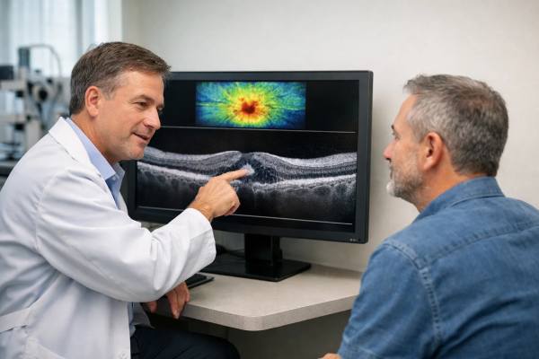
Uveitic macular edema (UME) is a common and potentially blinding complication of uveitis, an inflammatory condition that affects the uveal tract of the eye. The uveal tract consists of the iris, ciliary body, and choroid, and inflammation can cause a variety of complications, including fluid accumulation in the macula, the central part of the retina responsible for sharp, detailed vision. UME is defined as swelling or thickening of the macula caused by fluid leakage from blood vessels in the retina as a result of the inflammatory processes associated with uveitis.
Understanding Uveitis’ Impact on the Macula
To fully comprehend UME, we must first understand uveitis and its ability to disrupt normal ocular function. Uveitis is a broad term that encompasses several types of inflammation within the eye, which can be classified based on where the inflammation occurs:
- Anterior Uveitis: Inflammation primarily affects the eye’s anterior segment, which includes the iris and the anterior chamber. This is the most common form of uveitis, and it is frequently associated with autoimmune diseases like ankylosing spondylitis and juvenile idiopathic arthritis.
- Intermediate Uveitis: Inflammation occurs primarily in the vitreous and peripheral retina, and is often associated with systemic conditions such as multiple sclerosis and sarcoidosis.
- Posterior Uveitis: This type of uveitis affects the choroid and retina, causing inflammation in the posterior segment of the eye. Posterior uveitis is frequently associated with infections (e.g., toxoplasmosis) and autoimmune diseases (e.g., Behçet syndrome).
- Panuveitis: Panuveitis is an inflammation of the entire uveal tract, including the anterior and posterior segments. It is common in systemic inflammatory conditions like Vogt-Koyanagi-Harada disease.
Pathophysiology of Uveitic Macular Edema
Uveitic macular edema develops when uveitis-related inflammation causes a breakdown of the blood-retinal barrier, which normally prevents fluid and large molecules from leaking into the retinal tissue. The retinal pigment epithelium (RPE) and endothelial cells of the retinal blood vessels make up this barrier. Inflammation compromises the integrity of this barrier, allowing fluid to leak from the retinal capillaries into the macular tissue, causing swelling and thickening of the macula.
Several mechanisms aid in the development of UME:
- Cytokine Release: Uveitis triggers the release of inflammatory cytokines like TNF-α, IL-6, and VEGF. These cytokines increase vascular permeability, which causes fluid and protein leakage into the retinal layers, particularly the macula.
- Increased Vascular Permeability: The inflammation in uveitis makes the blood vessels in the retina more permeable, allowing fluid to seep into the surrounding retinal tissue. This causes an accumulation of extracellular fluid in the macula, which results in macular edema.
- Disruption of the Blood-Retinal Barrier: Uveitis disrupts the blood-retinal barrier, which is necessary for maintaining retinal homeostasis. This disruption is the result of an inflammatory process that affects both the endothelial cells of retinal capillaries and the RPE. As the barrier weakens, fluid leakage into the retinal tissue increases, causing macular edema.
- Cellular Infiltration: In severe cases of uveitis, inflammatory cells can infiltrate the retinal tissue, accelerating the breakdown of the blood-retinal barrier and promoting fluid accumulation in the macula.
- Chronic Inflammation: In cases of chronic uveitis, persistent inflammation causes long-term changes in the retinal microvasculature, such as capillary dropout and ischemia. These changes contribute to chronic fluid leakage and macular edema.
Clinical Features of Uveitic Macular Edema
The clinical presentation of UME varies according to the severity of the macular edema and the cause of the uveitis. However, a few key symptoms and signs are frequently associated with this condition:
- Decreased Visual Acuity: Visual acuity loss is a common and significant symptom of UME. Patients frequently report blurred or distorted vision, especially when reading or doing tasks that require fine detail. The swelling of the macula, which disrupts the normal arrangement of photoreceptor cells and impairs the eye’s ability to focus on fine details, is directly related to this vision loss.
- Metamorphopsia: Another common UME symptom is metamorphopsia, also known as visual distortion. Patients may describe straight lines as wavy or objects as larger or smaller than they actually are. The uneven swelling of the macula causes this symptom, which distorts the retinal architecture and, as a result, the visual image.
- Central Scotomas: Patients with UME may develop central scotomas, which are dark or blind areas in the center of their visual field. These scotomas are caused by a disruption in photoreceptor function in the swollen macula, which results in impaired vision.
- Color Vision Changes: Some UME patients experience changes in color vision, such as colors appearing washed out or less vibrant. This change is due to macula swelling, which affects the cone cells responsible for color perception.
- Photopsia: Photopsia, also known as the perception of light flashes, can occur in UME. This symptom may indicate the presence of retinal traction or inflammation near the macula.
- Floaters: In some cases, patients with uveitis may experience floaters, which are small, dark spots or threads that move across their field of vision. While floaters are not specific to UME, their presence may indicate concurrent vitritis (vitreous inflammation) or other inflammatory changes in the eye.
Risk Factors for Uveitic Macular Edema
Several factors raise the risk of developing UME, especially in people with uveitis:
- Chronic or Recurrent Uveitis: Patients with chronic or recurrent uveitis are more likely to develop UME due to ongoing inflammation and frequent episodes of blood-retinal barrier disruption.
- Underlying Systemic Diseases: Certain systemic inflammatory or autoimmune diseases, such as sarcoidosis, Behçet’s disease, and multiple sclerosis, are associated with an increased risk of uveitis and, as a result, UME.
- Infectious Causes of Uveitis: Infections like toxoplasmosis, herpesvirus, and tuberculosis can cause uveitis and increase the risk of developing macular edema, especially if the infection is not treated properly.
- Corticosteroid Use: Corticosteroids are commonly used to treat uveitis, but long-term or high doses can increase the risk of UME due to corticosteroid-induced changes in the blood-retinal barrier and increased vascular permeability.
- Ocular Surgery: Previous ocular surgery, particularly those involving the posterior segment of the eye, may increase the risk of UME by inducing inflammation and disrupting the normal ocular environment.
Risks of Uveitic Macular Edema
If not properly managed, UME can result in a number of serious complications, including permanent vision loss.
- Chronic Visual Impairment: Prolonged macular edema can cause long-term damage to the macula, resulting in chronic visual impairment. Even if the edema resolves, structural damage to the macula may cause irreversible vision loss.
- Macular Hole Formation: In severe cases of UME, continued swelling and traction on the macula can result in the formation of a macular hole, which is a full-thickness defect in the macula that significantly reduces central vision.
- Epiretinal Membrane: Chronic inflammation and macular edema can cause the formation of an epiretinal membrane, which is a layer of fibrous tissue on the retina’s surface. This membrane can exert additional traction on the macula, resulting in increased visual distortion and impaired vision.
- Secondary Glaucoma: Chronic uveitis and corticosteroid use can result in secondary glaucoma, a condition characterized by increased intraocular pressure that can impair vision further.
- Retinal Detachment: In rare cases, severe or chronic macular edema can lead to retinal detachment, a sight-threatening condition that necessitates immediate surgery.
Diagnostic Tools for Uveitic Macular Edema
To determine the underlying cause of uveitis, a combination of clinical evaluation, advanced imaging techniques, and, in some cases, laboratory tests are required. Accurate diagnosis is critical for effective treatment and avoiding long-term complications.
Clinical Evaluation
- Slit-Lamp Biomicroscopy: While the slit-lamp examines the anterior segment of the eye, it can also provide indirect evidence of macular edema by revealing signs of active inflammation in the uvea. Furthermore, using a special lens during a slit-lamp examination allows the clinician to see the posterior segment of the eye, which can sometimes reveal direct signs of macular edema, such as retinal thickening and the presence of fluid.
- Fundoscopy: Direct and indirect ophthalmoscopy (fundoscopy) is an essential component of the clinical evaluation, allowing the ophthalmologist to examine the retina and optic nerve head. During this examination, the clinician can directly visualize the macula and look for signs of edema like retinal thickening, cystic changes, or subretinal fluid. Fundoscopy is required to identify the characteristic “petaloid” pattern of macular edema, which appears as a flower-like pattern as fluid accumulates in the macular region.
Imaging Studies
- Optical Coherence Tomography (OCT) is the gold standard for detecting and monitoring uveitic macular edema. This non-invasive imaging technique produces high-resolution cross-sectional images of the retina, allowing for precise measurement of macular thickness and the detection of fluid accumulation. OCT can detect intraretinal cysts, subretinal fluid, and other structural changes related to macular edema. OCT can also detect even subtle changes in retinal thickness over time, making it invaluable for monitoring treatment response.
- Fluorescein Angiography (FA) is an imaging technique that involves injecting a fluorescent dye into the bloodstream and photographing the retina in sequence as the dye circulates through the retinal vasculature. FA in uveitic macular edema typically shows dye leakage from retinal blood vessels into the surrounding tissue, indicating a breakdown of the blood-retinal barrier. FA can also detect areas of capillary non-perfusion, neovascularization, and other vascular abnormalities linked to chronic inflammation. However, due to its invasive nature, FA is frequently reserved for cases in which the diagnosis is unclear or detailed vascular imaging is required.
- Indocyanine Green Angiography (ICGA): ICGA is similar to FA but uses indocyanine green dye to provide a more detailed view of the choroidal circulation. ICGA is especially useful in cases of posterior uveitis with suspected choroidal involvement. It aids in detecting choroidal neovascularization and other choroidal abnormalities that may contribute to macular edema. ICGA is frequently used in conjunction with FA and OCT to provide a thorough evaluation of the retinal and choroidal vasculatures.
- Fundus Autofluorescence (FAF): FAF is a non-invasive imaging technique that detects the natural fluorescence of lipofuscin, a byproduct of retinal metabolism. In uveitic macular edema, FAF can detect areas of retinal pigment epithelium (RPE) damage or dysfunction, which is frequently associated with chronic inflammation. FAF is especially useful for determining the long-term impact of macular edema on the RPE and detecting subtle changes in retinal health that may not be visible with other imaging modalities.
Lab Tests
- Blood Tests: Because uveitic macular edema is frequently associated with systemic inflammatory or infectious diseases, laboratory tests are required to determine the root cause of uveitis. Common tests include a complete blood count (CBC), erythrocyte sedimentation rate (ESR), C-reactive protein (CRP), and specific serological tests for autoimmune markers like antinuclear antibodies (ANA), rheumatoid factor (RF), and HLA-B27. Depending on the clinical situation, testing for infectious agents such as toxoplasmosis, tuberculosis, and herpesvirus may be necessary.
- Aqueous or Vitreous Sampling: When the cause of uveitis is unknown or an infectious etiology is suspected, the aqueous humor (from the anterior chamber) or vitreous humor (from the posterior segment) may be collected. These samples can be examined for the presence of infectious organisms, inflammatory cells, or other biomarkers, which can aid in determining the underlying cause of uveitis and guiding treatment decisions.
- Systemic Imaging: To assess for systemic involvement, patients with uveitis associated with systemic disease may require imaging studies such as chest X-rays, computed tomography (CT) scans, or magnetic resonance imaging (MRI). For example, chest X-rays can detect sarcoidosis, whereas MRI can assess neurological involvement in conditions such as multiple sclerosis. These imaging studies are critical for making an accurate diagnosis and managing the underlying systemic condition.
Uveitic Macular Edema Treatment
Managing uveitic macular edema (UME) necessitates a multifaceted approach that addresses both the underlying uveitis and the macular edema. The treatment aims to reduce inflammation, control macular edema, maintain visual acuity, and prevent recurrences. The management strategy is unique to each patient, depending on the severity of the condition, the underlying cause of the uveitis, and the patient’s overall health. Medical therapy, intravitreal injections, and, in some cases, surgical intervention are common treatments.
Medical Management
- Corticosteroids: Corticosteroids are the primary treatment for UME due to their potent anti-inflammatory properties. They can be administered in a variety of ways, depending on the severity and location of the inflammation.
- Topical Corticosteroids: For anterior uveitis, corticosteroid eye drops such as prednisolone acetate are commonly prescribed. Topical steroids, on the other hand, are generally insufficient as primary treatment for UME due to their limited ability to penetrate the posterior segment of the eye.
- Systemic Corticosteroids: Oral corticosteroids such as prednisone are frequently used to treat more severe or widespread uveitis, including cases with macular edema. Systemic corticosteroids are effective in reducing inflammation throughout the body, including the eyes, but long-term use has serious side effects such as weight gain, osteoporosis, hypertension, and an increased risk of infection.
- Periocular and Intravitreal Corticosteroids: Corticosteroid injections into or around the eye (periocular or intravitreal injections) are especially effective in treating UME. Agents such as triamcinolone acetonide can be injected into the vitreous cavity or the posterior sub-Tenon’s space, delivering a high dose of the drug directly to the site of inflammation. This method of managing UME is frequently preferred because it reduces systemic exposure and side effects while providing targeted treatment. However, multiple injections may be required, and there is a risk of complications such as elevated intraocular pressure (IOP) and cataract formation.
- Immunomodulatory Therapy: Immunomodulatory agents may be used when UME is associated with chronic or recurrent uveitis, or when corticosteroids are ineffective or have unacceptable side effects. This includes:
- Antimetabolites: Drugs such as methotrexate, azathioprine, and mycophenolate mofetil suppress the immune response and are used to manage chronic uveitis and reduce relapses.
- T-Cell Inhibitors: Cyclosporine and tacrolimus are calcineurin inhibitors that specifically target T-cell function, thereby reducing the immune-mediated inflammation that causes UME.
- Biologic Agents: TNF-α inhibitors, like adalimumab and infliximab, target specific inflammatory pathways in uveitis. These medications are especially effective in treating non-infectious uveitis associated with systemic autoimmune diseases.
- Non-Steroidal Anti-Inflammatory Drugs (NSAIDs): NSAIDs, either as topical eye drops or systemic medications, can be used as a supplement to corticosteroids. They help reduce inflammation and are especially beneficial in cases of mild to moderate uveitis. However, NSAIDs are less effective than corticosteroids in controlling uveitic macular edema and are usually used in conjunction with other treatments.
Intravitreal Injections
- Anti-VEGF Therapy: Vascular endothelial growth factor (VEGF) is a key factor in increasing vascular permeability and fluid leakage in the retina. Anti-VEGF agents, including bevacizumab (Avastin), ranibizumab (Lucentis), and aflibercept (Eylea), are injected directly into the vitreous cavity to reduce macular edema by inhibiting VEGF activity. While anti-VEGF therapy is commonly used to treat diabetic macular edema and age-related macular degeneration, it can also help manage UME, especially when inflammation is under control.
- Steroid Implants: Sustained-release intravitreal steroid implants, such as the dexamethasone implant (Ozurdex) and the fluocinolone acetonide implant (Iluvien), offer long-term macular edema relief with a single injection. These implants release corticosteroids over several months, reducing the need for frequent injections and providing long-term relief from macular edema. However, they carry risks such as increased IOP and cataract development, so careful patient selection and monitoring are required.
Surgical Management
In some cases, surgical intervention may be required to manage complications of UME or to treat refractory cases that do not respond to medical therapy.
- Vitrectomy: Pars plana vitrectomy is a surgical procedure that removes the vitreous gel from the eye. It is considered in cases of persistent macular edema caused by vitreomacular traction, epiretinal membrane, or significant vitreous opacification that contributes to inflammation. Vitrectomy can reduce traction on the macula and improve visual outcomes, especially in patients with complex uveitis.
- Cataract Surgery: Cataract formation is a common side effect of long-term corticosteroid use and chronic uveitis. Cataract surgery may be required to restore vision in UME patients who develop significant cataracts. However, managing inflammation before and after surgery is critical to lowering the risk of exacerbating uveitis and worsening macular edema.
Monitoring and Follow-up
Long-term monitoring is required for patients with UME because the condition is frequently chronic and prone to relapse. Regular follow-up appointments are required to assess visual acuity, monitor macular thickness with OCT, and determine treatment efficacy. Adjustments to therapy may be required depending on disease activity and the patient’s response to treatment. Managing UME frequently necessitates a multidisciplinary approach that includes ophthalmologists, rheumatologists, and other specialists who address both the ocular and systemic aspects of the disease.
Trusted Resources and Support
Books
- “Uveitis: Fundamentals and Clinical Practice” by Robert B. Nussenblatt and Scott M. Whitcup: This authoritative book provides a comprehensive overview of uveitis, including detailed discussions on the complications such as uveitic macular edema. It is an essential resource for clinicians managing patients with ocular inflammation.
- “Retinal Pharmacotherapy” edited by Quan Dong Nguyen and Diana V. Do: This book covers the pharmacological management of various retinal conditions, including uveitic macular edema. It offers insights into the latest therapies and treatment strategies, making it a valuable reference for ophthalmologists.
Organizations
- American Uveitis Society (AUS): The AUS is a professional organization dedicated to advancing the understanding and treatment of uveitis and related conditions, including uveitic macular edema. They provide resources for both patients and healthcare professionals, including access to research, clinical guidelines, and educational materials.
- The Foundation Fighting Blindness: This organization offers support and resources for individuals affected by various vision disorders, including uveitic macular edema. They provide information on the latest research, clinical trials, and support networks to help patients and their families manage the challenges associated with vision loss.


