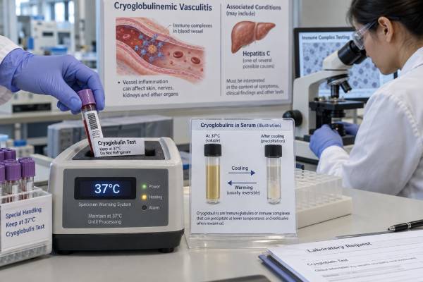
A cryoglobulin test checks serum for immunoglobulins that precipitate when cooled and dissolve again when warmed. These proteins may be present without symptoms, but they can also thicken or obstruct blood flow, form immune complexes, and inflame small vessels. The result is especially relevant when a person has palpable purpura, joint pain, weakness, peripheral neuropathy, kidney inflammation, Raynaud-type symptoms, skin ulcers, or signs of hyperviscosity. Hepatitis C is a major cause of mixed cryoglobulinemia, although autoimmune diseases and B-cell or plasma-cell disorders are also important. The test is unusually sensitive to specimen handling: blood must remain warm until serum is separated, then the serum is refrigerated long enough for slow-forming cryoglobulins to appear. A negative result can therefore be false if the tube cooled too early. Interpretation depends on the cryoglobulin type, cryocrit or concentration, complement C4, rheumatoid factor, hepatitis testing, monoclonal-protein studies, symptoms, and sometimes biopsy.
- Cryoglobulins are antibodies or antibody complexes that precipitate below body temperature and re-dissolve with warming.
- Blood must stay near 37°C until serum is separated to reduce false-negative results.
- Type I is usually monoclonal and linked to a blood-cell disorder; types II and III are mixed immune-complex forms.
- Low C4 and a positive rheumatoid factor strongly support mixed cryoglobulinemia but are not diagnostic alone.
- Coughing blood, rapidly worsening kidney function, digital ischemia, or hyperviscosity symptoms require urgent care.
Table of Contents
- Why the Cryoglobulin Test Is Ordered
- How the Sample Must Be Collected
- What Positive, Negative, and Cryocrit Results Mean
- Type I, Type II, and Type III Cryoglobulins
- Symptoms and Organ-Damage Patterns
- Hepatitis C, Autoimmune Disease, and Blood Disorders
- Next Tests, Treatment Context, and Urgent Care
Why the Cryoglobulin Test Is Ordered
Cryoglobulins are immunoglobulins—antibodies—or complexes containing immunoglobulins that become insoluble as temperature falls below 37°C. In the laboratory, they may produce a haze, gel, flakes, or a compact precipitate in refrigerated serum. When warmed back to body temperature, a true cryoglobulin should dissolve. The tendency to precipitate varies among proteins, so some form quickly while others require several days.
The test is ordered when symptoms suggest a cryoprotein is damaging circulation or triggering immune-complex vasculitis. Common reasons include recurrent palpable purpura, livedo, cold-sensitive color changes, unexplained ulcers, joint pain, fatigue, peripheral nerve symptoms, glomerulonephritis, or a combination of these findings. It may also be used when laboratory clues—such as low C4, high rheumatoid factor, a monoclonal protein, or hepatitis C infection—raise suspicion before the full syndrome appears.
A detected cryoglobulin does not automatically equal cryoglobulinemic vasculitis. Some people have circulating cryoglobulins without attributable tissue injury. The term cryoglobulinemia describes the laboratory finding, while cryoglobulinemic disease means the protein is producing clinical effects. Cryoglobulinemic vasculitis usually refers to immune-complex small-vessel inflammation caused by mixed type II or type III cryoglobulins.
Type I cryoglobulinemia behaves differently. A large monoclonal immunoglobulin load can increase blood viscosity or obstruct small vessels without classic immune-complex vasculitis. This can cause acrocyanosis, Raynaud phenomenon, retinal changes, digital ischemia, or neurologic symptoms. The test therefore helps identify a mechanism, but the type and clinical setting determine what the mechanism means.
Cryoglobulin testing is not a general screening test for vague fatigue or nonspecific pain. It is most useful when the pretest probability is meaningful and the laboratory can meet strict temperature requirements. A broad vasculitis blood test panel may be more appropriate when the initial question is simply whether systemic vessel inflammation is present.
How the Sample Must Be Collected
Correct collection is essential because cryoglobulins can disappear from the liquid portion of the sample before the laboratory examines it. If blood cools while cells and clot are still present, the proteins may precipitate onto the clot or tube wall. The separated serum can then look negative even though cryoglobulins were present in the patient.
Laboratories use their own validated protocols, but the core process usually follows these steps:
- The laboratory prepares prewarmed collection tubes, transport materials, and equipment.
- Blood is drawn into a plain serum tube and kept close to 37°C immediately after collection.
- The sample clots while warm and is centrifuged without allowing it to cool prematurely.
- Serum is separated from cells and clot before refrigeration.
- The serum is stored at about 4°C and inspected repeatedly, often for seven days.
- Any precipitate is washed, measured or described, and tested to confirm that it dissolves at 37°C.
- Immunofixation or immunotyping may identify whether the protein is monoclonal, polyclonal, or mixed.
The patient usually does not need to fast. The difficult preparation belongs to the collection site, not the person being tested. Because many routine draw stations cannot maintain warm transport and centrifugation, the clinician should arrange the test with the performing laboratory in advance. A tube sent through standard room-temperature transport may not be acceptable.
Type I cryoglobulins often precipitate within hours, while small mixed cryoglobulins can take several days. Reading the sample only after 24 or 48 hours can miss type II or type III disease. Seven-day observation improves sensitivity but also requires careful handling to distinguish true precipitate from fibrin, lipids, or contamination.
A paired plasma specimen may be collected when cryofibrinogen is in the differential. Cryoglobulins are identified in serum; cryofibrinogen is a fibrinogen-containing precipitate found in plasma but not serum. The cryofibrinogen test therefore answers a related but separate question.
False-negative results are more common than many patients realize. If the first test was drawn or transported incorrectly, or if the symptoms and supporting labs are highly characteristic, repeating the test at an experienced center can be reasonable. The new specimen should be collected before high-dose immunosuppression or plasma exchange when clinically feasible, because treatment may reduce the circulating protein.
What Positive, Negative, and Cryocrit Results Mean
A positive result means the laboratory observed a reversible cold precipitate in serum. The next questions are how much was present, what proteins made up the precipitate, and whether it explains the person’s illness. A report that says only “positive” provides less clinical value than one that includes cryocrit or concentration, immunochemical type, and comments about specimen quality.
Cryocrit is the percentage of the serum column occupied by the packed cryoprecipitate after centrifugation. For example, a 2% cryocrit means the visible precipitate occupies about 2% of the measured serum volume. Cryocrit is simple but imperfect. Results vary with tube shape, cooling duration, centrifugation, washing, and the density of the precipitate. It is not standardized well enough to serve as a universal severity scale.
A small cryocrit can still be pathogenic, particularly in mixed cryoglobulinemia with kidney, nerve, or skin disease. A high cryocrit may increase concern for type I disease or hyperviscosity, but symptoms do not rise in a straight line with the number. The temperature at which the protein precipitates, known as its thermal range, can matter as much as quantity. A protein that precipitates near room temperature may cause more trouble than one that forms only at refrigerator temperature.
A negative result means no qualifying precipitate appeared during the laboratory’s observation period. It does not exclude disease when handling was poor, the amount was low, the sample was obtained after treatment, or the protein remained attached to blood cells or the clot. A credible negative result requires a credible specimen.
The result should be interpreted with supporting markers:
- Complement C4: often substantially reduced in mixed cryoglobulinemia because classical-pathway activation consumes C4.
- Rheumatoid factor: commonly positive in type II disease because monoclonal IgM often binds the Fc portion of IgG.
- C3: may be normal or reduced; a low C4 out of proportion to C3 is a familiar pattern.
- Urinalysis and creatinine: reveal glomerular inflammation or loss of kidney function.
- Serum immunofixation and free light chains: help identify a monoclonal B-cell or plasma-cell process.
- Hepatitis C RNA: confirms active viral infection even when an antibody test documents only past exposure.
The C3 and C4 pattern is a clue, not a substitute for the cryoglobulin test. Similarly, a high rheumatoid factor result does not mean the person necessarily has rheumatoid arthritis.
Type I, Type II, and Type III Cryoglobulins
The Brouet classification divides cryoglobulins by immunoglobulin composition. This classification is clinically useful because each type points toward different causes and patterns of injury.
| Type | Typical composition | Common associations | Typical clinical pattern |
|---|---|---|---|
| Type I | One monoclonal immunoglobulin, often IgM or IgG | Multiple myeloma, Waldenström macroglobulinemia, lymphoma, monoclonal gammopathy | Hyperviscosity, acrocyanosis, livedo, ulcers, digital ischemia, thrombosis |
| Type II | Monoclonal IgM with rheumatoid-factor activity plus polyclonal IgG | Hepatitis C, Sjögren disease, B-cell disorders, other chronic infections | Immune-complex vasculitis affecting skin, nerves, joints, and kidneys |
| Type III | Polyclonal IgM with rheumatoid-factor activity plus polyclonal IgG | Autoimmune disease, infection, inflammatory conditions | Immune-complex vasculitis, often similar to type II |
Type I disease usually reflects a single abnormal clone of B cells or plasma cells. The monoclonal immunoglobulin may precipitate, increase viscosity, or obstruct microcirculation. Symptoms can include blurred vision, headache, dizziness, confusion, nosebleeds, mucosal bleeding, cold painful fingers, and skin necrosis. The underlying clone may be small and initially detectable only by sensitive protein testing.
Types II and III are called mixed cryoglobulins because more than one immunoglobulin component is involved. They form immune complexes that deposit in vessel walls and activate complement. Type II is more likely than type III to contain a clearly monoclonal component and may carry a stronger connection to B-cell clonal expansion.
Classification is not always clean. A specimen can contain more than one clone, a weak monoclonal band may be missed, or the composition may change over time. Immunofixation of both serum and the washed cryoprecipitate is more informative than visual inspection alone. The immunofixation blood test can help define the immunoglobulin and light-chain pattern.
Symptoms and Organ-Damage Patterns
Mixed cryoglobulinemic vasculitis most often affects the skin, joints, peripheral nerves, and kidneys. The classic triad of purpura, weakness, and joint pain is helpful when present, but many patients do not have all three features.
Palpable purpura usually appears as raised, nonblanching red-purple spots on the lower legs. Crops may recur and leave brown hemosiderin staining. More severe disease can cause blisters, ulcers, livedo, or necrosis. A skin biopsy of a fresh lesion often shows leukocytoclastic vasculitis, and direct immunofluorescence may demonstrate immunoglobulin and complement deposits.
Peripheral nerve injury can produce burning pain, numbness, tingling, weakness, or an asymmetric pattern called mononeuritis multiplex. New foot drop or hand weakness suggests more severe nerve ischemia and needs prompt assessment. Electromyography and nerve-conduction studies can document the pattern, but the laboratory and clinical context help identify the cause.
Kidney involvement commonly takes the form of membranoproliferative glomerulonephritis. Urine may contain blood, protein, and cellular casts. Patients may develop swelling, high blood pressure, reduced filtration, or rapidly progressive kidney failure. Low C4, rheumatoid factor positivity, active urine sediment, and detectable mixed cryoglobulins form a strong pattern, but kidney biopsy often provides the decisive diagnosis.
Lung involvement is uncommon but dangerous. Diffuse alveolar hemorrhage can cause coughing blood, falling hemoglobin, low oxygen, and widespread lung opacities. Gastrointestinal ischemia, heart involvement, and central nervous system disease are also rare but potentially life-threatening.
Type I symptoms may look more occlusive than inflammatory. Retinal vessel congestion can blur vision. Hyperviscosity may cause headache, dizziness, confusion, hearing changes, or bleeding. Fingers and toes may become blue, painful, ulcerated, or gangrenous. These presentations require urgent hematology assessment because rapid protein removal may be needed.
Cold exposure can worsen symptoms, but cryoglobulinemic vasculitis is not simply an allergy to cold. Many patients have active disease without an obvious temperature trigger, and normal indoor temperatures may be enough for a protein with a broad thermal range to precipitate in peripheral tissues.
Hepatitis C, Autoimmune Disease, and Blood Disorders
Hepatitis C virus has a special relationship with mixed cryoglobulinemia. Chronic viral stimulation can drive B-cell expansion and production of rheumatoid-factor-active IgM, which combines with IgG and viral antigens to form immune complexes. Widespread use of direct-acting antivirals has reduced many HCV-related cases, but hepatitis C remains a crucial cause to test for.
An HCV antibody result shows exposure at some point; it does not prove current infection. HCV RNA testing determines whether virus is actively circulating. Immunosuppressed patients can occasionally have atypical serology, so RNA testing is important when suspicion remains. Treating active HCV can improve cryoglobulinemic vasculitis, although cryoglobulins, B-cell clones, or vasculitic symptoms can persist or recur after virologic cure.
Other infections include hepatitis B, HIV, infective endocarditis, and selected chronic bacterial or parasitic diseases. Infection must be considered before immunosuppression because untreated infection can worsen dramatically when immune defenses are suppressed. Fever, a heart murmur, embolic findings, positive blood cultures, or risk factors for endocarditis may redirect the entire evaluation.
Autoimmune associations include Sjögren disease, systemic lupus erythematosus, rheumatoid arthritis, and other connective-tissue diseases. Sjögren disease deserves particular attention because cryoglobulinemic vasculitis can mark a more systemic phenotype and may coincide with increased lymphoma risk. Dry eyes or mouth, gland swelling, low C4, purpura, neuropathy, and monoclonal B-cell findings should prompt a careful assessment.
Type I cryoglobulins and some type II patterns point toward B-cell or plasma-cell disorders. Testing may include a complete blood count, blood smear, quantitative immunoglobulins, serum protein electrophoresis, immunofixation, serum free light chains, urine protein studies, and imaging. Bone marrow biopsy may be needed if a monoclonal protein, cytopenia, lymph node enlargement, organ enlargement, or other suspicious finding is present.
When no infection, autoimmune disease, or clonal disorder is found, the condition may be called essential cryoglobulinemia. That label should be revisited over time because an underlying B-cell disorder can become apparent later.
Next Tests, Treatment Context, and Urgent Care
A cryoglobulin result should launch a focused search for organ injury and cause. The usual next steps depend on the type and symptoms, but often include complete blood count, metabolic and liver panels, urinalysis with microscopy, urine protein quantification, creatinine, C3 and C4, rheumatoid factor, hepatitis B and C testing, HIV testing when appropriate, ANA and related autoimmune markers, serum electrophoresis, immunofixation, and free light chains.
Biopsy is useful when the diagnosis or injury mechanism remains uncertain. Skin biopsy should target a new purpuric lesion, ideally within 24 to 48 hours, because older lesions lose diagnostic detail. Kidney biopsy can separate cryoglobulinemic immune-complex glomerulonephritis from ANCA-associated pauci-immune disease, lupus nephritis, infection-related glomerulonephritis, or another process. A positive ANCA test does not cancel a low-complement cryoglobulinemic pattern or exclude infection.
Treatment is based on severity and cause rather than the cryocrit alone. Active hepatitis C is treated with direct-acting antivirals. A B-cell or plasma-cell disorder may require clone-directed therapy. Autoimmune mixed cryoglobulinemic vasculitis may be treated with corticosteroids, rituximab, or other immunosuppressive approaches. Plasma exchange can rapidly remove circulating cryoproteins in selected organ-threatening or hyperviscosity presentations, but it does not remove the source of production.
Mild skin or joint symptoms may improve with treatment of the underlying cause and protection from cold. Severe kidney disease, pulmonary hemorrhage, progressive neuropathy, gastrointestinal ischemia, or widespread skin necrosis usually requires urgent specialist care and faster immune-directed treatment. Antiviral therapy alone may not act quickly enough for immediately threatened organs.
Monitoring should follow clinical endpoints: fewer purpura flares, healing ulcers, improving nerve function, stable creatinine, less urine protein and blood, restored complement, and control of the underlying infection or clone. Cryoglobulin levels may fall slowly, fluctuate, or stay detectable after symptoms improve. Conversely, relapse can occur after an HCV cure, so persistent symptoms should not be dismissed solely because RNA remains negative.
Seek emergency care for coughing blood, severe shortness of breath, rapidly reduced urine output, sudden swelling with dark urine, new foot or hand weakness, confusion, vision loss, chest pain, severe abdominal pain, a cold blue digit, rapidly spreading skin necrosis, or uncontrolled bleeding. These signs can indicate pulmonary hemorrhage, rapidly progressive glomerulonephritis, nerve infarction, hyperviscosity, major-vessel occlusion, or tissue-threatening vasculitis.
A well-performed cryoglobulin test can be decisive, but it is only the beginning of interpretation. The most reliable diagnosis joins correct warm collection, cryoglobulin classification, complement and rheumatoid-factor patterns, testing for hepatitis C and monoclonal proteins, and direct evidence of organ injury.
References
- Practical Details for the Detection and Interpretation of Cryoglobulins 2022 (Review)
- Laboratory Practices for the Detection and Characterization of Cryoglobulins and Cryofibrinogen 2025 (Laboratory Study)
- Cryoglobulinemic vasculitis: a 2023 update 2024 (Review)
- Clinical and Serological Profiles in Cryoglobulinemia: Analysis of Isotypes and Etiologies 2024 (Clinical Study)
- Persistence of Cryoglobulinemic Vasculitis after DAA Induced HCV Cure 2022 (Systematic Review)
- Hepatitis C 2026 (Official Fact Sheet)
Disclaimer
This article is educational and cannot diagnose cryoglobulinemia or determine whether a positive test is causing organ damage. Specimen handling, cryoglobulin type, infection status, autoimmune findings, and biopsy results must be reviewed by a qualified clinician. Seek urgent care for breathing difficulty, coughing blood, severe kidney symptoms, neurologic deficits, vision changes, digital ischemia, or rapidly spreading skin necrosis.