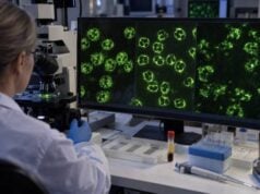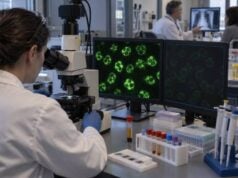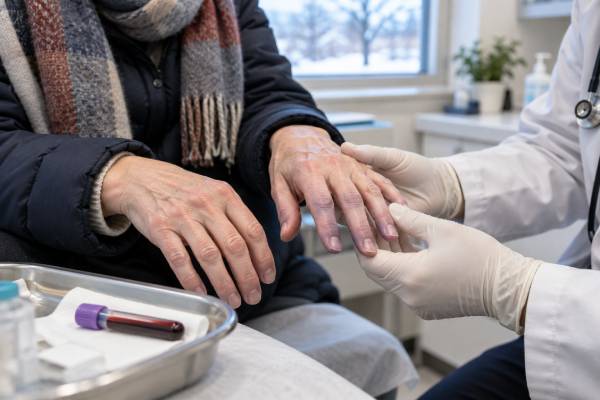
A cryofibrinogen test looks for a cold-precipitable protein complex in plasma. The complex usually contains fibrinogen, fibrin, fibronectin, and smaller amounts of other proteins. It can form a visible deposit when plasma cools and dissolve again when warmed. In some people, this finding is incidental. In others, cryofibrinogen contributes to small-vessel blockage, painful purpura, livedo, ulcers, tissue necrosis, Raynaud-type symptoms, thrombosis, nerve injury, or rare kidney disease. The test is technically demanding because the blood must stay warm until plasma is separated; cooling too early can remove the target from the liquid sample and produce a misleading result. Cryofibrinogen is also different from a cryoglobulin: cryofibrinogen appears in plasma but not serum, while cryoglobulins can be detected in serum. A useful interpretation therefore connects the laboratory finding with symptoms, biopsy results, clotting history, medications, infection testing, autoimmune evaluation, and screening for blood or solid-organ cancers.
- Cryofibrinogen is detected in anticoagulated plasma, not in serum.
- The blood tube must usually remain at about 37°C until cells are separated from plasma.
- A positive result may be incidental and does not by itself diagnose vasculitis or a clotting disorder.
- Painful cold-triggered purpura, livedo, ulcers, or digital ischemia make the finding more clinically important.
- Rapidly spreading skin necrosis, a cold painful limb, chest pain, or neurologic deficits require urgent assessment.
Table of Contents
- What the Cryofibrinogen Test Detects
- Collection, Processing, and False Results
- Positive, Negative, and Quantitative Results
- Symptoms and Types of Vessel Injury
- Primary and Secondary Cryofibrinogenemia
- Cryofibrinogen Versus Cryoglobulins and Other Mimics
- Follow-Up, Treatment Context, and Urgent Signs
What the Cryofibrinogen Test Detects
Cryofibrinogen is the name given to a cold-precipitable complex found in plasma. Despite the name, it is not simply an abnormal fibrinogen molecule. The precipitate can contain fibrinogen, fibrin, fibronectin, factor VIII, and other plasma proteins. When the specimen is cooled to roughly 4°C, the material may become cloudy, form flocculent strands, gel, or settle as a deposit. A true cryoprecipitate should dissolve again when warmed.
The plasma requirement is fundamental. Plasma comes from blood collected into an anticoagulant tube, so it still contains fibrinogen and other clotting proteins. Serum comes from blood that has clotted, which removes most fibrinogen. Cryofibrinogen should therefore appear in plasma but not in the paired serum specimen. A cold precipitate found in serum suggests a cryoglobulin or another protein rather than isolated cryofibrinogen.
The laboratory finding is called cryofibrinogenemia. The term does not automatically mean that the person has cryofibrinogenemic disease. Small amounts have been reported in people without attributable symptoms and in hospitalized patients whose illness has another cause. Clinicians reserve stronger causal language for cases in which the result fits the clinical picture and other evidence—such as skin or kidney biopsy—supports cryoprotein-related vascular injury.
Cryofibrinogen can harm tissue in more than one way. Cold-dependent precipitation may obstruct small vessels and reduce blood flow. The complex may also promote clot formation, inhibit normal clot breakdown, injure endothelium, or trigger inflammation. As a result, biopsy findings vary. Some samples show bland thrombi with little vessel-wall inflammation; others show leukocytoclastic vasculitis, in which neutrophils damage small vessels. Calling every positive case “vasculitis” misses this important distinction.
A standard fibrinogen blood test measures the circulating level or function of fibrinogen and cannot substitute for cryofibrinogen testing. A person may have a normal routine fibrinogen result but still form a cold precipitate in plasma.
Collection, Processing, and False Results
The preanalytic phase—the period between blood collection and laboratory separation—is the most vulnerable part of this test. The ordering clinician should contact the laboratory before the draw because laboratories differ in tube type, minimum volume, transport process, incubation time, and reporting method. Many use EDTA lavender-top tubes; some accept citrate tubes. A serum tube is often collected at the same time so cryofibrinogen and cryoglobulins can be compared.
The usual process includes these critical steps:
- The correct tubes and collection equipment are warmed according to the laboratory protocol.
- Blood is drawn and immediately kept near 37°C.
- The specimen remains warm during transport and centrifugation until plasma is separated from blood cells.
- The separated plasma is refrigerated, commonly at 4°C, and inspected over several days.
- Any precipitate is tested for re-dissolution on warming and may be characterized with staining, immunofixation, electrophoresis, or other protein methods.
Many laboratories observe the specimen for at least 72 hours; some continue for seven days because a low-level precipitate can develop slowly. Turnaround may therefore be a week or longer. The exact schedule matters less than following the performing laboratory’s validated protocol.
Premature cooling can cause a false-negative result. Cryofibrinogen may precipitate before plasma is separated and become trapped with the cells or clot. The final plasma then appears negative even though the protein was present in the original blood. A tube left at room temperature, transported without a warm carrier, or processed in a refrigerated centrifuge may be unacceptable. Laboratories may reject specimens that arrive cold.
False-positive or nonspecific findings are also possible. Fibrin strands from incomplete separation, lipids, cellular debris, heparin-related material, or other proteins may resemble a cryoprecipitate. Confirming that the deposit disappears with warming and is present in plasma but absent from serum improves specificity. Protein characterization can help when the appearance is unclear.
A negative result is most reassuring when collection was documented as correct and the clinical suspicion is low. If symptoms strongly suggest a cold-sensitive occlusive disorder, the clinician may repeat the test through a center experienced with cryoproteins. Repetition is especially reasonable when the first specimen cooled before separation or when only serum—not plasma—was tested.
No fasting or special diet is usually required. However, the patient should not independently warm an arm with unsafe heat or change anticoagulant medication before the draw. The laboratory and clinical team should coordinate the collection conditions.
Positive, Negative, and Quantitative Results
Most reports begin with a qualitative result: detected or not detected. “Positive” means a reversible cold precipitate consistent with cryofibrinogen was seen in plasma and not in serum. “Negative” means no qualifying precipitate was found during the laboratory’s observation period. Neither word should be interpreted without knowing whether the sample met temperature and processing requirements.
Some laboratories report a cryocrit, the percentage of the plasma column occupied by the packed precipitate. Others measure protein concentration or describe the deposit as trace, low, moderate, or high. These approaches are not fully standardized. A cryocrit from one laboratory may not be directly comparable with a value from another because tube dimensions, cooling time, centrifugation, washing, and reading methods differ.
There is no universal level that separates harmless positivity from symptomatic disease. A small amount can be relevant when it matches cold-triggered ischemic lesions and biopsy findings. A larger amount can be incidental, secondary to another illness, or poorly correlated with day-to-day symptoms. The clinical effect also depends on the temperature at which the material precipitates, how quickly it forms, its protein composition, blood flow, and other clotting risks.
A useful report may include:
- specimen type and whether paired serum was tested;
- duration and temperature of cold incubation;
- visual description of the precipitate;
- evidence that it re-dissolved with warming;
- cryocrit or protein concentration, if the laboratory validates that measurement;
- immunochemical characterization, when performed; and
- comments about preanalytic limitations.
A newly positive result should be compared with the reason the test was ordered. In a person with no cold sensitivity, skin lesions, thrombosis, neuropathy, or kidney abnormalities, the result may be an associated laboratory finding rather than the explanation for illness. In someone with painful retiform purpura and biopsy-proven vessel thrombosis, the same result carries much more weight.
Serial values are sometimes used during follow-up, but symptom control and organ function matter more than a numeric cryocrit alone. A falling result can support improvement when the skin, nerves, kidneys, and circulation improve at the same time. Persistent positivity without active injury does not always require treatment escalation.
Symptoms and Types of Vessel Injury
Skin is the most commonly affected organ because surface tissues and fingers or toes cool more readily than the body’s core. Symptoms may worsen in winter, air-conditioned environments, or after direct contact with cold objects. They can also occur without an obvious cold trigger.
Typical findings include painful purpura, bluish discoloration, livedo reticularis or retiform purpura, Raynaud-type color changes, tender nodules, nonhealing ulcers, blisters, and black necrotic tissue. Lesions often involve the legs, feet, toes, fingers, ears, or nose. Pain that seems out of proportion to the visible skin change may signal deeper ischemia.
Two broad injury patterns can look similar at the bedside:
| Pattern | What is happening | Clues |
|---|---|---|
| Inflammatory small-vessel injury | Immune and inflammatory cells damage vessel walls | Palpable purpura, swelling, inflammatory biopsy findings, possible systemic vasculitis |
| Occlusive or thrombotic injury | Cold precipitate and clot obstruct blood flow with limited vessel-wall inflammation | Retiform pattern, sharply painful ischemia, ulcers, necrosis, thrombosis on biopsy |
| Mixed injury | Inflammation and thrombosis occur together | Overlapping purpura, vessel-wall damage, fibrin thrombi, severe tissue loss |
This distinction affects treatment. Immunosuppression may help a true inflammatory vasculitis but can be ineffective or risky when the dominant problem is thrombosis, infection, or an untreated malignancy. Anticoagulation may be considered for documented thrombosis, yet it is not automatically appropriate for every skin lesion or positive test. Biopsy of a fresh, active lesion can clarify the mechanism.
Other reported manifestations include joint pain, muscle pain, peripheral neuropathy, thrombophlebitis, arterial thrombosis, and constitutional symptoms. Kidney disease is rare but potentially serious. It may cause blood or protein in the urine, swelling, high blood pressure, or declining filtration. Modern kidney series describe membranoproliferative patterns, thrombotic microangiopathy, ischemic injury, and fibrinogen-containing deposits.
Cryofibrinogenemia may coexist with another vasculitis rather than cause every symptom. A broader vasculitis laboratory evaluation helps identify ANCA-associated disease, immune-complex disease, infection, or another mechanism when the presentation is systemic.
Primary and Secondary Cryofibrinogenemia
Primary, or essential, cryofibrinogenemia is diagnosed only after a reasonable search finds no associated condition. It often presents with skin-limited cold sensitivity, recurrent ulcers, purpura, livedo, or thrombosis. Because evidence comes mainly from small series and case reports, the true frequency and natural history remain uncertain.
Secondary cryofibrinogenemia occurs with another disorder. The association does not always prove causation, but it can reveal an illness that needs separate treatment. Important categories include:
- Autoimmune and inflammatory disease: systemic lupus erythematosus, rheumatoid arthritis, Sjögren disease, systemic sclerosis, inflammatory myopathies, Behçet disease, inflammatory bowel disease, and other vasculitides.
- Infection: bacterial infections, mycobacterial disease, hepatitis viruses, HIV, and other persistent infections have been reported.
- Cancer and clonal blood disorders: lymphoma, leukemia, plasma-cell disorders, monoclonal gammopathy, and solid tumors may be associated.
- Thrombotic or vascular conditions: antiphospholipid syndrome and other prothrombotic states can overlap with the same clinical picture.
- Tissue injury or severe systemic illness: trauma, surgery, critical illness, and inflammatory states may produce transient findings in some patients.
The workup should be tailored rather than indiscriminate. A complete blood count and smear may reveal anemia, abnormal white cells, or thrombocytopenia. Metabolic and liver panels assess organ function. ESR and CRP describe inflammation but are not specific. Urinalysis, urine protein measurement, and creatinine screen for kidney injury. Serum protein electrophoresis, immunofixation, quantitative immunoglobulins, and free light chains look for a monoclonal protein.
Infection testing depends on risk and symptoms. It may include hepatitis B, hepatitis C, HIV, blood cultures, tuberculosis testing, or targeted studies. Autoimmune tests can include ANA, complement C3 and C4, ANCA, rheumatoid factor, and antiphospholipid antibodies. The antiphospholipid antibody panel is especially relevant when unexplained venous or arterial thrombosis, pregnancy morbidity, or livedo is present.
Age-appropriate cancer screening remains important. New weight loss, night sweats, enlarged lymph nodes, abnormal blood counts, a monoclonal protein, or unexplained organ enlargement may justify hematology or oncology assessment. The goal is not merely to label the cryofibrinogen result, but to find a treatable driver.
Cryofibrinogen Versus Cryoglobulins and Other Mimics
Cryofibrinogen and cryoglobulins both precipitate in the cold, but they are not interchangeable. Cryoglobulins are immunoglobulins that remain available in serum after clotting. Cryofibrinogen depends on plasma clotting proteins and should disappear from serum. Testing paired plasma and serum is the clearest laboratory distinction.
| Feature | Cryofibrinogen | Cryoglobulins |
|---|---|---|
| Specimen that should be positive | Plasma only | Serum; may also be seen in plasma |
| Main protein class | Fibrinogen/fibrin/fibronectin complex | Immunoglobulins, sometimes with complement |
| Common injury pattern | Thrombotic occlusion, sometimes vasculitis | Hyperviscosity or immune-complex vasculitis, depending on type |
| Common associated disorders | Autoimmune disease, infection, cancer, monoclonal gammopathy | Hepatitis C, autoimmune disease, B-cell or plasma-cell disorders |
The cryoglobulin test may be ordered at the same draw because the two abnormalities can coexist. Low C4, positive rheumatoid factor, palpable purpura, neuropathy, and glomerulonephritis favor mixed cryoglobulinemic vasculitis, but none is perfectly specific.
Other mimics include antiphospholipid syndrome, cholesterol emboli, calciphylaxis, thrombotic thrombocytopenic purpura, disseminated intravascular coagulation, heparin-induced thrombocytopenia, cold agglutinin disease, peripheral arterial disease, embolic disease, chilblains, severe Raynaud phenomenon, and medication-induced vasculopathy. Skin infection and pyoderma gangrenosum can also cause painful ulcers.
Routine coagulation tests may be normal in cryofibrinogenemia. Prothrombin time, activated partial thromboplastin time, platelet count, D-dimer, and routine fibrinogen help evaluate other clotting conditions but do not confirm or exclude a cryofibrinogen precipitate. Vascular imaging may be necessary when a larger artery or vein appears obstructed.
A skin biopsy should ideally sample a new, active lesion and extend deeply enough to evaluate small and medium vessels. The pathologist may use routine stains, direct immunofluorescence, and fibrin or fibrinogen staining. Because lesions can evolve, an old ulcer edge may show only nonspecific scarring and necrosis.
Follow-Up, Treatment Context, and Urgent Signs
Management starts by deciding whether cryofibrinogen is actually causing disease. An asymptomatic positive result may require observation and investigation for an associated condition rather than immediate therapy. Symptomatic cases need coordinated care from dermatology, rheumatology, hematology, nephrology, vascular medicine, or another specialty based on the organs involved.
Practical measures often include keeping the whole body and affected limbs warm, using layered clothing and insulated gloves or footwear, avoiding sudden temperature shifts, protecting ulcers, and stopping tobacco or nicotine exposure. Direct high heat should not be applied to numb or ischemic tissue because burns can occur. Wound care and prompt treatment of secondary infection are important when ulcers are present.
Treatment evidence is limited, and no single regimen fits every case. Clinicians may treat an underlying infection, autoimmune disease, malignancy, or monoclonal gammopathy. Antiplatelet or anticoagulant therapy may be used for selected thrombotic presentations after bleeding risk and the mechanism are assessed. Anti-inflammatory or immunosuppressive drugs may be considered when biopsy and systemic findings show inflammatory disease. Fibrinolytic agents, plasma exchange, and other therapies have been reported in severe or refractory cases, but decisions rely on specialist judgment rather than a standardized cryocrit threshold.
Follow-up focuses on outcomes that matter: healing of ulcers, reduced pain, restored warmth and color, fewer new lesions, stable nerve function, absence of new clots, and preserved kidney function. Photographs can document skin evolution. Urinalysis, creatinine, blood pressure, blood counts, and protein studies may be repeated according to the suspected cause. Cryofibrinogen levels can supplement this assessment but should not replace it.
Urgent evaluation is needed for a suddenly cold, pale, blue, or intensely painful limb or digit; rapidly spreading black skin; new weakness, facial droop, speech difficulty, or severe headache; chest pain or shortness of breath; coughing blood; one-sided leg swelling; marked reduction in urine; or fever with an infected ulcer. These findings can indicate limb-threatening ischemia, stroke, pulmonary embolism, pulmonary hemorrhage, severe kidney injury, or sepsis.
The central interpretation is simple: the test identifies a cold-precipitable plasma complex, but symptoms and tissue injury determine its importance. Correct specimen handling establishes whether the laboratory finding is real; clinical evaluation establishes whether it is responsible for disease.
References
- Laboratory Practices for the Detection and Characterization of Cryoglobulins and Cryofibrinogen 2025 (Laboratory Study)
- Cryoproteins in Non-HCV-Related Autoimmune Disorders: A Serious Cold-Induced Problem 2025 (Review)
- Cryfibrinogen-Associated Glomerulonephritis and Monoclonal Gammopathy of Renal Significance—Case Report and Literature Review 2025 (Review)
- Clinicopathological Spectrum and Treatment Outcomes of Cryofibrinogen-Associated Nephropathies 2026 (Clinical Study)
- Cryofibrinogen 2021 (Laboratory Test Guide)
- Cryofibrinogenemia 2013 (Review)
Disclaimer
This article is for education and cannot determine whether a cryofibrinogen result is causing symptoms. Collection errors, other clotting disorders, infection, autoimmune disease, and cancer can change the interpretation, so results require review by a qualified clinician. Seek urgent care for sudden limb ischemia, rapidly spreading skin necrosis, chest pain, breathing difficulty, neurologic deficits, or severe kidney symptoms.