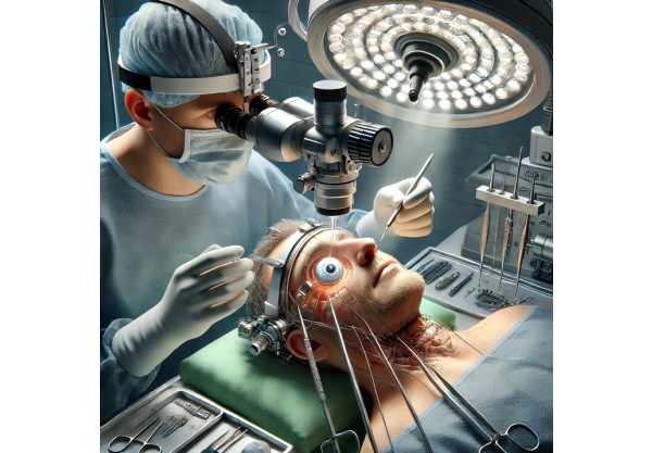
Retinal detachment is a serious ocular condition in which the retina, the light-sensitive layer of tissue in the back of the eye, separates from the supporting tissue. If not treated promptly, this separation can cause vision loss. The retina converts light into neural signals, which are then sent to the brain to enable vision. When the retina detaches, it loses blood supply and stops working properly, resulting in a rapid deterioration in vision.
There are three types of retinal detachments: rhegmatogenous, tractional, and exudative. Rhegmatogenous retinal detachment is the most common type, which occurs when a tear or break in the retina allows fluid to seep underneath and separate it from the underlying tissue. Tractional retinal detachment occurs when scar tissue on the retina’s surface contracts and pulls the retina away from the back of the eyeball. Exudative retinal detachment is less common and occurs when fluid accumulates beneath the retina without a tear or break, which is frequently caused by inflammation, injury, or vascular abnormalities.
Symptoms of retinal detachment include sudden flashes of light, a significant increase in the number of floaters (small specks or strands in the field of vision), a shadow or curtain effect over a portion of the visual field, and a significant decrease in visual acuity. Early detection and treatment are critical for avoiding permanent vision loss.
Retinal Detachment Management Strategies
To effectively treat retinal detachment, prompt medical intervention is required to reattach the retina and restore normal vision. The type, location, and severity of the detachment, as well as the patient’s overall eye health, determine the appropriate treatment.
Initial Assessment and Diagnosis
The first step in treating retinal detachment is a thorough eye examination by an ophthalmologist. This includes:
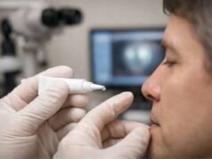
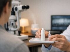
- Dilated Fundus Examination: The ophthalmologist uses special eye drops to dilate the pupils before examining the retina and optic nerve with an ophthalmoscope.
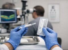
- Optical Coherence Tomography (OCT): This imaging technique generates detailed cross-sectional images of the retina, assisting in determining the extent of the detachment and any associated conditions.
- Ultrasound Imaging: When the retina is obscured by bleeding or other obstructions, ultrasound can be used to visualize the retina and assess the presence and extent of detachment.
Treatment Methods
Retinal detachment is treated using a variety of surgical and non-surgical methods, depending on the circumstances of each case.
Non-Surgical Treatments
- Laser Photocoagulation: Small retinal tears or breaks that have not yet progressed to full detachment can be treated with laser photocoagulation to create burns around them. This secures the retina to the underlying tissue, preventing fluid from entering and causing detachment.
- Cryopexy: Similar to laser photocoagulation, cryopexy involves applying a freezing probe to the outside of the eye near the tear. This freezes the area, leaving a scar that seals the tear and aids in the reattachment of the retina.
Surgical Treatments
- Pneumatic Retinopexy: This procedure treats certain types of retinal detachment. The eye’s vitreous cavity receives a small gas bubble injection. The patient is then positioned so that the bubble presses against the point of detachment, flattening the retina. The tear is then sealed using either laser photocoagulation or cryopexy.
- Scleral Buckling: This surgical procedure involves wrapping a flexible band (scleral buckle) around the circumference of the eye. This indents the eye wall, reducing the force of the vitreous pulling on the retina and allowing the retina to reattach to the underlying tissue. It is frequently paired with cryopexy or laser photocoagulation.
- Vitrectomy: Vitrectomy is a more complex surgery for severe or complicated retinal detachments. It involves removing the vitreous gel from the eye and replacing it with a gas bubble, silicone oil, or saline solution. This procedure enables the retina to flatten and reattach. The use of laser photocoagulation or cryopexy ensures that the retina stays attached.
Post-operative Care
Postoperative care is critical to the success of retinal detachment surgery. This includes:
- Medications: Patients are frequently given antibiotic and anti-inflammatory eye drops to help prevent infection and inflammation.
- Positioning: In some cases, patients must maintain a specific head position to ensure that the gas bubble or silicone oil used during surgery remains in the proper position to support the retina.
- Follow-Up Visits: Regular follow-up visits are required to monitor healing, evaluate surgical success, and detect any signs of complications or recurrence.
Retinal Detachment: Advanced Treatment Innovations
Recent advances in retinal detachment treatment have significantly improved patient outcomes, providing new hope through novel technologies and techniques. These cutting-edge innovations are transforming the management of retinal detachment, providing patients with more effective, less invasive, and faster recovery options.
Advanced Surgical Techniques
The most recent surgical innovations prioritize precision, minimal invasiveness, and faster recovery times.
- Microincision Vitrectomy Surgery (MIVS): MIVS uses smaller gauge instruments (23, 25, or 27-gauge) than traditional vitrectomy tools. These tiny instruments allow for smaller incisions, reducing surgical trauma, accelerating recovery, and lowering the risk of complications. MIVS’ precision allows surgeons to perform delicate maneuvers within the eye, resulting in more effective retinal reattachment.
- Robotic-Assisted Retinal Surgery: Retinal surgery is increasingly incorporating robotics, which provides unprecedented precision and control. Robotic systems can help surgeons perform extremely delicate procedures while reducing hand tremors and improving outcomes. These systems are particularly useful in complex cases where precise manipulation of retinal tissues is required.
- Intraoperative OCT: Intraoperative optical coherence tomography (OCT) provides real-time imaging during surgery, allowing surgeons to see the retina and underlying structures clearly. This technology improves the precision of surgical interventions, ensuring complete reattachment and lowering the risk of postoperative complications.
Advanced Imaging and Diagnostics
Enhanced imaging techniques have transformed the diagnosis and management of retinal detachment.
- Ultra-Widefield Imaging: Ultra-widefield imaging systems can take detailed images of up to 200 degrees of the retina in a single shot. This comprehensive view enables early detection of retinal tears and detachments, improving diagnostic accuracy and allowing for timely intervention.
- Adaptive Optics Imaging: This technology creates high-resolution images of the retina at the cellular level. This enables detailed visualization of retinal structures, which aids in the early detection of subtle changes that may signal the onset of detachment.
Pharmaceutical Innovations
Innovative pharmacological approaches are being developed to promote retinal health and improve surgical outcomes.
- Adjunctive Therapies: Anti-VEGF (vascular endothelial growth factor) agents, which are commonly used to treat age-related macular degeneration, are being tested as adjunctive treatments for retinal detachment. These agents can reduce intraocular inflammation and neovascularization, improving surgical outcomes and lowering the risk of recurrence.
- Neuroprotective Agents: Research into neuroprotective drugs seeks to protect retinal neurons from ischemic damage during detachment. These agents may preserve visual function by protecting retinal cells from apoptosis (cell death) during periods of low blood flow.
Gene Therapy & Regenerative Medicine
Gene therapy and regenerative medicine are leading research into retinal detachment treatment.
- Gene Editing: Technologies such as CRISPR-Cas9 have the potential to correct genetic mutations that predispose individuals to retinal detachment. Gene editing, which targets specific genes involved in retinal health, may prevent the onset of detachment in genetically susceptible individuals.
- Stem Cell Therapy: Stem cell research shows promise in regenerating damaged retinal tissues. Stem cells can differentiate into a variety of retinal cell types, potentially restoring function and structure in detached retinas. Clinical trials are currently underway to determine the safety and efficacy of stem cell-based therapies for retinal diseases.
Personalized Medical Approaches
Personalized medicine is changing the way we treat retinal detachment, with therapies tailored to individual patient profiles based on genetic, molecular, and clinical data.
- Genetic Profiling: Genetic testing can identify people who are more likely to develop retinal detachment, which can help guide preventive measures and personalized treatment plans. Understanding the genetic basis of detachment can lead to more targeted treatments that address specific underlying causes.
- Biomarker Analysis: Advances in biomarker research allow for the identification of specific molecules associated with retinal health and disease. Biomarker analysis can help track disease progression, predict treatment outcomes, and tailor therapies to individual patients.
Future Directions
The future of retinal detachment treatment appears bright, with ongoing research and technological advancements paving the way for even more effective and minimally invasive options. Continued research into advanced surgical techniques, innovative pharmacological treatments, and regenerative medicine approaches is likely to yield new breakthroughs. As our understanding of the underlying mechanisms of retinal detachment grows, targeted treatments that address the root causes of the condition will become more feasible, providing hope for long-term improvements in patient outcomes.
Innovative Alternatives for Retinal Detachment
While traditional surgical treatments for retinal detachment, such as vitrectomy, scleral buckling, and pneumatic retinopexy, are well known for their effectiveness, there is growing interest in alternative treatments. These methods can provide additional options for patients, particularly those who are not good candidates for traditional surgeries or prefer less invasive procedures. Here, we look at some of the most promising alternative treatments for retinal detachment, including their mechanisms, applications, and results.
Hypobaric Oxygen Therapy
In hypobaric oxygen therapy (HBOT), patients breathe pure oxygen in a pressurized environment. This method significantly increases oxygen delivery to tissues, including the retina. HBOT has shown potential benefits for retinal detachment, despite its traditional use for conditions such as decompression sickness and wound healing.
The Mechanism of Action:
HBOT increases blood oxygen saturation, which improves oxygen delivery to ischemic retinal tissues. This can reduce hypoxia-induced damage and improve retinal health. Improved oxygenation reduces retinal swelling and may aid reattachment efforts.
Clinical Applications:
Patients undergoing HBOT are typically treated in a hyperbaric chamber, where the pressure exceeds normal atmospheric levels. Sessions can last between 30 minutes and two hours, depending on the protocol.
Outcomes:
Studies have shown that HBOT can improve visual outcomes in patients with retinal detachment, especially when combined with other treatments. It can help to recover retinal function and reduce the likelihood of complications. However, larger clinical trials are required to develop standardized protocols and confirm long-term benefits.
Acupuncture and TCM
Acupuncture and other modalities in Traditional Chinese Medicine (TCM) provide a comprehensive approach to treating a variety of ocular conditions, including retinal detachment. These practices aim to restore balance and promote healing by stimulating specific points on the body.
The Mechanism of Action:
Acupuncture is the practice of inserting thin needles into the skin at specific acupoints. This is thought to increase the body’s energy flow (Qi) and trigger natural healing processes. Acupunctures around the eyes and along meridians related to eye health are used to treat retinal detachment.
Clinical Applications:
Acupuncture sessions are usually 30 to 60 minutes long and take place over several visits. TCM practitioners may also employ herbal medicine, dietary changes, and lifestyle advice to promote overall eye health and recovery.
Outcomes:
While scientific evidence on the efficacy of acupuncture for retinal detachment is limited, some case reports and small-scale studies indicate that it can improve symptoms and aid in recovery when used in conjunction with conventional treatments. Patients frequently report relief from symptoms such as eye pain and discomfort, though the impact on the reattachment process is still being investigated.
Microcurrent Stimulation Therapy
Microcurrent stimulation therapy employs low-level electrical currents to stimulate retinal cells and promote healing. This non-invasive treatment has sparked interest due to its potential to improve retinal function and facilitate reattachment.
The Mechanism of Action:
Microcurrent therapy sends tiny electrical impulses to the retina, boosting cellular activity and increasing blood flow. These impulses are thought to improve the metabolic function of retinal cells, assisting in their repair and maintenance.
Clinical Applications:
Patients typically have sessions with a handheld device that applies microcurrent to the eye area. Treatment protocols vary, but sessions are usually about 20 minutes long and repeated several times per week.
Outcomes:
Preliminary research and anecdotal evidence indicate that microcurrent stimulation can help stabilize retinal function and improve visual outcomes in patients with retinal detachments. However, larger-scale clinical trials are required to determine its efficacy and develop standardized treatment guidelines.
Dietary Supplements and Nutrition Therapy
Nutritional therapy, including the use of dietary supplements, is important for maintaining eye health and may aid in the treatment of retinal detachment. Specific nutrients are required for retinal function and can help the healing process.
Key Nutrients & Supplements:
- Omega-3 Fatty Acids: Fish oil contains omega-3 fatty acids, which have anti-inflammatory properties and are essential for retinal health. Omega-3 supplements may help to reduce retinal inflammation and promote overall eye health.
- Antioxidants: Vitamins C and E, as well as minerals like zinc and selenium, help protect retinal cells from oxidative stress. Antioxidant supplements can help protect the retina from free radical damage and promote healing.
- Carotenoids: The retina contains carotenoids such as lutein and zeaxanthin, which play a protective role. These nutrients can be obtained as supplements or from leafy greens. They help to filter harmful blue light and maintain retinal integrity.
- Vitamin A: Vitamin A is essential for retinal health and helps photoreceptors function. Adequate intake can help prevent further deterioration and promote recovery.
Clinical Applications:
Nutritional therapy entails making dietary changes to include foods high in these essential nutrients or taking specific supplements prescribed by a healthcare provider.
Outcomes:
Proper nutrition appears to benefit retinal health and recovery in patients with retinal detachment. While not a stand-alone treatment, nutritional therapy can enhance other medical or surgical interventions and improve overall results.
Herbal Remedies
Herbal remedies have been used for centuries to promote eye health and treat a variety of ocular ailments. Specific herbs are thought to have properties that can help with the treatment of retinal detachment.
Key Herbal Remedies
- Ginkgo Biloba: Ginkgo Biloba, known for its antioxidant properties and ability to improve blood circulation, may help maintain retinal health and reduce ischemic damage.
- Bilberry Extract: Containing antioxidants, bilberry extract is thought to strengthen retinal tissues and improve microcirculation in the eyes.
- Curcumin: Curcumin, the active ingredient in turmeric, has strong anti-inflammatory and antioxidant properties. It may reduce retinal inflammation and promote healing.
Clinical Applications:
Herbal remedies can be taken as supplements or added to the diet. It is critical to consult with a healthcare provider before beginning any herbal regimen, particularly for patients with retinal detachment.
Outcomes:
While there is limited scientific evidence to support the use of herbal remedies for retinal detachment, some studies and anecdotal reports indicate potential benefits. These remedies can supplement traditional treatments and improve overall eye health.
Hyperbaric Medicine
Hyperbaric medicine, which involves the use of hyperbaric oxygen chambers, has shown promise as an additional treatment for retinal detachment.
The Mechanism of Action:
Hyperbaric oxygen therapy (HBOT) increases the amount of oxygen in the blood, which improves oxygen delivery to ischemic retinal tissue. This can help to reduce hypoxia-induced damage while also promoting healing.
Clinical Applications:
HBOT involves breathing pure oxygen in a pressurized chamber. Treatment protocols vary, but typically consist of 60 to 90-minute sessions spread out over several weeks.
Outcomes:
Studies show that HBOT can improve visual outcomes in patients with retinal detachment, especially when combined with other treatments. It can help to recover retinal function and reduce the likelihood of complications.
Ayurvedic Medicine.
Ayurvedic medicine, an ancient system of natural healing from India, provides holistic approaches to treating eye conditions such as retinal detachment.
Main Ayurvedic Treatments:
- Triphala: Triphala, a traditional Ayurvedic herbal formulation for eye health, can help detoxify and rejuvenate the eyes.
- Netra Tarpana is a therapeutic procedure that involves pouring medicated ghee (clarified butter) over the eyes. This treatment is thought to nourish and strengthen retinal tissues.
- Diet and Lifestyle Changes: Ayurveda emphasizes the role of diet and lifestyle in maintaining eye health. Dietary recommendations and lifestyle practices are tailored to each individual’s constitution (Prakriti) and health status.
Clinical Applications:
Trained practitioners administer Ayurvedic treatments, which may include herbal remedies, dietary changes, and specialized therapies such as Netra Tarpana.
Outcomes:
While there is limited scientific evidence to support Ayurvedic treatments for retinal detachment, many patients report improved symptoms and overall eye health. Ayurvedic approaches can supplement conventional treatments and provide comprehensive retinal health care.


