What is Dacryocele?
Dacryocele, also known as lacrimal sac cyst, is a rare congenital or acquired condition in which the nasolacrimal duct becomes obstructed and distended, resulting in the formation of a cystic swelling at the eye’s medial canthus. This condition is usually distinguished by a bluish, cystic swelling caused by the accumulation of tears and mucus in the lacrimal sac. It is most commonly seen in neonates due to congenital obstruction, but it can also occur in adults due to chronic dacryocystitis or trauma. Dacryocele can be unilateral or bilateral, and it may be associated with other ocular or systemic conditions. Early detection and treatment are critical to avoiding complications like infection or the development of secondary dacryocystitis.
Dacryocele insights
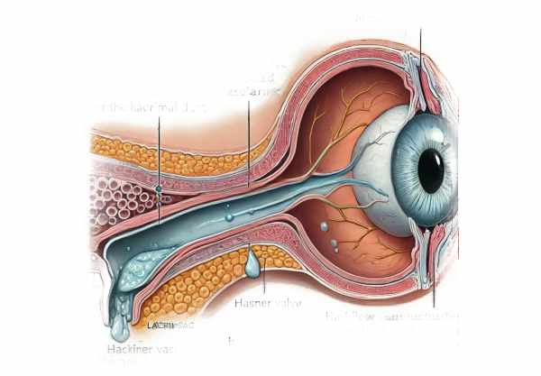
Dacryocele, also known as lacrimal sac cyst, is a distinct entity among lacrimal system disorders. The condition is caused primarily by an obstruction at the distal end of the nasolacrimal duct, specifically the Hasner valve, which does not open properly. This obstruction causes a backflow of tears and mucus into the lacrimal sac, causing it to distend and form a cyst.
Etiology and Pathophysiology
The primary cause of dacryocele is a congenital obstruction of the nasolacrimal duct. During fetal development, the nasolacrimal duct is formed by canalizing epithelial cords to form a functional duct. In some cases, the canalisation process is incomplete, resulting in a congenital dacryocele. The obstruction is typically found at the Hasner valve, a mucosal fold at the distal end of the duct that should open into the nasal cavity’s inferior meatus.
Acquired dacryocele, on the other hand, is less common and is typically caused by chronic dacryocystitis, trauma, or surgical interventions that disrupt the normal anatomy of the nasolacrimal duct. Chronic inflammation causes ductal fibrosis and stenosis, which results in secondary obstruction and cyst formation.
Clinical Presentation
Dacryocele presents differently in neonates than in adults. It is frequently observed in newborns shortly after birth. The defining feature is a bluish, cystic swelling at the medial canthus, which may be accompanied by epiphora (excessive tearing) and mucoid discharge. Except for secondary infections, the swelling is usually non-tender. In some cases, swelling can cause nasal obstruction, resulting in respiratory distress, especially in neonates who breathe solely through their noses.
Adults with dacryocele may experience similar cystic swelling and epiphora. However, it is frequently associated with chronic dacryocystitis symptoms such as recurrent infections, pain, and redness in the medial canthus area. If left untreated, the cystic mass can become inflamed, resulting in the formation of an abscess or fistula.
Complications
The majority of dacryocele complications are caused by secondary infections. In neonates, the dacryocele can become infected, resulting in acute dacryocystitis, which is characterized by erythema, tenderness, and purulent discharge. If not treated promptly, it can lead to cellulitis or even orbital cellulitis, which is a serious condition that necessitates immediate medical attention.
Another significant complication is the possibility of respiratory distress in newborns. Large dacryoceles can cause nasal obstruction, making it difficult for the infant to breathe, especially while eating. This can result in poor nutrition, weight loss, and other systemic complications.
Diagnostics
Dacryocele is primarily diagnosed clinically, with the characteristic swelling at the medial canthus serving as the primary indicator. It is critical in neonates to distinguish dacryocele from other causes of medial canthal swelling, such as encephalocele, hemangioma, or dermoid cyst. A thorough clinical examination and history are required for an accurate diagnosis.
Imaging studies, such as ultrasound or MRI, can help confirm the diagnosis and determine the size of the cyst. Ultrasound typically reveals a cystic mass with internal echoes, indicating the presence of mucus and debris. MRI can provide detailed information about the anatomy of the nasolacrimal duct and its surrounding structures, which is especially useful in complex cases or when surgical intervention is required.
Prognosis
The prognosis for dacryocele is generally favorable, particularly with early diagnosis and appropriate treatment. Most neonates respond well to conservative treatment or probing, resulting in cyst and symptom resolution. Adult surgical outcomes are also favorable, especially with advancements in endoscopic techniques.
Prevention Tips
Preventive measures for dacryocele concentrate primarily on lowering the risk of obstruction and managing predisposing factors:
- Prenatal Care: Providing adequate prenatal care and monitoring can aid in the early detection of congenital anomalies. Regular ultrasounds can reveal abnormalities in the fetal nasolacrimal duct.
- Proper Hygiene: Practicing good facial and ocular hygiene can help prevent infections that can lead to dacryocele. This includes regularly cleansing the eyelids and face.
- Early Intervention: Early treatment of congenital nasolacrimal duct obstruction in infants can prevent the development of a dacryocele. Parents should be educated about the value of early intervention.
- Avoidance of Trauma: Protecting the eye and surrounding areas from trauma can lower the risk of developing a dacryocele. Wear protective eyewear when participating in activities that may cause injury.
- Management of Chronic Conditions: Proper treatment of chronic conditions such as sinusitis and dacryocystitis is critical in preventing secondary dacryocele. Regular follow-ups with a healthcare provider can help you manage these conditions more effectively.
- Prophylactic Antibiotics: In cases of recurring dacryocystitis, prophylactic antibiotics may be prescribed to prevent infections that could result in the formation of a dacryocele.
Diagnostic Techniques for Dacryocele
Dacryocele is diagnosed using a combination of clinical and imaging techniques to determine the presence and size of the lacrimal sac cyst. Here are the main diagnostic methods:
Clinical Examination
The first step in diagnosing dacryocele is a thorough clinical examination. The presence of a bluish, cystic swelling at the medial canthus is often enough to suspect dacryocele in newborns. Except for secondary infections, the swelling is usually non-tender. In adults, the presence of a similar cystic swelling, which is frequently accompanied by chronic dacryocystitis symptoms such as recurrent infections and epiphora, may indicate dacryocele.
Nasolacrimal Duct Probing
In some cases, a diagnostic probe of the nasolacrimal duct can be used. This entails carefully inserting a fine probe through the puncta and canaliculi into the nasolacrimal duct. The ease or difficulty of passing the probe can reveal the location and severity of the obstruction.
Imaging Studies: Ultrasound
Ultrasound is a non-invasive imaging technique commonly used to confirm the diagnosis of dacryocele. It can detect a cystic mass in the medial canthus, which is usually filled with anechoic or hypoechoic fluid and contains accumulated tears and mucus. Ultrasound is especially beneficial in neonates due to its safety and lack of radiation exposure.
Magnetic Resonance Imaging (MRI)
MRI provides detailed images of the lacrimal system and surrounding structures, which is especially useful in complex cases or when planning surgical intervention. An MRI can reveal the anatomy of the nasolacrimal duct, pinpoint the exact location of the obstruction, and evaluate any associated anomalies. It is especially useful in distinguishing dacryocele from other conditions that can cause similar swelling, such as encephaloceles and dermoid cysts.
Computed Tomography (CT) Scan
CT scans are less commonly used, but they can be useful in cases requiring detailed bone anatomy, such as trauma or chronic infection that has caused bony changes. CT scans can aid in determining the extent of the disease and developing surgical strategies.
Dacryocystography
Dacryocystography is a specialized radiographic technique that involves injecting contrast material into the lacrimal sac via the puncta, followed by imaging to reveal the nasolacrimal duct system. This technique can provide precise information about the obstruction’s location and the anatomy of the lacrimal drainage system. It is especially useful for assessing complex cases and planning surgical interventions.
Differential Diagnosis
It is critical to distinguish dacryocele from other sources of medial canthal swelling. Conditions to consider include dermoid cysts, encephaloceles, hemangiomas, and nasolacrimal duct cysts (which are frequently associated with intranasal cysts). A thorough clinical history, examination, and appropriate imaging studies help to distinguish these conditions.
Dacryocele Treatment Approaches
Dacryocele treatment includes a variety of options, ranging from conservative management to surgical intervention, depending on the patient’s age, severity of symptoms, and complications.
Conservative Management
In neonates, conservative management is frequently the first line of treatment. This includes:
Lacrimal Sac Massage
Parents are taught to gently massage the lacrimal sac several times daily. This technique increases pressure within the sac, causing the nasolacrimal duct to open and drain its contents. Typically, the massage involves pressing down and outward on the medial canthal area.
Investigating
If conservative measures fail, or the neonate has significant nasal obstruction or recurring infections, the nasolacrimal duct should be probed. Probing is a minor procedure that uses general anesthesia to pass a fine probe through the puncta and into the nasolacrimal duct to relieve obstruction. This procedure is highly successful in neonates and infants.
Antibiotic Therapy
In cases of secondary infection, antibiotic therapy is required. Broad-spectrum antibiotics are administered, with adjustments based on culture and sensitivity results. Oral antibiotics are usually adequate, but severe cases may necessitate intravenous antibiotics and hospitalization.
Surgical Intervention
For persistent or complicated dacryocele, surgical options are considered.
Dacryocystorhinostomy(DCR)
DCR is the standard surgical procedure for dacryocele, particularly in adults. This procedure opens up a new drainage pathway between the lacrimal sac and the nasal cavity, bypassing the obstructed nasolacrimal duct. DCR can be performed using either external or endoscopic approaches, with the latter being less invasive and resulting in faster recovery.
Endoscopic DCR
Endoscopic DCR has grown in popularity due to its minimally invasive nature. Endoscopic instruments are used to create a drainage pathway that can be seen directly. This technique minimizes the risk of scarring and has a high success rate.
Balloon Dacryoplasty
Balloon dacryoplasty is a newer, less invasive technique that is primarily used on children. A small balloon catheter is inserted into the nasolacrimal duct and inflated to dilate it and relieve the obstruction. This procedure is typically carried out under general anesthesia and has shown promising results in pediatric patients.
Innovative and Emerging Therapies
Emerging therapies for dacryocele include the use of adjunctive medications like mitomycin C during DCR to reduce scar formation and increase success rates. Furthermore, advances in endoscopic techniques and instrumentation continue to improve the safety and efficacy of surgical procedures.
Early detection and appropriate treatment are critical in preventing complications and ensuring positive outcomes for patients with dacryocele. With advances in both conservative and surgical treatments, the prognosis for dacryocele remains favorable.
Trusted Resources
Books
- “Diseases of the Lacrimal System” by Adam J. Cohen and Michael Mercandetti
- “Principles and Practice of Lacrimal Surgery” by Mohammad Javed Ali


