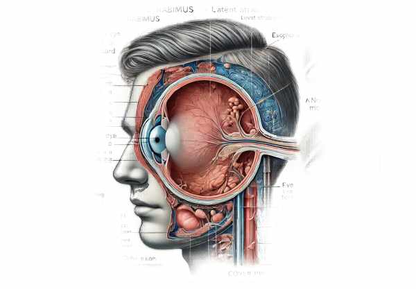
What is exophoria?
Exophoria is an ocular condition in which one eye moves outward while focusing on an object. Exophoria, as opposed to exotropia, is typically latent and only becomes apparent under certain conditions, such as stress, fatigue, or disruption of binocular vision. This condition is part of a larger group of eye alignment disorders known as heterophoria, in which the eyes drift out of alignment while maintaining overall binocular vision. Exophoria can affect people of any age and may be asymptomatic or cause symptoms such as eye strain, headaches, and difficulty concentrating.
Comprehensive Investigation of Exophoria
Exophoria is a type of latent strabismus in which one eye moves outward intermittently. This deviation occurs when binocular vision is interrupted, such as during an eye exam’s cover test. Unlike more visible forms of strabismus, such as exotropia, the deviation in exophoria is not constant and frequently goes unnoticed by the affected person.
Epidemiology
Exophoria is a common condition that affects a large proportion of the population in varying degrees. According to studies, its prevalence varies greatly, with some estimating that it affects up to 10% of the general population. The condition can appear at any age, but it is usually first noticed in childhood or adolescence. Gender does not appear to have a significant influence on the prevalence of exophoria, which can be found in a variety of ethnic and demographic groups.
Symptoms and Presentation
Many people with exophoria are asymptomatic, especially if the deviation is minor and the person possesses strong compensatory mechanisms to maintain binocular vision. However, symptomatic cases can present with a variety of issues, including:
- Eye strain (asthenopia): Typically occurs after prolonged periods of close work, such as reading or using a digital device.
- Headache: Commonly occurs following tasks that require sustained focus.
- Blurred vision: Most noticeable during activities that require intense concentration.
- Diplopia (double vision): Intermittent, often accompanied by fatigue or stress.
- Difficulty concentrating: Especially during reading or other near-vision activities.
Causes and Risk Factors
The exact cause of exophoria is multifactorial, involving both genetic and environmental factors. Some key causes and risk factors are:
- Genetics: A family history of strabismus or other binocular vision disorders may increase the risk of developing exophoria.
- Refractive Errors: Uncorrected hyperopia (farsightedness) can cause accommodative convergence issues, which contribute to exophoria.
- Vision Stress: Prolonged near work without breaks, which is common in modern digital device use, can aggravate the condition.
- Neurological Factors: Specific neurological conditions affecting the cranial nerves can impair eye movement coordination, resulting in exophoria.
Pathophysiology
Exophoria results from an imbalance in the neuromuscular control of the eye muscles. The binocular vision system is based on precise coordination of each eye’s six extraocular muscles. When the neural mechanisms that control these muscles are disrupted, one eye may drift outward if binocular vision is not actively maintained. This disruption could be caused by a variety of factors, including muscle weakness, nerve dysfunction, or impaired neurological control.
Impact on Daily Life
Many people with exophoria are unaware of their condition, but those who do can have a significant impact on their daily lives. Symptoms like eye strain and headaches can have a negative impact on productivity and quality of life, especially in environments that require prolonged near vision tasks, such as academia or the workplace. Furthermore, people with severe symptoms may struggle with tasks requiring precise visual coordination, such as sports or driving.
Variability in Presentation
Exophoria can have a wide range of presentation and severity. Some people may only experience occasional symptoms, whereas others may have more frequent and severe problems. Fatigue, stress, and visual demands can all have an impact on how severe your symptoms are. In some cases, exophoria can progress over time, especially if underlying risk factors are not addressed.
Psychological and Social Concerns
Living with exophoria can have psychological and social consequences, particularly for those who exhibit noticeable symptoms. Dealing with chronic headaches and visual discomfort can be frustrating and have a negative impact on quality of life. In children, undiagnosed exophoria can impair academic performance and participation in activities, potentially leading to social withdrawal or anxiety.
Clinical Examination
During a clinical examination, exophoria is typically detected using a cover test, in which one eye is covered while the patient focuses on a target. The movement of the uncovered eye is examined for indications of outward deviation. Additional tests, such as the Maddox rod test or the prism cover test, may be used to determine the degree of deviation and the patient’s ability to maintain binocular vision.
Differential Diagnosis
Differentiating exophoria from other types of strabismus is critical for effective treatment. Conditions that may present similarly are:
- Exotropia: Unlike exophoria, exotropia causes a constant outward deviation.
- Intermittent Exotropia: Is distinguished by periods of normal alignment interspersed with outward deviation.
- Convergence Insufficiency: This term often overlaps with exophoria, but it specifically refers to the difficulty of maintaining convergence for nearby tasks.
Exophoria Diagnostic Techniques
Exophoria is diagnosed using a combination of clinical evaluation and specialized tests that assess eye alignment and coordination. The goal is to identify the presence and extent of the deviation, as well as any underlying causes of the condition.
Clinical Evaluation
A thorough clinical evaluation starts with a detailed patient history. This includes questions about the frequency and severity of symptoms, as well as any factors that exacerbate or alleviate them. The clinician will also ask about the patient’s family history of strabismus or other ocular conditions, as well as their overall health and any medications they are currently taking.
Cover Test
The cover test is the primary diagnostic tool for detecting exophoria. During this test, the patient is instructed to focus on a target while one eye is covered. The clinician watches the movement of the uncovered eye as the cover is placed and removed. Exophoria causes the covered eye to move outward when uncovered, indicating a latent outward deviation.
Prism Coverage Test
The prism cover test is used to quantify the degree of exophoria. Prisms of varying strengths are placed in front of the eye to balance the deviation. The strength of the prism required to properly align the eyes indicates the degree of exophoria. This test yields useful information for developing management strategies.
Maddox Rod Test
The Maddox rod test is another method for assessing eye alignment. A Maddox rod, a cylindrical lens that produces a streak of light, is placed in front of one eye while the patient gazes at a point light source. The orientation and position of the streak relative to the light source influence the presence and severity of exophoria.
Hirschberg Test
The Hirschberg test consists of shining a light into the patient’s eyes and observing the reflection on the corneas. The position of the reflections can help determine whether the eyes are properly aligned. In exophoria, reflections may be slightly off-center, indicating an outward deviation.
Near-Point of Convergence (NPC) Test
The NPC test determines the closest point where the eyes can maintain convergence while focusing on an object. Exophoria patients may have difficulty maintaining convergence, resulting in symptoms such as double vision or eye strain while performing near tasks.
Binocular Vision Assessment
Assessing binocular vision function is critical in the diagnosis of exophoria. The Worth 4-dot test and the Randot Stereoacuity test assess the patient’s ability to use both eyes to create a single, three-dimensional image. Difficulty with these tasks may indicate a problem with binocular vision caused by exophoria.
Refraction Test
A refraction test determines whether refractive errors, such as hyperopia or myopia, are causing the exophoria. Correcting these errors with glasses or contact lenses can help relieve symptoms and improve alignment.
Additional Imaging
In some cases, additional imaging studies, such as MRI or CT scans, may be required to rule out underlying neurological or structural abnormalities that are causing the exophoria. These tests are typically reserved for cases with unusual presentations or when other neurological symptoms exist.
Comprehensive Eye Examination
A thorough eye examination, including pupil dilation to examine the retina and optic nerve, is required to rule out other ocular conditions that may mimic or exacerbate exophoria. This examination provides a thorough assessment of overall eye health.
Managing Exophoria Effectively
Standard Treatment Options:
Exophoria treatment aims to improve binocular vision while also reducing symptoms. Standard treatments include:
- Prescription glasses or contact lenses: Correcting underlying refractive errors, such as hyperopia, can significantly reduce eye strain while improving alignment. Specialized lenses, such as prism glasses, can also be prescribed to help with eye alignment.
- Vision Therapy: – Exercises to improve eye coordination and focus. These exercises are tailored to each patient’s specific needs and are usually carried out under the supervision of an optometrist. Vision therapy aims to strengthen the eye muscles and improve the brain’s ability to control eye movements, thereby alleviating exophoria.
- Orthoptic exercises: Orthoptic exercises are a type of vision therapy that specifically targets the muscles that control eye movements. These exercises may include pencil push-ups, in which the patient focuses on a small target moving closer to the eyes, or computer-based programs designed to train convergence.
- Use of Prisms: – Eyeglasses with prisms can align images seen by each eye, allowing the brain to fuse them into a single image. This can be especially beneficial for patients with severe exophoria who experience double vision.
Innovative and Emerging Therapies
- Neuro-Visual Training: Technological advancements have enabled sophisticated neuro-visual training programs. These programs employ virtual reality and interactive software to create immersive environments that test and train the visual system. Neuro-visual training is designed to improve the brain’s ability to control eye movements and overall visual function.
- Biofeedback Therapy: – This therapy provides real-time feedback on eye movement and alignment to help patients gain control over their eye muscles. This method can be especially useful for patients who struggle with traditional vision therapy exercises.
- Pharmaceutical Interventions: – Research on pharmacological treatments for exophoria is ongoing. Certain neurotransmitter-affecting medications may help improve eye movement coordination. However, these treatments are still experimental and not widely available.
- Surgical Options: – If conservative treatments fail to treat severe exophoria, surgery may be a viable option. Surgical procedures usually involve adjusting the length or position of the eye muscles to improve alignment. This option is typically reserved for cases involving significant functional impairment.
Tips to Avoid Exophoria
- Schedule regular eye exams at least once a year to identify and treat vision issues early.
- Correct Refractive Errors: – Correct any refractive errors, like myopia or hyperopia, with glasses or contact lenses.
- Maintain Good Posture: – Practice good posture, especially during activities that require prolonged near vision, to reduce eye strain.
- Take Breaks: – Use the 20-20-20 rule: every 20 minutes, look at something 20 feet away for at least 20 seconds to rest your eyes.
- Use proper lighting to reduce eye strain while reading or working.
- To reduce glare on digital screens, adjust brightness and contrast and use blue light filters.
- Regular eye exercises, such as focusing on a distant object or performing convergence exercises, can help strengthen eye muscles.
- Stay Hydrated: – Stay hydrated to avoid dry eyes, which can worsen symptoms of exophoria.
- Manage Stress: – Reduce stress through techniques like meditation or yoga, as it can worsen exophoria symptoms.
- Maintain Proper Nutrition: – Consume a well-balanced diet rich in vitamins and minerals, especially those that promote eye health, like vitamins A, C, and E, and omega-3 fatty acids.
Trusted Resources
Books
- “Clinical Management of Binocular Vision” by Mitchell Scheiman and Bruce Wick
- “Binocular Vision and Ocular Motility: Theory and Management of Strabismus” by Gunter K. Von Noorden and Emilio C. Campos


