What is Goldenhar syndrome?
Goldenhar Syndrome, also known as the oculo-auriculo-vertebral (OAV) spectrum, is a rare congenital condition marked by craniofacial anomalies that primarily affect the development of the eyes, ears, and vertebrae. The ocular manifestations of Goldenhar Syndrome are particularly significant because they can cause visual impairment and other complications. These manifestations may include epibulbar dermoids, colobomas, microphthalmia, and other eye structural abnormalities. Understanding the ocular aspects of Goldenhar Syndrome is critical for early diagnosis and treatment, resulting in better outcomes for those affected.
Detailed Examination of Goldenhar Syndrome Ocular Manifestations
Goldenhar Syndrome causes a wide range of ocular manifestations, which can have a significant impact on a patient’s vision and quality of life. These ocular anomalies frequently coexist with other craniofacial and systemic abnormalities, complicating the clinical picture. We will look at the various ocular features associated with Goldenhar Syndrome, their pathophysiology, and implications.
Epibulbar Dermoids
Epibulbar dermoids are benign congenital growths made up of choristomatous tissue, which is not typically found at the site. These growths are one of the most common ocular manifestations of Goldenhar Syndrome, appearing on the eye’s surface, usually at the junction of the cornea and sclera.
Characteristics:
- Location: Typically found at the limbus, but can also occur on the cornea or conjunctiva.
- Appearsance: They appear as white or yellowish, well-defined lesions.
- Impact on Vision: Depending on their size and location, epibulbar dermoids can impair vision, cause astigmatism, and progress to amblyopia (lazy eye).
Microphthalmia and anophthalmia
Microphthalmia refers to abnormally small eyes, whereas anophthalmia means the complete absence of one or both eyes. These conditions are quite common in Goldenhar Syndrome and can have serious consequences for visual function.
Characteristics:
- Microphthalmia: The affected eye is smaller than normal and may have other structural abnormalities.
- Anophthalmia: The absence of an eye(s), usually accompanied by underdeveloped or absent ocular adnexa, such as the orbit, eyelids, and lacrimal apparatus.
- Impact on Vision: Both conditions cause severe visual impairment or blindness in the affected eye (s).
Colobomas
Colobomas are congenital defects caused by incomplete closure of the embryonic fissure. Colobomas in Goldenhar Syndrome can affect a variety of eye structures, including the iris, retina, choroid, and optic nerve.
Characteristics:
- Location: Can occur in the iris, creating a keyhole appearance, or in the retina and optic nerve, causing significant visual deficits.
- Appearance: Iris colobomas appear as notches or gaps in the iris. Ophthalmoscopic examination detects retinal colobomas.
- The Impact on Vision: The visual impact varies according to the size and location of the coloboma. Colobomas affecting the retina or optic nerve typically cause more severe visual impairment.
Other Structural Abnormalities
Goldenhar Syndrome may also include other structural abnormalities of the eye and its adnexa, resulting in a wide range of clinical presentations.
Characteristics: – Strabismus: Misalignment of the eyes can cause double vision and amblyopia if not corrected.
- Ptosis: Drooping of the upper eyelid, which can obstruct vision and lead to amblyopia.
- Lacrimal System Anomalies: Abnormalities in the lacrimal apparatus can interfere with tear production and drainage, resulting in dry eyes or chronic tearing.
- Upper Eyelid Colobomas: Notches or gaps in the eyelid that can cause corneal damage and infection.
Pathophysiology
The exact pathogenesis of Goldenhar Syndrome and its ocular manifestations is unknown, but it is thought to involve disruptions in the development of the first and second branchial arches during embryogenesis. These developmental anomalies are likely caused by a combination of genetic and environmental factors. Genetic mutations and chromosomal abnormalities are thought to play a role in Goldenhar Syndrome, but no specific gene has been definitively linked to it.
Implications
The ocular manifestations of Goldenhar Syndrome can have significant consequences for the affected individual:
- Visual Impairment: Depending on the severity and combination of anomalies, patients may experience a variety of visual impairments, ranging from mild to total blindness.
- Psychosocial Impact: The visibility of ocular and craniofacial abnormalities can have an impact on patients’ self-esteem and social interactions, particularly in children.
- Comprehensive Management: Effective management necessitates a multidisciplinary approach that includes ophthalmologists, geneticists, pediatricians, and other specialists to address all aspects of the syndrome and its manifestations.
Understanding the ocular manifestations of Goldenhar Syndrome is critical for developing effective treatment plans and providing comprehensive care to improve the quality of life for those affected.
Diagnostic methods
A thorough clinical evaluation is required to diagnose the ocular manifestations of Goldenhar Syndrome, followed by specialized imaging and genetic testing to confirm the diagnosis and determine the extent of ocular involvement.
Clinical Evaluation
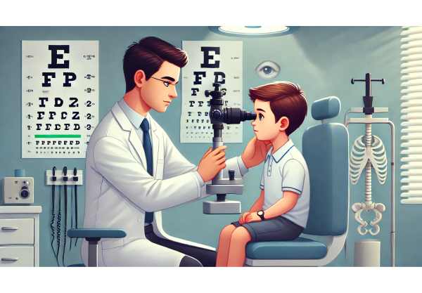
- Comprehensive Eye Exam: A thorough eye examination is required to detect and document the presence of epibulbar dermoids, colobomas, microphthalmia, anophthalmia, and other structural abnormalities. Visual acuity testing, slit-lamp examination, and fundoscopy are all necessary components of this assessment.
- Ophthalmoscopy: Direct and indirect ophthalmoscopy provide detailed views of the retina, optic nerve, and other posterior segment structures. This is critical for detecting retinal and optic nerve colobomas and determining their effects on vision.
- Assessment of Ocular Motility: Assessing eye movements aids in the diagnosis of strabismus and other motility disorders associated with Goldenhar Syndrome.
Imaging Studies
- Ultrasound Biomicroscopy (UBM): UBM generates high-resolution images of the anterior segment, which can be used to characterize epibulbar dermoids, assess microphthalmia, and evaluate other anterior segment anomalies.
- Magnetic Resonance Imaging (MRI): MRI of the orbits and brain is useful for determining the severity of ocular and orbital abnormalities, especially in cases of microphthalmia and anophthalmia. MRI can also be used to detect associated craniofacial and intracranial anomalies.
- Computed Tomography (CT) Scan: CT scans provide detailed images of the orbit’s bony structures, which can aid in surgical planning for eyelid and orbital anomalies.
Genetic Testing
- Chromosomal Analysis: Karyotyping and chromosomal microarray analysis can reveal chromosomal abnormalities and genetic mutations linked to Goldenhar Syndrome.
- Molecular Genetic Testing: Advanced genetic testing, such as whole-exome sequencing, can identify specific genetic mutations associated with the syndrome, facilitating diagnosis and genetic counseling.
Ancillary tests
- Visual Field Testing: Visual field tests are useful in determining the functional impact of colobomas and other retinal abnormalities on the patient’s peripheral vision.
- Electroretinography (ERG): ERG measures the retina’s electrical responses to light stimuli, providing information on retinal function that can be impacted by colobomas and other retinal abnormalities.
Treatment
The treatment of ocular manifestations in Goldenhar Syndrome is multifaceted, with surgical, medical, and supportive interventions tailored to each patient’s specific needs. The primary goals are to improve visual function, correct structural abnormalities, and avoid complications.
Standard Treatment Options
- Surgical Intervention: – Epibulbar Dermoids: Surgical excision is commonly used to remove dermoids that impair vision or cause significant astigmatism. The timing of surgery is critical; it is usually delayed until the child is older to reduce the risk of anesthesia and improve surgical outcomes.
- Colobomas: Surgical repair of colobomas is difficult and usually limited to eyelid colobomas to protect the cornea. Instead of direct surgical intervention, intraocular colobomas are frequently treated with monitoring and supportive care.
- Microphthalmia/Anophthalmia: Orbital expansion procedures, such as the insertion of conformers or expanders, are used to stimulate orbital growth and support prosthetic eye placement, resulting in a better cosmetic appearance and normal facial development.
- Medical Management: – Amblyopia Treatment: Early intervention with patching or atropine drops is crucial for managing amblyopia, especially in cases with significant anisometropia or strabismus.
- Strabismus Management: Strabismus can be treated with glasses, prisms, or surgical correction to align the eyes and improve binocular vision.
- Vision Rehabilitation: – Low Vision Aids: Magnifiers, specialized glasses, and adaptive technologies can improve visual function and quality of life for patients with significant visual impairments.
- Occupational Therapy: Participating in occupational therapy can help children develop important skills and adapt to visual limitations, promoting independence and improving developmental outcomes.
Innovative and Emerging Therapies
- Stem Cell Therapy: Researchers are working to regenerate damaged ocular tissues and improve outcomes for patients with microphthalmia or colobomas. While still experimental, these therapies show promise for future treatment strategies.
- Gene Therapy: Advances in gene therapy show promise for correcting genetic mutations linked to Goldenhar Syndrome. These treatments are still in the early stages of development, but they may offer targeted interventions for the underlying genetic causes.
- 3D Printing: Advancements in 3D printing technology are being used to create personalized ocular prostheses and surgical models. These advancements enable better fitting prosthetics and more precise surgical planning.
- Regenerative Medicine: Regenerative medicine techniques, such as tissue engineering, are being investigated for the development of bioengineered corneal and scleral tissues. These approaches aim to improve surgical outcomes while lowering the risk of complications.
To effectively treat the ocular manifestations of Goldenhar Syndrome, a multidisciplinary team of ophthalmologists, surgeons, geneticists, and vision rehabilitation specialists must collaborate. Continuous advances in medical technology and research are increasing the possibility of better outcomes and quality of life for affected people.
Best Practices to Avoid Complications of Goldenhar Syndrome
- Regular Eye Examinations: Schedule comprehensive eye exams beginning in infancy to monitor and detect any ocular abnormalities early. Regular follow-up visits are essential for continuous evaluation and management.
- Early Intervention: Address amblyopia and strabismus as soon as possible with appropriate treatments such as patching, corrective lenses, or surgery to avoid long-term visual impairment.
- Protective Eyewear: Encourage the use of protective eyewear when participating in activities that pose a risk of eye injury to avoid further trauma to already vulnerable eyes.
- Genetic Counseling: For families with a history of Goldenhar Syndrome, genetic counseling can provide important information about the risk of recurrence in future pregnancies and help guide early intervention strategies.
- Holistic Care: Collaborate with a multidisciplinary team to address not only the ocular but also the craniofacial and systemic symptoms of the syndrome. This comprehensive care strategy ensures that all aspects of the condition are effectively managed.
- Parental Education: Educate parents on the signs and symptoms of Goldenhar Syndrome-related ocular issues, as well as the importance of following treatment plans and follow-up appointments.
- Environmental Safety: Maintain a safe home environment to avoid accidents that could worsen ocular conditions. Ensure adequate lighting and remove any hazards that could cause falls or injuries.
- Nutritional Support: Eat a well-balanced diet high in essential vitamins and minerals to promote overall eye health and development.
- Adaptive Learning Tools: Use adaptive learning tools and technologies to support the educational needs of children with visual impairments, ensuring they have access to appropriate resources to promote learning and development.
Individuals with Goldenhar Syndrome can reduce the risk of complications while also improving their overall ocular health and quality of life by following these preventive measures.
Trusted Resources
Books
- “Ocular and Craniofacial Disorders” by A. W. van der Meulen and J. A. de Vries
- “Genetic Diseases of the Eye” by Elias I. Traboulsi
- “Pediatric Ophthalmology: Current Thought and A Practical Guide” by Edward M. Wilson, Richard A. Saunders, and Trivedi Rupal


