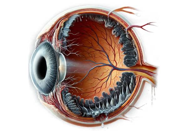
Retinal astrocytic hamartoma is a rare, benign retinal tumor that develops from astrocytes, which are a type of glial cell within the retina. These tumors are usually non-cancerous and linked to genetic conditions, particularly tuberous sclerosis complex (TSC) and, less frequently, neurofibromatosis type 1 (NF1). Despite their benign nature, retinal astrocytic hamartomas can cause visual disturbances, particularly if they become large enough to interfere with retinal architecture or cause complications such as retinal detachment or hemorrhage.
Understanding the Retina’s Cellular Composition
The retina is a thin layer of tissue located at the back of the eye that converts light into neural signals sent to the brain for visual recognition. The retina is made up of several layers, each containing a different type of cell, such as photoreceptors (rods and cones), bipolar cells, ganglion cells, and glial cells (including astrocytes).
Astrocytes provide structural support to retinal neurons, regulate blood flow, and maintain the blood-retinal barrier, all of which are critical to their health and functionality. They are a type of glial cell that includes Müller cells and microglia and contributes to the overall homeostasis of the retinal environment.
Pathophysiology Of Retinal Astrocytic Hamartoma
Retinal astrocytic hamartomas develop as a result of abnormal astrocyte growth and proliferation within the retina. This proliferation causes the formation of a hamartoma, which is a benign tumor-like growth made up of an overgrowth of normal tissue elements arranged abnormally. The overgrowth in retinal astrocytic hamartomas involves astrocytes and the extracellular matrix.
These hamartomas are usually congenital, which means they are present at birth, but they may not be diagnosed until later in life when symptoms appear or during routine eye exams. These hamartomas grow slowly and can remain stable for years. However, in some cases, they may enlarge or change in appearance, especially in the presence of underlying genetic conditions such as TSC or NF1.
Relationship with Tuberous Sclerosis Complex and Neurofibromatosis Type 1
Tuberous sclerosis complex (TSC) is a genetic disorder characterized by the development of benign tumors in various organs such as the skin, brain, kidneys, heart, and eye. Mutations in the TSC1 and TSC2 genes, which regulate cell growth and proliferation, are to blame. Individuals with TSC are more likely to develop retinal astrocytic hamartomas, which can occur in up to 50% of patients with TSC. The presence of retinal astrocytic hamartomas in a patient can be a strong clinical indicator of TSC, especially when combined with other disease-specific features such as facial angiofibromas, cortical tubers, or renal angiomyolipomas.
Neurofibromatosis type 1 (NF1) is another genetic condition that can cause retinal astrocytic hamartomas, though less frequently than TSC. NF1 causes neurofibromas, café-au-lait spots on the skin, and Lisch nodules in the eyes. Retinal astrocytic hamartomas in NF1 patients are typically less prominent and less frequently symptomatic than those in TSC.
Clinical Features and Symptoms
The clinical presentation of retinal astrocytic hamartomas varies greatly depending on the tumor’s size, location, and the presence of any associated complications. In many cases, these hamartomas are asymptomatic and only discovered during routine eye examinations. When symptoms do occur, they are usually caused by the hamartoma’s size and growth, as well as its effect on the surrounding retinal tissue.
Patients with retinal astrocytic hamartomas may have the following symptoms:
- Visual Disturbances: Examples include blurring of vision, visual field defects, and scotomas (blind spots) in the visual field. The location of the hamartoma within the retina has a strong correlation with the degree of visual impairment. If the hamartoma is located close to the macula, the central part of the retina responsible for sharp, detailed vision, it is more likely to cause noticeable visual symptoms.
- Photopsia: Some patients may experience flashes of light, a symptom known as photopsia, which occurs when the hamartoma causes traction or irritation of the retinal tissue.
- Floaters: Floaters, which are small, shadowy shapes that appear in the field of vision, can occur if the hamartoma is associated with vitreous changes or bleeding into the vitreous humor.
- Complications: In rare cases, retinal astrocytic hamartomas can result in more serious complications like retinal detachment or vitreous hemorrhage. Retinal detachment occurs when the retina separates from the underlying supportive tissue, posing a risk of permanent vision loss if not treated immediately. Vitreous hemorrhage, which occurs when blood enters the vitreous cavity of the eye, can also impair vision and may necessitate surgical intervention.
Morphological Feature and Growth Patterns
During an eye examination, retinal astrocytic hamartomas show distinct morphological characteristics. These tumors frequently present as white, yellowish, or grayish lesions on the retina, with a distinct, nodular appearance. They may have a mulberry-like surface texture in some cases because calcium deposits are present within the tumor.
There are three primary types of retinal astrocytic hamartomas based on their appearance and growth patterns:
- Classic Type: This is the most common type, and it appears as a well-defined, elevated lesion with a nodular surface. These tumors may contain calcifications, giving them a distinct appearance in imaging studies.
- Translucent Type: These hamartomas are less dense, presenting as semi-transparent lesions with minimal elevation. They may be harder to detect during a routine examination.
- Calcified Type: This form is distinguished by extensive calcification within the hamartoma, which is visible on imaging and gives the tumor a more opaque appearance. Calcified hamartomas are more likely to cause visual symptoms because they are denser and may have an impact on surrounding retinal structures.
Retinal astrocytic hamartomas typically grow slowly, and many lesions remain stable for years with no significant changes. However, in some cases, particularly in patients with TSC, hamartomas can grow in size or number over time, potentially increasing the risk of complications.
Differential Diagnosis
Retinal astrocytic hamartomas must be distinguished from other retinal lesions that may present with similar features. This includes:
- Retinoblastoma: A malignant retinal tumor that mainly affects young children. Unlike retinal astrocytic hamartomas, retinoblastoma is a cancerous tumor that requires immediate treatment. Differentiating between the two is critical, as their management strategies are vastly different.
- Retinal Hemangioblastoma: Another benign retinal tumor linked to von Hippel-Lindau disease. This tumor also requires close monitoring because it has the potential to cause retinal detachment or vitreous hemorrhage.
- Choroidal Osteoma: A benign, bone-like tumor of the choroid that resembles a calcified retinal astrocytic hamartoma. Imaging tests can aid in distinguishing between these lesions.
Accurate diagnosis is critical for effective management and monitoring for potential complications associated with retinal astrocytic hamartomas.
Techniques for Diagnosing Retinal Astrocytic Hamartoma
Diagnosing retinal astrocytic hamartoma requires a combination of clinical evaluation, imaging studies, and, in some cases, genetic testing. Accurate diagnosis is critical for distinguishing this benign condition from other potentially malignant or vision-threatening retinal lesions.
Clinical Evaluation
The diagnostic process begins with a thorough clinical evaluation, which includes a detailed patient history as well as a comprehensive eye examination. During the history-taking process, the clinician will inquire about the presence of visual symptoms such as blurred vision, photopsia, or floaters, as well as any prior history of genetic conditions such as tuberous sclerosis complex (TSC) or neurofibromatosis type 1. The patient’s family history may also provide useful information, especially if there is a known predisposition to these genetic conditions.
Fundus Examination
A fundus examination is an essential part of the diagnostic process. The ophthalmologist can examine the retina and detect any lesions using an ophthalmoscope or slit lamp with a fundus lens. Retinal astrocytic hamartomas usually appear as white, yellowish, or greyish nodules on the retina. The distinctive nodular surface and possible presence of calcifications can help distinguish these hamartomas from other retinal lesions.
Optical Coherence Tomography(OCT)
Optical coherence tomography (OCT) is a non-invasive imaging technique that produces high-resolution cross-sections of the retina. OCT is especially useful for determining the structural characteristics of retinal astrocytic hamartomas. It can reveal the lesion’s thickness and extent, as well as any retinal edema or layer disruption. OCT can also help distinguish between the various types of hamartomas, including classic, translucent, and calcified forms.
Fundal Fluorescein Angiography (FFA)
Fundus fluorescein angiography (FFA) entails injecting a fluorescent dye into the bloodstream and then photographing the retina in rapid succession as the dye circulates through the retinal blood vessels. FFA is especially useful for determining the vascular characteristics of retinal astrocytic hamartomas. These lesions usually have little or no dye leakage, which distinguishes them from other retinal tumors like retinoblastoma and retinal hemangioblastoma, which can have more pronounced leakage. FFA can also help identify any associated retinal or choroidal abnormalities that may affect the overall management strategy.
Autofluorescence Imaging
Autofluorescence imaging is another useful diagnostic tool for assessing retinal astrocytic hamartomas. This method makes use of the natural fluorescence emitted by certain retinal structures when exposed to specific wavelengths of light. Retinal astrocytic hamartomas frequently exhibit distinct autofluorescence patterns, which aid in their identification and differentiation from other retinal lesions. Calcified hamartomas, in particular, may exhibit increased autofluorescence due to calcium deposits.
Ultrasound Imaging
Ocular ultrasound, specifically B-scan ultrasonography, can be used to assess the internal characteristics of retinal astrocytic hamartomas, particularly if the lesions are large or have a significant impact on retinal architecture. Ultrasound can help determine the size of the lesion, its echogenicity (the amount of sound waves that bounce off the tumor), and any associated complications, such as vitreous hemorrhage or retinal detachment. This imaging modality is especially useful when media opacities such as cataracts or vitreous hemorrhage obscure the view of the retina.
Genetic Testing
Genetic testing may be recommended for retinal astrocytic hamartomas that are suspected of being associated with genetic conditions such as tuberous sclerosis complex (TSC) or neurofibromatosis type 1 (NF1). Genetic testing can confirm the presence of mutations in the TSC1, TSC2, or NF1 genes, which cause these conditions. Identifying a genetic predisposition is critical for the patient’s overall management because these conditions can affect multiple organ systems and necessitate multidisciplinary treatment.
Approaches to Managing Retinal Astrocytic Hamartoma
The management of retinal astrocytic hamartoma is primarily concerned with monitoring the lesion for changes, managing any associated symptoms, and dealing with complications as they arise. Because retinal astrocytic hamartomas are benign and frequently asymptomatic, treatment is not always required. However, if the hamartoma causes visual disturbances or poses a risk of complications, intervention may be necessary.
Observation and Monitoring
In many cases, especially when the hamartoma is small, stable, and asymptomatic, regular observation is the best approach. This includes regular eye exams to check for changes in the lesion’s size, appearance, or associated retinal effects. Regular monitoring is especially important for patients with genetic conditions such as tuberous sclerosis complex (TSC) or neurofibromatosis type 1 (NF1), as these patients are more likely to develop multiple hamartomas or other ocular and systemic complications.
During these follow-up visits, ophthalmologists will perform a thorough eye examination, including visual acuity testing, fundus examination, and possibly imaging studies such as optical coherence tomography (OCT) or fundus fluorescein angiography (FFA), to determine the stability of the hamartoma.
Symptom Management
If the retinal astrocytic hamartoma causes symptoms like visual disturbances, photopsia, or floaters, symptomatic treatment may be required. The nature and severity of the symptoms will determine the appropriate treatment.
- Vision Correction: If the hamartoma impairs vision, corrective lenses or glasses may be required to improve visual acuity. However, this is usually only effective if the hamartoma does not cause significant structural damage to the retina.
- Laser Photocoagulation: If the hamartoma is accompanied by retinal edema or exudation that impairs vision, laser photocoagulation may be used to stabilize the retina. This procedure uses a laser to create small burns around the lesion, which can help seal leaking blood vessels and reduce retinal swelling.
- Vitreous Hemorrhage Management: If the hamartoma causes a vitreous hemorrhage, which occurs when blood leaks into the vitreous cavity, treatment may consist of watchful waiting if the hemorrhage is minor and likely to resolve on its own. In more severe cases, a vitrectomy may be necessary. Vitrectomy is a surgical procedure that removes the vitreous gel from the eye, allowing for blood removal and improved access to repair the retina if necessary.
- Management of Retinal Detachment: Retinal detachment is a serious complication that can occur if the hamartoma causes significant traction on the retina. Surgical intervention, such as scleral buckling or vitrectomy in conjunction with laser therapy, may be required to reattach the retina and prevent permanent vision loss.
Genetics and Multidisciplinary Management
For patients with retinal astrocytic hamartomas caused by genetic conditions such as TSC or NF1, treatment frequently extends beyond the ocular issues. These patients usually require multidisciplinary care, which includes collaboration with neurologists, dermatologists, nephrologists, and genetic counselors to address the broader implications of their condition.
Genetic counseling is an essential part of treating patients with TSC or NF1. Counseling can help patients and their families understand the genetic nature of their condition, the likelihood of passing it down to offspring, and the implications for other family members. Surveillance for other manifestations of the disease, such as brain or kidney tumors, is critical for TSC patients and may influence the treatment of their retinal hamartomas.
Long-term Follow-up
Even when retinal astrocytic hamartomas are stable and asymptomatic, long-term monitoring is required. Regular eye exams are recommended to detect any changes in the hamartoma or the onset of new symptoms early. This proactive approach helps to avoid complications and ensures that any necessary interventions are carried out in a timely manner.
Trusted Resources and Support
Books
- “Clinical Atlas of Retinal Diseases” by Jack J. Kanski and Brad Bowling – This comprehensive atlas provides detailed visual documentation of various retinal conditions, including retinal astrocytic hamartomas, with diagnostic images and treatment options.
- “Retinal and Choroidal Manifestations of Selected Systemic Diseases” by J. Fernando Arévalo – A valuable resource for understanding the ocular manifestations of systemic diseases like tuberous sclerosis complex and neurofibromatosis, with specific sections on retinal astrocytic hamartomas.
Organizations
- American Academy of Ophthalmology (AAO) – The AAO offers extensive resources, including clinical guidelines, educational materials, and patient support for a wide range of ocular conditions, including retinal tumors and related genetic disorders.
- Tuberous Sclerosis Alliance (TS Alliance) – A dedicated organization providing support, education, and research funding for patients with tuberous sclerosis complex, offering resources on the management of retinal astrocytic hamartomas and other related conditions.


