Introduction
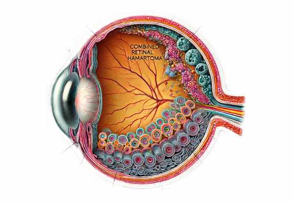
Combined Hamartoma of the Retina and Retinal Pigment Epithelium (CHRRPE) is a rare, benign ocular condition marked by abnormal proliferation of retinal and retinal pigment epithelial cells. This congenital anomaly typically manifests as a grayish or pigmented lesion on the retina, which is frequently accompanied by retinal distortion, traction, and gliosis. CHRRPE can impair visual acuity and cause a variety of visual disturbances, depending on its size and location. Although CHRRPE is generally non-progressive, it requires close monitoring to manage potential complications and differentiate it from other retinal pathologies with similar presentations.
Insights into Combined Retinal Hamartoma and Retinal Pigment Epithelium
CHRRPE is a unique ocular condition characterized by abnormal growth of both retinal and retinal pigment epithelial (RPE) cells. This section discusses the pathophysiology, clinical presentation, complications, and overall impact of the condition on patients’ vision and quality of life.
Pathophysiology
CHRRPE is a congenital malformation caused by the abnormal proliferation of cells from the retina and the RPE. The term “hamartoma” refers to a benign, focal malformation that resembles a neoplasm but is caused by the overgrowth of normal tissue components. This overgrowth in CHRRPE includes retinal glial cells, RPE cells, and occasionally fibrovascular tissue.
The exact cause of CHRRPE is unknown, but it is thought to be the result of developmental disruptions during retina and RPE formation. These disruptions could be caused by genetic mutations, but specific genetic markers have not been consistently identified. The lesion typically appears in early childhood and remains stable throughout life.
Clinical Presentation
CHRRPE manifests as a pigmented or grayish lesion on the retina that is frequently discovered during routine ophthalmic examinations or while investigating visual disturbances. The clinical characteristics of CHRRPE vary greatly depending on the size, location, and extent of the lesion. Common manifestations include:
- Visual Disturbances: Patients with CHRRPE may experience a variety of visual symptoms, such as blurred vision, visual field defects, metamorphopsia (distorted vision), and decreased visual acuity. The severity of these symptoms is determined by how close the lesion is to the macula and how it affects retinal structure.
- Retinal Traction and Distortion:** The hamartomatous growth frequently exerts traction on the surrounding retinal tissue, resulting in retinal distortion and possible tractional detachment. This can result in symptoms like photopsia (light flashes) and floaters.
- The Epiretinal Membrane (ERM): An ERM is common in CHRRPE and is caused by glial proliferation on the retina’s surface. ERMs can also contribute to retinal traction and visual disturbances.
- Gliosis: Reactive gliosis, which is defined by the proliferation of glial cells, is common in CHRRPE. Gliosis can cause scarring and fibrosis, compromising retinal function.
Differential Diagnosis: CHRRPE should be distinguished from other retinal conditions with similar clinical features. Differential diagnosis includes:
- Retinoblastoma is a malignant retinal tumor that primarily affects children. Unlike CHRRPE, retinoblastoma frequently presents with leukocoria (a white pupillary reflex) and necessitates immediate treatment.
- Congenital Hypertrophy of the Retinal Pigment Epithelium (CHRPE): This benign condition involves hypertrophic RPE cells and manifests as flat, pigmented lesions that lack the tractional and gliotic changes seen in CHRRPE.
- Choroidal Nevus: A benign pigmented lesion of the choroid that is usually asymptomatic and stable, as opposed to CHRRPE, which is proliferative and tractional.
- Toxocariasis: An ocular parasitic infection that can cause granulomas and retinal detachment, similar to the appearance of CHRRPE.
Complications
While CHRRPE is generally benign and non-progressive, it can cause a number of complications, the majority of which are related to its effects on retinal structure and function.
- Retinal Detachment: Tractional forces exerted by the hamartomatous lesion can cause retinal detachment, a serious condition that necessitates immediate surgical intervention to avoid permanent vision loss.
- Macular Involvement: A lesion near or involving the macula can have a significant impact on central vision, resulting in profound visual impairment.
- Secondary Glaucoma: In rare cases, extensive retinal involvement and associated vascular changes can result in high intraocular pressure and secondary glaucoma.
Effects on Quality of Life
The impact of CHRRPE on a patient’s quality of life is determined by the severity of their visual impairment and the presence of complications. Key features include:
- Visual Function: Patients with significant retinal involvement may struggle with reading, driving, and other tasks that require fine visual acuity. Central vision loss can be especially debilitating.
- Psychosocial Impact: Being diagnosed with a rare ocular condition can cause anxiety and stress for both patients and their families. Concerns about potential vision loss, as well as the need for ongoing monitoring, can have an impact on one’s mental health.
- Educational and Professional Challenges: Children with CHRRPE may experience difficulties at school due to visual disturbances, necessitating special educational support. Adults with significant visual impairments may require vocational rehabilitation and job modifications.
Prevention Tips
Preventing CHRRPE is difficult due to its congenital nature and unknown etiology. However, there are several measures that can help manage the condition and reduce the risk of complications:
- Schedule regular eye exams, particularly for children and those with a family history of ocular conditions. Early detection of CHRRPE enables timely intervention and monitoring.
- Awareness of Symptoms: Be aware of symptoms such as blurred vision, visual field defects, flashes of light, and floaters. Early detection of these symptoms can prompt a comprehensive ophthalmic examination.
- Protective Eyewear: – Wear protective eyewear for activities that may cause eye injury, such as sports or hazardous work environments. Preventing eye trauma can lower the risk of exacerbated retinal conditions.
- Maintain a healthy lifestyle through a balanced diet high in antioxidants and omega-3 fatty acids. These nutrients promote overall eye health and may help to reduce the effects of retinal conditions.
- Genetic Counseling: – Families with a history of congenital retinal conditions should seek genetic counseling to better understand the risks and inheritance patterns. Genetic counseling can offer useful information for family planning and early intervention.
- Avoid smoking, as it increases the risk of ocular diseases. Avoiding smoking can help protect the retina and lower the risk of complications.
- Manage Systemic Conditions: – Control diabetes and hypertension, which can harm retinal health. Regular medical check-ups and effective treatment for these conditions are critical.
- Regular monitoring by an ophthalmologist is crucial for patients with CHRRPE. Ongoing surveillance can detect changes in the lesion and provide timely intervention to avoid complications.
- Educational Support: – Help children with CHRRPE overcome visual challenges at school. Early intervention and accommodations can improve learning and development.
- Visual Aids and Rehabilitation: – Use low vision aids and rehabilitation services to maximize remaining vision and independence.
Diagnostic methods
The diagnosis of Combined Hamartoma of the Retina and Retinal Pigment Epithelium (CHRRPE) is based on a combination of clinical examination and advanced imaging techniques to accurately characterize the lesion and distinguish it from other ocular conditions.
Fundoscopy Examination
A comprehensive fundoscopic examination is the first step in diagnosing CHRRPE. To visualize the retina, ophthalmologists use slit-lamp biomicroscopes and indirect ophthalmoscopy. CHRRPE manifests as a greyish or pigmented lesion with an irregular surface, frequently involving the retinal vasculature and resulting in retinal folds or traction changes.
Optical Coherence Tomography(OCT)
Optical Coherence Tomography (OCT) is a non-invasive imaging technique that produces detailed cross-sectional images of the retina. OCT is useful in determining the structural characteristics of a hamartoma, such as its size, extent, and effect on the retinal layers. It aids in the identification of associated characteristics such as retinal edema, traction, and macular involvement. OCT is especially useful for tracking the lesion over time and detecting subtle changes that may require intervention.
Fluorescein Angiography(FA)
Fluorescein Angiography (FA) entails injecting a fluorescent dye into the bloodstream and taking a series of photographs of the retina while it circulates. FA aids in assessing the vascular characteristics of CHRRPE by identifying abnormal blood vessels, leakage, and capillary non-perfusion areas. This technique is useful for distinguishing CHRRPE from other retinal conditions that have similar appearance.
B-scan ultrasonography
B-Scan ultrasonography is an effective diagnostic tool for determining the internal reflectivity and size of CHRRPE lesions, particularly when the fundus view is obscured by media opacities or significant elevation. This modality provides information about the lesion’s acoustic properties, which can help differentiate CHRRPE from other intraocular tumors.
Genetic Testing
Genetic testing may be recommended when CHRRPE is suspected of being linked to genetic syndromes such as neurofibromatosis type 2 or tuberous sclerosis complex. Identifying genetic mutations can help confirm a diagnosis and guide treatment of systemic manifestations.
Treatment
Treatment for Combined Hamartoma of the Retina and Retinal Pigment Epithelium (CHRRPE) is tailored to each patient’s symptoms, lesion characteristics, and overall health. While many cases of CHRRPE are stable and do not require intervention, treatment may be required in cases with significant visual impairment or complications.
Observation
In asymptomatic patients or those with minor visual disturbance, regular observation is frequently the preferred management strategy. Periodic fundoscopic examinations and OCT imaging are used to monitor the lesion’s size, appearance, and associated retinal complications. This approach helps to avoid unnecessary interventions while also ensuring early detection of any progression.
Surgical Intervention
Patients with significant retinal traction, macular involvement, or retinal detachment may require surgical intervention. Vitrectomy, a surgical procedure that removes the vitreous gel from the eye, can reduce tractional forces and treat retinal detachment. During vitrectomy, the surgeon may perform membrane peeling to remove epiretinal membranes that contribute to traction. Surgical outcomes are determined by the extent of retinal involvement and the presence of pre-existing retinal damage.
Laser Photocoagulation
Laser photocoagulation may be used when there is vascular leakage or proliferative changes associated with CHRRPE. The laser treatment is intended to seal leaking blood vessels and reduce the risk of additional complications such as vitreous hemorrhage or exudative retinal detachment. This procedure is usually performed under local anesthesia and may require several sessions, depending on the size of the lesion.
Anti-VEGF Therapies
Anti-vascular endothelial growth factor (anti-VEGF) therapy is a new treatment option for CHRRPE patients with severe macular edema or neovascularization. Intravitreal injections of anti-VEGF agents, such as bevacizumab or ranibizumab, can help reduce vascular leakage while improving macular anatomy. While anti-VEGF therapy is primarily used to treat conditions such as age-related macular degeneration, it has shown promise in the management of CHRRPE-related complications.
Innovative Therapies
Gene therapy and targeted molecular treatments are two innovative therapies for CHRRPE that are currently being investigated. These approaches aim to address the underlying genetic and cellular mechanisms that cause hamartoma formation. While still in the experimental stage, these therapies have the potential to provide more effective and personalized treatment options in the future.
Trusted Resources
Books
- “Retinal Vascular Disease” by A.M. Joussen, T.W. Gardner, B. Kirchhof, S.J. Ryan
- “Clinical Ophthalmic Oncology” by Jesse L. Berry and Bertil Damato
- “Retina” by Stephen J. Ryan


