Keratoconus is a progressive eye disorder that weakens the cornea, causing it to bulge into a cone-like shape. This abnormal curvature distorts vision and often leads to severe astigmatism, light sensitivity, and other vision challenges. For many patients, spectacles or even specialized contact lenses may no longer adequately correct eyesight as the disease advances. Traditional treatments like corneal cross-linking, intrastromal ring segments, or even corneal transplantation can help. However, each of these carries limitations or varying degrees of invasiveness.
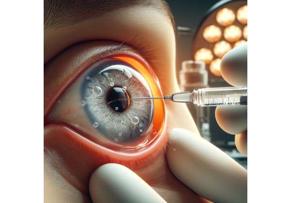
A newly approved hydrogel injection therapy is now emerging as a game-changer in keratoconus management. Unlike some other procedures requiring substantial incisions or the removal of tissue, this injectable technology is minimally invasive and aims to reinforce the corneal framework from within. The specialized hydrogel stabilizes the thinning cornea, slowing or halting disease progression and improving functional vision. This innovative approach has excited corneal specialists worldwide, offering a novel blend of safety, convenience, and efficacy for patients struggling with keratoconus.
A Pioneering Approach to Strengthening the Cornea
In the quest to address keratoconus—characterized by thinning and protrusion of the corneal stroma—clinicians have leveraged solutions ranging from contact lenses to major surgery. While corneal cross-linking (CXL) has become a standard to halt progression by chemically strengthening collagen fibers, it requires UV light exposure and riboflavin drops, and its effects can be unpredictable in advanced or extremely thin corneas. Hydrogel injections represent an alternative or complement, introducing a stable, biocompatible material directly into the cornea’s mid-layers.
Rationale and Underlying Technology
In keratoconus, the stroma progressively weakens due to biochemical imbalances in collagen cross-link formation. The idea behind hydrogel therapy is to add volume and mechanical support to the cornea from within, thereby re-establishing a more normal shape and thickness profile. This novel hydrogel is engineered to integrate seamlessly with collagen lamellae, effectively “bulking up” compromised areas without interfering with corneal physiology. Unlike rigid ring segments, which physically push or pull the cornea into a better contour, the hydrogel gently coalesces with existing tissue.
Researchers developed the hydrogel formula after extensive trials in materials science, focusing on transparency, elasticity, and durability. To ensure that the injected material would not impede vision or trigger immunologic rejection, the hydrogel’s constituents are derived from inert polymers or naturally occurring substances known for minimal inflammatory responses. They also ensure strong adhesion so that it does not migrate or degrade prematurely.
Ideal Candidates for This Injectable
Hydrogel injection therapy is best suited for patients with mild to moderate keratoconus who still have sufficient corneal thickness to sustain the procedure. It may help individuals who:
- Show early signs of disease progression: The cornea might not be extremely thin yet, but topographical changes indicate the condition is worsening.
- Are not optimal for cross-linking alone: Perhaps they have borderline thickness or other complicating factors that might compromise standard CXL success.
- Desire a minimally invasive approach: People looking for a less disruptive intervention compared to ring implantation or corneal transplant.
- Seek synergy with cross-linking: Some specialists consider combining hydrogel injection with CXL for added structural fortification.
That said, the therapy might be less effective in advanced keratoconus where significant scarring has developed or the cornea’s shape has progressed too far to salvage with an injectable. In such cases, more extensive procedures may be necessary, but there remains interest in using hydrogel for partial or interim improvements. Ultimately, careful corneal mapping, pachymetry (thickness measurement), and clinical evaluations guide the selection process.
Comparisons to Conventional Methods
Traditional interventions each have strengths and drawbacks:
- Corneal Cross-Linking: Renowned for halting disease progression by stiffening corneal collagen. However, it doesn’t add thickness and may be less effective if the cornea is extremely thin or the disease is advanced.
- Intrastromal Ring Segments: Help flatten the steep cornea by exerting mechanical pressure, though they require incisional channels and can sometimes induce irregular astigmatism or extrusion.
- Contact Lenses: Scleral or rigid lenses correct vision but do not slow progression or strengthen the cornea.
- Penetrating or Lamellar Transplantation: Effective for very advanced disease but carries higher risk, including graft rejection, and requires a lengthier recovery.
Hydrogel injections fill a niche: they are relatively low-risk compared to surgery, have the potential to simultaneously thicken and stabilize corneal tissue, and can integrate well with other treatments. For these reasons, specialists anticipate a broad role for hydrogel therapy in bridging the gap between mild disease amenable to cross-linking alone and severe disease requiring transplantation.
Steps to a Successful Injectable Procedure
Although hydrogel injections may appear straightforward, they demand precision, specialized tools, and a nuanced understanding of corneal anatomy. The surgical steps aim to ensure even distribution of the hydrogel within targeted layers, minimal stress to surrounding tissues, and optimal shaping to enhance vision. Here is a closer look at how clinicians approach the procedure and the protocols that support patient safety and success.
Preoperative Planning and Diagnostic Imaging
As with any corneal intervention, accurate assessment lays the foundation for good outcomes. Key elements include:
- Corneal Topography or Tomography: Provides a detailed map of curvature, local steepness, and morphological irregularities. The intended injection site and volume can be planned around these measurements.
- Pachymetry: Determining thickness at various points ensures that the cornea can tolerate the injected material, avoiding potential bulges or tears.
- Slit-Lamp Examination: Assesses the corneal surface, detecting scarring, epithelial defects, or other conditions that might complicate injection.
- Overall Ocular Health Check: Screening for concurrent diseases like cataracts or retinal conditions is crucial, as they might affect visual outcomes or indicate more pressing issues.
Based on these investigations, the eye care team decides the approximate volume of hydrogel to inject and the depth of placement. If cross-linking is planned in tandem—either simultaneously or sequentially—coordinating each step with imaging data is paramount.
Local Anesthesia and Injection Delivery
On the day of the procedure, patients typically receive topical anesthetic drops, or in some cases, mild sedation or local infiltration anesthesia. Once the eye is numb:
- Incision or Micro-Needle Entry: A small, precisely placed incision or needle entry port is created. Modern micro-needles are extremely fine, minimizing trauma to corneal layers.
- Hydrogel Injection: Using a controlled syringe or micro-injector system, the surgeon slowly introduces the pre-prepared hydrogel into the mid-stromal plane. Good technique ensures the gel spreads evenly, reinforcing the structural lamellae without air pockets or lumps.
- Volume and Distribution Control: Real-time imaging or careful visual observation helps track where the gel is migrating. Surgeons may gently manipulate the cornea to ensure uniform layering.
- Incision Closure or Self-Seal: In many cases, the tiny port self-seals, or a suture may be used if necessary. The surgeon checks for any leaks or bulges before concluding.
Because the injection is minimally invasive, operative times are relatively short—often under an hour. Patients can typically return home the same day, though they must follow postoperative guidelines and schedule follow-up visits for corneal checks.
Potential Pairing with Cross-Linking
While hydrogel therapy can work alone, some doctors integrate corneal cross-linking to further stabilize the collagen framework:
- Concurrent Procedure: Immediately following injection, the surgeon may apply riboflavin and UV light to the cornea, initiating cross-links around the hydrogel-lamellae interface. This approach could boost synergy—mechanical filling combined with biochemical stiffening.
- Staged Intervention: Alternatively, surgeons may allow the hydrogel to settle for several weeks, ensuring it has integrated well, and then perform cross-linking. This spacing might reduce the risk of excessive swelling or complicating factors.
Each strategy depends on the patient’s corneal thickness, disease severity, and the surgeon’s familiarity with combining these modalities. While the synergy approach is promising, it also raises the importance of thorough follow-up imaging to verify the final corneal shape and confirm stable outcomes.
Postoperative Care and Monitoring
After the procedure, patients can expect a regimen of antibiotic and anti-inflammatory drops to prevent infection and control mild inflammation. Specialists check corneal healing periodically, looking for:
- Epithelial Integrity: Ensuring the surface remains healthy with no persistent defects.
- Structural Stability: Monitoring curvature through topography to confirm that the cornea remains stable and that the hydrogel does not shift.
- Clarity and Visual Acuity: Evaluating improvements in best-corrected visual acuity, changes in astigmatism, or scarring.
Unlike major surgeries, downtime is relatively brief. Many patients resume normal activities within days, although contact lens fittings may be postponed until the corneal shape stabilizes. If cross-linking or other enhancements are planned, the timeline is determined by how quickly the cornea recovers from the injection.
By incorporating advanced imaging, meticulous injection methods, and thoughtful postoperative management, hydrogel therapy has the potential to deliver stable, long-lasting corneal support with minimal trauma—an attractive proposition for those seeking an intermediate solution between non-surgical measures and corneal transplant.
New Data and Clinical Findings on Hydrogel Efficacy
As with any emerging medical technology, clinical studies and research findings provide the backbone for validating hydrogel injections in keratoconus. Although the field is still maturing, a growing pool of published data underscores the approach’s capacity to stabilize corneal shape, improve visual function, and reduce the rate of disease progression. This overview highlights key findings and ongoing investigation areas, offering a window into why hydrogel therapy is capturing widespread interest.
Initial Trials and Feasibility Studies
Early-stage clinical trials typically evaluated safety, tolerability, and the basic biomechanical effects:
- Safety Profile: Patients in pilot studies generally experienced mild, transient discomfort or redness at the injection site, with minimal risk of infection. No major immunologic or toxic reactions to the hydrogel material were reported.
- Corneal Thickness and K-Values: Data indicated modest increases in central or mid-peripheral thickness, aligning with the injection zone. Corneal topography often revealed flatter curvatures (lower K-values), signifying a partial correction of the keratoconic cone.
- Early Efficacy Indicators: Although many patients still required contact lenses for optimum vision, some reported improved uncorrected visual acuity or decreased astigmatism. These benefits often became more pronounced over weeks or months as the cornea adapted and epithelial remodeling smoothed the surface.
Such pilot successes paved the way for expanded trials, which are more rigorously designed, randomizing patients to different volumes of hydrogel, different injection depths, or comparing injection plus cross-linking to cross-linking alone.
Mid- to Long-Term Observations
Researchers with longer follow-up periods—exceeding 12 to 24 months—began highlighting sustainability and additional benefits:
- Stable Corneal Topography: A majority of study participants showed minimal or no progression in the cone shape. This stability hints that hydrogel can prevent further thinning or warpage, akin to a scaffolding effect.
- Reduced Dependence on Hard Lenses: Some patients were able to switch to simpler lens designs, thanks to less severe irregular astigmatism.
- Better Tolerance vs. Other Procedures: Compared with intracorneal ring segments or repeated cross-linking attempts, patients generally reported fewer side effects and a gentler recovery.
While these results are promising, they emphasize the importance of refined surgical technique—particularly volume control, layering depth, and avoidance of injection near the corneal apex to minimize potential haze. Studies also stress that the best improvements came when disease progression was caught relatively early, underlining the well-known principle that earlier intervention yields better outcomes in keratoconus.
Ongoing Investigations and Future Directions
As the technology garners more interest, a host of new research initiatives are in motion:
- Composite Hydrogels and Enhanced Formulations: Scientists are tweaking polymer compositions to optimize clarity, longevity, and synergy with existing collagen. Some aim to introduce growth factors or cross-linking agents into the gel itself.
- Custom Injection Patterns: Emerging imaging software might help tailor injection volumes to each patient’s unique topographic map. Automated systems could deliver micro-doses at strategically mapped coordinates, akin to topography-guided partial corrections.
- Comparative Studies with Cross-Linking: Rigorous, large-scale randomized trials will help define which patients gain the most from stand-alone hydrogel vs. cross-linking vs. a combined approach.
- Integration with Tissue Engineering: A few advanced projects explore fusing hydrogel technology with corneal tissue or engineered scaffolds, aiming for partial or full corneal replacements that might obviate the need for donor grafts in advanced disease.
While it is still early days, the results so far suggest a powerful tool for bridging the gap between milder keratoconus treatments and more invasive surgeries. This “injectable solution” theme resonates with broader trends in regenerative medicine, wherein minimally invasive yet biologically integrative therapies are favored.
Real-World Cases and Expert Opinions
Though formal studies are crucial, anecdotal and real-world accounts from leading corneal specialists also play a role. Many have recounted individual successes: patients who had borderline corneal thickness for cross-linking or who disliked the risk profile of ring segments found hydrogel injections an appealing alternative. Some even note improved tolerance of contact lenses, highlighting a dual benefit: mechanical support plus a smoother corneal surface. However, experts caution that consistent outcomes hinge on thorough training in injection technique and patient selection.
Overall, the research trajectory for hydrogel injections in keratoconus shows encouraging momentum. As trials expand, new data will refine best practices—pinpointing which injection strategies yield the most stable, comfortable, and enduring results. For patients, these developments represent a leap forward in preserving vision against a notoriously progressive disease.
Balancing Pros and Cons of a Novel Solution
Any emerging therapy must balance excitement over potential benefits with a clear-eyed appraisal of risks and limitations. Hydrogel injections are no exception. While the approach offers a compelling alternative to more invasive procedures, understanding its safety profile, indications, and real-world efficacy is vital for clinicians and patients alike. Here is an overview of the key points to consider when evaluating hydrogel therapy for keratoconus.
Documented Advantages
Many specialists speak positively about hydrogel injections for reasons including:
- Minimally Invasive Nature: Compared to corneal transplant or intrastromal ring segments, injection typically involves only a small needle incision. This often translates into reduced surgical trauma, faster recovery, and fewer complications like infection or scarring.
- Structural Augmentation: By physically adding volume to thinning stromal layers, the hydrogel helps restore a more physiologic corneal curvature. This approach can complement other treatments rather than replacing them.
- Potential for Early Intervention: Because it is relatively conservative, hydrogel therapy might be offered at earlier stages of disease progression. Early stabilization can spare patients from advanced vision loss or more radical surgery.
- Tailored Approach: Surgeons can modulate the injection volume or location. If needed, additional injections may be done to refine the result or address new focal weak spots.
Potential Drawbacks and Risks
That said, no therapy is devoid of concerns. Hydrogel injection for keratoconus must address issues such as:
- Long-Term Durability: While short-to-mid-term results are positive, the full longevity is still under scrutiny. Researchers want to see if injected hydrogel eventually degrades or shifts, especially under chronic biomechanical stress from blinking or eye rubbing.
- Operator Technique Sensitivity: Small differences in injection depth or volume can influence the outcome. Insufficient coverage might yield limited effect, while over-injection can cause local bulges or interface haze.
- Risk of Ectatic Progression Elsewhere: The hydrogel supports the treated area, but if the disease remains active, new bulges could appear adjacent to or away from the injection site. This underscores the need for ongoing disease monitoring and possible complementary interventions like cross-linking.
- Cost and Accessibility: As a new technology, the therapy might not be widely available, and specialized training or equipment is required. Pricing can be high initially, limiting broad adoption.
Managing Adverse Effects
In published trials and preliminary use, common side effects included mild, short-lived corneal edema or localized irritation. Rarely, if the injection was misdirected or an infection set in, more serious complications could arise. Thorough preoperative checks, sterile technique, and vigilance in follow-up help minimize these risks. Should the hydrogel dislocate or cause irregular astigmatism, partial removal or revision may be necessary.
Maximizing Safety Through Protocols
Clinicians employing hydrogel therapy often incorporate the following measures:
- Initial Trial in One Eye: For bilateral keratoconus, many surgeons prefer sequential treatment, verifying safe outcomes before proceeding to the second eye.
- Close Monitoring of Inflammatory Markers: They watch for subtle signs of corneal infiltration or unusual haze, which might suggest an immune response or infection.
- Gradual Taper of Eye Drops: If postoperative steroids or antibiotics are used, a careful taper reduces rebound inflammation while minimizing extended steroid-related side effects.
- Patient Education: Encouraging patients to avoid eye rubbing, follow up promptly if vision changes, and maintain routine check-ups fosters better, safer results.
In sum, hydrogel injections demonstrate a favorable safety and efficacy profile, especially in properly selected cases. However, like any advanced intervention, success depends on rigorous technique, prudent patient choice, and coordinated follow-up. For those facing the threat of progressive corneal deformation, the therapy can be an invaluable step in maintaining functional eyesight.
The Financial Outlook for This Advanced Therapy
As with many state-of-the-art medical procedures, hydrogel injection therapy for keratoconus may have higher upfront costs than well-established techniques. The specialized nature of the hydrogel material, the surgical devices used for injection, and the required imaging can all contribute to the total price. In different clinical centers, fees might range from around \$3,000 to \$5,000 per eye, though regional variations can exist. Some insurers may partially cover it if they classify it as medically necessary to prevent corneal transplant. Still, coverage remains inconsistent across plans. Patients interested in exploring this option are advised to request detailed cost breakdowns and discuss possible payment plans or financing with their provider, especially if they need bilateral treatment.
Disclaimer: This article is for informational purposes only and does not replace professional medical advice, diagnosis, or treatment. Always consult a qualified healthcare provider regarding any questions you may have about a medical condition or treatment.
We encourage you to share this article with friends, family, or online groups that might benefit from knowing about hydrogel injections for keratoconus. Just use our Facebook and X (formerly Twitter) share buttons—or your favorite social platforms—to spread the word and help more people discover this new path toward maintaining corneal health!


