
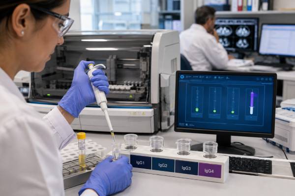
An IgG4 blood test measures one of the four subclasses of immunoglobulin G, the most abundant antibody class in blood. The test is best known for helping clinicians evaluate IgG4-related disease, an uncommon immune-mediated condition that can cause swelling, masses, inflammation, and progressive scarring in organs such as the pancreas, bile ducts, salivary glands, kidneys, lungs, or tissues around the aorta. A high result can support that diagnosis, but it cannot establish it. Many people with elevated IgG4 have another explanation, and some people with confirmed IgG4-related disease have a normal blood level. Interpretation therefore depends on the degree of elevation, symptoms, organ pattern, imaging, other laboratory results, and often a tissue biopsy. This article explains what the number means, why it may be high, how clinicians separate IgG4-related disease from cancer and other mimics, and how results are used during treatment and follow-up.
- IgG4 is a small fraction of total IgG, so the result must be read with the laboratory’s own reference interval.
- A mildly high level is nonspecific and occurs in allergic, infectious, autoimmune, liver, and malignant conditions.
- A markedly high result raises suspicion for IgG4-related disease but still does not prove it.
- Normal serum IgG4 does not completely exclude organ-limited or fibrotic IgG4-related disease.
- Diagnosis usually requires clinical, imaging, laboratory, and pathology findings to agree.
Table of Contents
- What the IgG4 blood test measures
- Normal range and how results are reported
- Why IgG4 levels rise
- Organ patterns of IgG4-related disease
- Why high IgG4 does not confirm the diagnosis
- Diagnostic workup, imaging, and biopsy
- Treatment, monitoring, and relapse
- When results or symptoms need prompt care
What the IgG4 blood test measures
Immunoglobulin G has four subclasses: IgG1, IgG2, IgG3, and IgG4. IgG4 normally makes up only a small proportion of total IgG. It is produced after repeated or prolonged immune stimulation and has unusual biological behavior. Compared with other antibody subclasses, it activates complement poorly and can exchange half-molecules with other IgG4 antibodies, creating functionally monovalent antibodies. These properties often make IgG4 relatively anti-inflammatory in ordinary immune responses. An elevated concentration is therefore a marker of immune activity, not proof that the IgG4 molecule itself is damaging tissue.
The test is usually performed on serum from a routine blood draw. A clinician may order IgG4 alone, but it is often included in an IgG subclass test. Total IgG, IgA, and IgM may also be measured with an immunoglobulin panel. These related tests show whether the abnormality is isolated to IgG4 or part of a broader increase in antibody production.
Common reasons to request serum IgG4 include:
- unexplained pancreatic enlargement, pancreatitis, or a pancreatic mass;
- narrowing or thickening of the bile ducts without a clear cause;
- persistent, usually painless enlargement of salivary or lacrimal glands;
- orbital swelling, eye protrusion, or an inflammatory orbital mass;
- kidney lesions, impaired kidney function, or low complement levels;
- retroperitoneal fibrosis, ureteral obstruction, or tissue surrounding the aorta;
- suspected multisystem IgG4-related disease; or
- follow-up of a person already diagnosed with the condition.
The blood test is not a population screening test. Ordering it without a compatible clinical question can identify incidental elevations that lead to anxiety and unnecessary testing. Its value is greatest when the organ presentation already resembles a recognized IgG4-related pattern.
IgG4-related disease is a systemic fibroinflammatory disorder. “Fibroinflammatory” means that immune-cell infiltration and inflammation occur together with fibrosis, or scar-like tissue deposition. The condition can be active and cellular early, then become more fibrotic over time. Once advanced scarring has damaged an organ, lowering the IgG4 level may not restore lost function. That is why the test is interpreted in the context of organ injury rather than as an isolated number.
Normal range and how results are reported
There is no single universal normal range for serum IgG4. Laboratories use different instruments, antibody reagents, calibration systems, populations, and units. One laboratory may report milligrams per deciliter (mg/dL), another milligrams per liter (mg/L), and another grams per liter (g/L). The conversions are straightforward: 1 g/L equals 100 mg/dL and 1 mg/dL equals 10 mg/L. Even so, the correct comparison is always the reference interval printed beside the patient’s result.
Adult upper limits commonly fall somewhere around 86 to 135 mg/dL, but the range can be wider depending on the laboratory. Age matters, particularly in children, because subclass concentrations change during immune development. A result should not be labeled high by comparing it with a range from a different laboratory or an adult internet reference.
The threshold of 135 mg/dL appears frequently in diagnostic literature and in the 2020 revised comprehensive diagnostic criteria for IgG4-related disease. It is a useful clinical reference point, not a universal definition of abnormality. A laboratory with an upper limit of 86 mg/dL, for example, would consider 120 mg/dL high even though it is below 135 mg/dL. Conversely, a laboratory whose validated upper limit is 140 mg/dL might not flag 135 mg/dL.
Clinicians often interpret the result by magnitude relative to the local upper limit:
- Within range: IgG4-related disease becomes less likely in some settings, but it is not excluded.
- Mildly elevated: This is common and nonspecific. The clinical context matters more than the flag.
- More than about twice the upper limit: The result is more supportive when typical organs are involved, although mimics remain possible.
- Very high or several-fold elevated: Suspicion increases further, especially with multisystem disease, but even extreme values are not diagnostic on their own.
The proportion of IgG4 relative to total IgG can add context. A person may have a high absolute IgG4 because all immunoglobulins are elevated, or may have a disproportionate IgG4 rise. Neither pattern is independently conclusive. Likewise, an apparently normal total IgG does not rule out an isolated IgG4 elevation.
Rarely, an extremely high concentration can cause antigen excess, or a prozone effect, and produce a falsely low assay result. When the clinical picture and number strongly conflict, the laboratory can retest the sample at additional dilutions.
A single value is only one point in time. Treatment and laboratory variation affect serial measurements, which should be interpreted with symptoms and organ-specific tests.
Why IgG4 levels rise
IgG4 can increase whenever the immune system is repeatedly stimulated in a way that favors class switching toward this subclass. The differential diagnosis is broad. The meaning of the result depends on whether the elevation is slight, persistent, disproportionate, associated with other immunoglobulin changes, or accompanied by a characteristic organ lesion.
Allergic and atopic disease can raise IgG4, often together with IgE and eosinophils. Asthma, eczema, allergic rhinitis, and chronic antigen exposure may produce mild or moderate elevations. An elevated IgG4 is not an allergy test and does not identify the trigger. Food-specific IgG4 testing is not a validated method for diagnosing food allergy or intolerance.
Chronic infection can stimulate polyclonal antibody production. Parasitic infections, chronic bacterial infections, and some fungal infections may increase IgG4. Travel history, exposure, immune status, fever, inflammatory markers, cultures, imaging, and pathogen-specific tests guide evaluation; the IgG4 number alone cannot distinguish infection from immune-mediated inflammation.
Autoimmune and inflammatory disorders can overlap with elevated IgG4. Examples include rheumatoid arthritis, systemic lupus erythematosus, inflammatory bowel disease, sarcoidosis, and some forms of vasculitis. Certain conditions may also contain increased IgG4-positive plasma cells in tissue, which is one reason pathology must be interpreted by an experienced pathologist rather than by cell counts alone.
Liver, biliary, and pancreatic disease may increase IgG4 even when classic IgG4-related disease is absent. Primary sclerosing cholangitis, cirrhosis, chronic pancreatitis, and pancreatic or biliary malignancies can produce elevations. This overlap is especially important because autoimmune pancreatitis and IgG4-related sclerosing cholangitis can closely resemble cancer or other biliary disorders.
Cancer and hematologic disease are less common but critical alternatives. Pancreatic adenocarcinoma, cholangiocarcinoma, lymphoma, plasma-cell disorders, and other malignancies may be associated with elevated serum IgG4 or IgG4-positive cells in a biopsy. When total protein or globulins are abnormal, clinicians may use serum protein electrophoresis and immunofixation to determine whether antibody production is broad and polyclonal or produced by a single clone.
IgG4-related disease becomes a stronger consideration when the elevation accompanies a typical combination of organ enlargement, wall thickening, mass-like lesions, fibrosis, or characteristic imaging. The disease often affects more than one site, sometimes at different times. A high value in a person with painless salivary swelling, autoimmune pancreatitis, and kidney lesions carries very different weight from the same value found incidentally in an otherwise well person with seasonal allergies.
Low IgG4 is usually less informative. Healthy people may have little measurable IgG4, and an isolated low value does not establish immune deficiency. Recurrent infections and functional antibody responses carry more weight.
Organ patterns of IgG4-related disease
IgG4-related disease can involve almost any organ, but several recurring patterns help clinicians recognize it. Symptoms are often subacute and surprisingly painless despite substantial enlargement or obstruction. Constitutional symptoms such as high fever and marked weight loss are less typical and should increase concern for infection, cancer, or another inflammatory disorder.
Pancreas and bile ducts. Type 1 autoimmune pancreatitis is the pancreatic form of IgG4-related disease. It may cause jaundice, abdominal discomfort, weight loss, new diabetes, or pancreatic enlargement discovered on imaging. Diffuse disease can create a “sausage-shaped” pancreas with a surrounding rim; focal disease can resemble pancreatic cancer. IgG4-related sclerosing cholangitis can narrow the bile ducts and cause jaundice, dark urine, pale stools, itching, or abnormal liver enzymes. Because pancreatic and biliary malignancies can appear similar, exclusion of cancer is essential before treatment.
Salivary, lacrimal, and orbital disease. Major salivary glands under the jaw or near the ears may enlarge, often symmetrically and without much pain. Lacrimal gland involvement can create swelling above or around the eyes. Orbital disease may involve extraocular muscles, nerves, or soft tissues, leading to eye protrusion, double vision, reduced vision, or pressure symptoms. Dry mouth or dry eyes can occur, but the pattern differs from classic Sjögren disease and requires targeted evaluation.
Kidneys. IgG4-related tubulointerstitial nephritis can cause impaired kidney function, patchy cortical lesions, renal enlargement, or protein in the urine. Complement C3 and C4 may be low during active renal disease. Because low complement has many causes, a C3 result or C4 result supports interpretation only when combined with kidney findings and the rest of the clinical picture.
Retroperitoneum, aorta, and arteries. Fibrous tissue can form around the abdominal aorta or ureters, a pattern called retroperitoneal fibrosis. Ureteral compression may silently obstruct urine flow and cause hydronephrosis or kidney failure. Inflammation may also occur around the aorta or other large arteries. These lesions can be discovered incidentally, but pain in the back, abdomen, or flank may occur.
Lungs and chest. Pulmonary involvement can produce nodules, airway changes, interstitial abnormalities, pleural disease, or enlarged lymph nodes. Many of these findings overlap with infection, sarcoidosis, lymphoma, and lung cancer. Symptoms range from none to cough, shortness of breath, or chest discomfort.
Lymph nodes and other sites. Lymphadenopathy is common but nonspecific. A lymph node biopsy alone is often difficult to interpret because many immune and malignant conditions can produce IgG4-positive plasma cells. Less frequent sites include the thyroid, meninges, pituitary gland, skin, prostate, mesentery, breast, and pericardium.
Organ patterns may cluster. One person may present with pancreatic and biliary disease; another with head-and-neck enlargement; another with retroperitoneal or vascular fibrosis. Serum IgG4 tends to be higher in extensive multisystem disease, while isolated fibrotic lesions may occur with a normal or only mildly elevated concentration. The organ map therefore matters as much as the blood value.
Why high IgG4 does not confirm the diagnosis
No serum IgG4 concentration has perfect sensitivity or specificity for IgG4-related disease. Sensitivity asks how often the test is high in people who truly have the disease; specificity asks how often it is not high in people who do not. Both vary across studies because patient populations, organ manifestations, thresholds, and assays differ.
A value just above the upper limit has a low positive predictive value when testing is performed broadly. In practical terms, most mildly high results do not turn out to represent IgG4-related disease. The probability rises as the concentration becomes more elevated and as typical organs are involved, but alternative diagnoses remain possible even at several times the upper limit.
A normal result also has limits. Serum IgG4 may be normal in a minority of people with confirmed disease, particularly when involvement is localized, predominantly fibrotic, or measured after corticosteroid treatment. A normal value should not end the evaluation when imaging and tissue findings strongly suggest the condition.
Tissue staining creates a related pitfall. Numerous IgG4-positive plasma cells can occur in inflammatory diseases, infections, and around tumors. The ratio of IgG4-positive to total IgG-positive plasma cells is useful, but neither the ratio nor an absolute cell count should be read in isolation. Required thresholds differ by organ and sample type. A small needle sample may miss diagnostic architecture, while lymph nodes often lack the more specific fibrotic changes.
Several mimics deserve deliberate exclusion:
- pancreatic cancer, cholangiocarcinoma, lymphoma, and other malignancies;
- primary sclerosing cholangitis and non-IgG4 chronic pancreatitis;
- Sjögren disease and other connective-tissue diseases;
- granulomatosis with polyangiitis and eosinophilic granulomatosis with polyangiitis;
- sarcoidosis, multicentric Castleman disease, and histiocytic disorders;
- bacterial, mycobacterial, fungal, or parasitic infection; and
- idiopathic or secondary retroperitoneal fibrosis.
Marked fever, neutrophilia, or a very high C-reactive protein concentration is not the usual pattern of IgG4-related disease and should prompt a search for infection or another inflammatory process. Likewise, night sweats, rapidly progressive weight loss, destructive bone lesions, or a single atypical mass may favor malignancy.
Classification criteria are designed mainly to create homogeneous groups for research. They can help clinicians organize evidence, but they are not substitutes for medical diagnosis. A person can have clinically confirmed IgG4-related disease without meeting a research classification score, and a person can resemble the criteria yet have a dangerous mimic. The safest interpretation asks whether all findings fit one coherent diagnosis and whether competing explanations have been adequately ruled out.
Diagnostic workup, imaging, and biopsy
Evaluation begins with the reason the test was ordered. A specialist reviews the time course, pain, gland swelling, jaundice, urinary symptoms, eye symptoms, breathing problems, allergic history, medication exposure, prior cancers, infections, and earlier imaging. Physical examination may identify salivary or lacrimal enlargement, lymph nodes, orbital changes, jaundice, or signs of organ dysfunction.
Laboratory testing is tailored to the affected site. It may include:
- complete blood count with eosinophils;
- metabolic panel, creatinine, and urinalysis;
- liver enzymes, bilirubin, alkaline phosphatase, amylase, or lipase;
- total IgG, IgG subclasses, IgE, and serum protein studies;
- C3 and C4 complement;
- C-reactive protein and erythrocyte sedimentation rate;
- autoantibodies for Sjögren disease or ANCA-associated vasculitis; and
- infection tests when exposure or imaging makes infection plausible.
Imaging defines the organ pattern and identifies a safe biopsy target. Ultrasound, CT, MRI, cholangiopancreatography, endoscopic ultrasound, or PET may be used. Broader imaging can reveal silent disease and a safer tissue target.
A biopsy is often central because it can support IgG4-related disease and, equally important, detect cancer or infection. The ideal target is an actively involved, accessible organ rather than an isolated lymph node. Core tissue is generally more informative than cytology because pathologists need to see architecture.
The classic microscopic pattern includes three major features:
- a dense lymphoplasmacytic infiltrate;
- fibrosis arranged in a whorled or “storiform” pattern; and
- obliterative phlebitis, in which inflamed fibrous tissue narrows or closes veins.
Immunostaining shows the number of IgG4-positive plasma cells and the proportion relative to all IgG-positive plasma cells. A ratio above about 40 percent can be supportive, but organ-specific standards and the overall tissue pattern determine its weight. Eosinophils may be present. Necrosis, prominent neutrophils, granulomas, or destructive vasculitis suggest other diagnoses.
Not every organ displays all three classic features. Early lesions may have inflammation with little fibrosis; late lesions may be scarred with few plasma cells. Salivary and lacrimal samples can look different from pancreatic or retroperitoneal tissue. Treatment before biopsy can reduce inflammatory findings. Expert pathology review is especially valuable when the sample is small or the location is unusual.
A “steroid trial” should not be used casually to diagnose a mass. Many inflammatory conditions improve with corticosteroids, and steroids can temporarily shrink lymphoma or obscure biopsy findings. When cancer or infection is possible, obtaining appropriate tissue and microbiological studies before immunosuppression is safer.
Treatment, monitoring, and relapse
Treatment is based on active disease, threatened organ function, accumulated damage, relapse risk, and treatment toxicity—not merely on whether serum IgG4 is high. Some asymptomatic, limited cases can be observed closely, but disease affecting the pancreas, bile ducts, kidneys, aorta, retroperitoneum, meninges, or orbit commonly requires therapy because obstruction or irreversible injury can develop.
Glucocorticoids have long been used to induce remission and often produce a rapid clinical and radiographic response. The dose and taper depend on organ severity, comorbidities, and local practice. Diabetes, osteoporosis, infection risk, cataracts, mood effects, hypertension, and other toxicities make prolonged steroid exposure undesirable.
B-cell-directed therapy is important because B cells and plasmablasts are central to the disease. Rituximab has been used as a steroid-sparing treatment, especially for relapsing or refractory disease, although regulatory status and access vary by country and indication. In 2025, the United States approved inebilizumab-cdon for adults with IgG4-related disease after a randomized trial showed a lower risk of treated flares than placebo. Treatment selection remains individualized and may involve rheumatology, gastroenterology, nephrology, hematology, ophthalmology, or other organ specialists.
Procedures may accompany immune therapy. Biliary or ureteral obstruction may need drainage or stenting, and severe vascular, orbital, or fibrotic complications may require surgical input. Immunosuppression may not reverse fixed scars.
Monitoring combines several domains:
- symptoms and physical examination;
- organ-specific laboratory markers, such as bilirubin, alkaline phosphatase, creatinine, and urine protein;
- repeat imaging when appropriate;
- serum IgG4 if it was elevated at baseline;
- IgE, eosinophils, and complement when initially abnormal; and
- medication safety tests and infection prevention.
Serum IgG4 often falls with successful therapy, but complete normalization is not required for remission. Some people remain clinically well with a persistently elevated level. Conversely, a rising concentration can precede relapse but does not prove that a flare is occurring. Escalating treatment solely because the number increased risks unnecessary toxicity. Clinicians look for corroborating symptoms, organ-function changes, examination findings, or imaging.
Relapse is common enough that follow-up continues after initial improvement. Higher baseline IgG4, elevated IgE, eosinophilia, low complement, and multiple involved organs have been associated with greater relapse risk, but none predicts an individual course with certainty. Early recognition matters because recurrent inflammation can add fibrosis and permanent damage.
Patients receiving immunosuppressive therapy may need vaccination review, screening for latent infections, bone protection, and monitoring for low immunoglobulins or infection complications. The plan should distinguish damage from active disease: persistent duct narrowing, gland atrophy, or kidney impairment may reflect old fibrosis rather than ongoing inflammation.
When results or symptoms need prompt care
A high IgG4 value by itself is rarely an emergency. Urgency comes from the symptoms and organs involved. Contact the ordering clinician promptly when a newly high result accompanies unexplained gland swelling, jaundice, worsening kidney function, a pancreatic or orbital mass, hydronephrosis, or imaging that shows inflammation around a major blood vessel.
Seek urgent medical assessment for:
- yellow skin or eyes with fever, chills, confusion, or severe abdominal pain;
- sudden or progressive loss of vision, severe eye pain, or rapidly increasing eye protrusion;
- inability to urinate, very low urine output, or severe flank pain;
- chest, abdominal, or back pain that is abrupt, intense, or associated with fainting;
- new weakness, severe headache, seizures, or other neurologic symptoms;
- shortness of breath, coughing blood, or low oxygen symptoms; or
- signs of serious infection while taking steroids or other immunosuppressive medicine.
Useful questions include how far the result exceeds the laboratory limit, whether total IgG is also high, which organ findings prompted testing, which mimics remain possible, whether biopsy is needed, and how improvement will be measured.
For an incidental mild elevation without symptoms or abnormal imaging, the next step is usually measured rather than urgent. The clinician may repeat the test, review allergies and infections, check total immunoglobulins and protein studies, or decide that no extensive workup is needed. Repeating the number without a clinical plan can create noise rather than clarity.
For a strongly compatible organ presentation, normal IgG4 should not provide false reassurance, and high IgG4 should not create diagnostic certainty. The central principle is clinicopathological correlation: the blood result, organ pattern, imaging, pathology, and exclusion of mimics must point in the same direction. That approach identifies people who need timely treatment while protecting others from an incorrect label and unnecessary immunosuppression.
References
- Current and future advances in practice: IgG4-related disease 2024 (Review)
- IgG4-Related Disease: Current and Future Insights into Pathological Diagnosis 2025 (Review)
- New Developments in the Treatment of IgG4-Related Disease 2025 (Review)
- Inebilizumab for Treatment of IgG4-Related Disease 2025 (Randomized Controlled Trial)
- Clinical Significance of Serum IgG4 in the Diagnosis and Therapeutic Monitoring of IgG4-Related Diseases 2023
- 2020 revised comprehensive diagnostic (RCD) criteria for IgG4-RD 2021 (Diagnostic Criteria)
Disclaimer
This article provides general educational information and does not diagnose IgG4-related disease or any other condition. Laboratory ranges, diagnostic pathways, and treatment options vary, so results should be interpreted by a qualified clinician who knows the symptoms, imaging, medications, and medical history. Seek urgent medical care for severe or rapidly worsening symptoms.





