Ptosis, or blepharoptosis, is a condition in which the upper eyelid droops. This drooping can occur in one or both eyes and can affect people of any age. Ptosis can be congenital (present at birth) or acquired later in life due to a variety of causes such as aging, trauma, or neurological disorders. The severity of ptosis varies, from a slight drooping to complete closure of the eyelid, which can impair vision and interfere with daily activities.
The condition results from dysfunction or weakness of the muscles responsible for lifting the eyelid, specifically the levator and Müller’s muscles. In some cases, nerve damage to the muscles’ control systems may be to blame. Patients with ptosis frequently have difficulty keeping their eyelids open, resulting in eye strain, fatigue, and, in severe cases, amblyopia (lazy eye) in children if left untreated.
Understanding ptosis is critical for accurate diagnosis and treatment. Identifying the underlying cause is critical because it influences the treatment plan. Early intervention can help people with this condition avoid complications and improve their quality of life. With advances in medical and surgical treatments, effective ptosis management is now possible, giving patients hope and better outcomes.
Standard Methods for Ptosis Management
The cause, severity, and age of the patient all influence the management and treatment of ptosis. The treatment aims to improve eyelid function, vision, and cosmetic appearance. There are both non-surgical and surgical options available, with treatments tailored to each individual’s needs.
Non-surgical Treatments
Nonsurgical approaches are typically considered for mild cases of ptosis or when surgery is not an option. Treatments include:
- Crutch Glasses: Special glasses with a bar or “crutch” attached can mechanically lift the eyelid, providing short-term relief from drooping. This is a non-invasive option, but it may not be appropriate for all patients.
- Eye Exercises: If ptosis is caused by muscle weakness or fatigue, specific eye exercises to strengthen the eyelid muscles may be recommended. However, the efficacy of this approach varies, and it is not a viable alternative to surgical intervention in moderate to severe cases.
Surgical Treatments
Surgery is the most common and effective treatment for ptosis, particularly when it impairs vision or appearance. There are several surgical techniques available, and the procedure used is determined by the severity of the ptosis and the underlying cause.
- Levator Resection: This procedure involves shortening or tightening the levator muscle to lift the eyelid. It is most commonly used when the levator muscle function is still partially intact. The surgery is performed through an incision in the eyelid crease, which reduces visible scarring.
- Müller’s Muscle-Conjunctival Resection (MMCR): This technique is used for mild to moderate ptosis in which the levator muscle function is reasonably good. Lifting the eyelid requires the removal of a portion of Müller’s muscle and the conjunctiva. This procedure is carried out from the inside of the eyelid, avoiding external incisions.
- Frontalis Sling: For severe ptosis or cases where the levator muscle is severely weak or non-functional, a frontalis sling procedure may be used. This technique connects the eyelid to the frontalis muscle (forehead muscle) with a sling made of autologous (patient’s own) tissue, synthetic material, or donor tissue. This allows the forehead muscle to lift the eyelid, making up for the weak levator muscle.
- Aponeurotic Repair: Aponeurotic repair is used to treat acquired ptosis caused by detachment or weakening of the levator aponeurosis (the tendon-like structure that connects the levator muscle to the eyelid). This procedure reattaches or tightens the levator aponeurosis to restore proper eyelid elevation.
Post-operative Care
Postoperative care is critical for achieving successful results after ptosis surgery. Patients are usually given antibiotic and anti-inflammatory eye drops to prevent infection and inflammation. Regular follow-up visits are scheduled to monitor healing and evaluate the surgery’s effectiveness. Temporary side effects such as swelling, bruising, and mild discomfort are common but usually go away within a few weeks.
Breakthrough Innovations in Ptosis Management
Recent advances in ptosis treatment have transformed the approach to treating this condition. Novel surgical techniques, advanced medical therapies, and improved diagnostic tools all contribute to increased treatment precision and effectiveness.
Minimal Invasive Surgical Techniques
Minimally invasive techniques have grown in popularity due to their shorter recovery times and lower risk of complications. These procedures use smaller incisions and advanced instruments to make precise surgical corrections with little tissue disruption.
- Endoscopic Ptosis Repair: Endoscopic procedures use small cameras and specialized instruments to perform surgery through small incisions. This method provides better visualization of the surgical area, allowing for more precise adjustments while lowering the risk of scarring. Endoscopic ptosis repair is especially useful for patients who are concerned about cosmetic results.
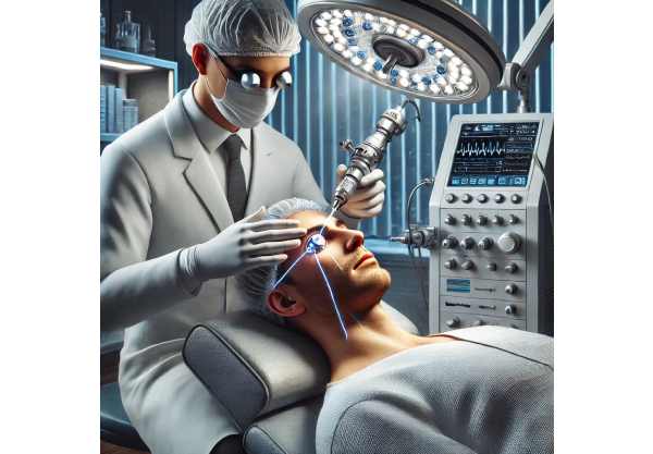
- Laser-Assisted Surgery: Ptosis surgery now uses laser technology to improve precision and reduce tissue damage. Laser-assisted techniques can be used to create incisions, coagulate blood vessels, and refine tissue removal, resulting in faster healing and better outcomes.
Advanced Eyelid Prosthetics
Eyelid prosthetic innovations have opened up new options for patients with severe ptosis or who are not candidates for surgery.
- Magnetic Eyelid Prosthetics: Small, discreet magnets have been used to help lift the eyelid. These systems can be integrated into glasses or custom-made prosthetic devices to provide a non-invasive solution for ptosis management.
- Electromechanical Eyelid Lifts: These devices use tiny motors and sensors to detect the position of the eyelid and provide mechanical assistance in lifting it. These devices are intended to be comfortable and simple to use, providing an alternative to traditional surgical procedures.
Gene Therapy & Regenerative Medicine
Emerging fields like gene therapy and regenerative medicine show promise for future ptosis treatments because they address the underlying causes of muscle and nerve dysfunction.
- Gene Editing Technologies: Techniques such as CRISPR-Cas9 are being investigated to correct genetic mutations that cause congenital ptosis. By targeting specific genes involved in muscle development and function, researchers hope to create therapies that restore normal muscle activity and prevent ptosis from occurring.
- Stem Cell Therapy: Stem cells can regenerate damaged muscle and nerve tissues. Experimental studies are looking into using stem cell injections to restore levator muscle function and improve eyelid elevation. While still in their early stages, these therapies may provide long-term solutions for ptosis management.
Pharmaceutical Advances
Pharmacological advances are opening up new possibilities for non-surgical ptosis treatment, particularly in cases caused by underlying medical conditions.
- Botulinum Toxin (Botox): Botox injections, which are commonly used cosmetically, have been shown to be effective in treating certain types of ptosis. Botox, which temporarily paralyzes specific muscles, can help lift the eyelid and improve its appearance. This treatment is commonly used for neurogenic ptosis caused by nerve dysfunction.
- Neuroprotective Agents: Neuroprotective drug research aims to prevent nerve damage while also promoting nerve regeneration. These agents could potentially be used to treat ptosis caused by nerve injuries or degenerative conditions, providing a non-surgical option for managing the condition.
Improved Diagnostic Tools
Accurate diagnosis is critical for successful ptosis management. Advances in diagnostic tools improve the ability to assess eyelid function and determine the best treatment approach.
- High-Resolution Imaging: Advanced imaging technologies such as optical coherence tomography (OCT) and high-resolution ultrasound allow for detailed views of the eyelid structures, including the levator muscle, aponeurosis, and surrounding tissues. These imaging techniques help with the precise diagnosis and planning of surgical procedures.
- Electrophysiological Testing: Electromyography (EMG) and nerve conduction studies are used to assess the function of the muscles and nerves that control eyelid movement. These tests help to determine the underlying cause of ptosis and guide treatment decisions.
Personalized Treatment Approaches
The trend toward personalized medicine is influencing ptosis treatment. Tailoring interventions to individual patient characteristics and needs improves outcomes and satisfaction.
- Customized Surgical Techniques: Surgeons are increasingly using patient-specific information, such as muscle strength and anatomical variations, to personalize surgical procedures. This approach ensures that the chosen technique is best suited to the patient’s specific condition.
- Patient-Centered Care Plans: Comprehensive care plans that consider patient preferences, lifestyle, and cosmetic concerns are becoming the norm. This comprehensive approach addresses not only the functional aspects of ptosis, but also the patient’s overall health and quality of life.


