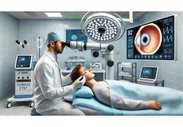
Dermoid cysts of the orbit are among the most common benign orbital tumors seen in children, though they may also present in adults. These slow-growing lesions arise from embryonic tissue trapped during facial development and can cause swelling, discomfort, or visual changes if left untreated. Early recognition and appropriate management are essential for preventing complications and ensuring the best possible outcomes. In this comprehensive, evidence-based guide, we explore everything from the nature and prevalence of orbital dermoid cysts to both conventional and advanced therapies, giving you the latest insights for optimal care and peace of mind.
Table of Contents
- Understanding Orbital Dermoid Cysts and Their Epidemiology
- Standard Non-Surgical and Medical Management Approaches
- Operative Procedures and Interventional Advances
- Cutting-Edge Treatments and Recent Technological Breakthroughs
- Upcoming Research and Future Directions
- Frequently Asked Questions
Understanding Orbital Dermoid Cysts and Their Epidemiology
Dermoid cysts in the orbit are benign, slow-growing, congenital tumors that develop when skin and skin structures (such as hair follicles or sebaceous glands) become trapped along the lines where embryonic facial bones fuse. While generally harmless, these cysts can gradually enlarge, leading to cosmetic concerns, pressure effects, or, in rare cases, vision changes.
Pathophysiology and Key Features:
- Origin: Dermoid cysts originate from ectodermal tissue, usually along bony sutures in the orbit.
- Location: Most frequently located in the superotemporal (outer upper) quadrant of the orbit, but can appear anywhere around the eye socket.
- Growth Pattern: Typically present at birth or early childhood, though some may remain unnoticed until adulthood due to slow expansion.
Epidemiology and Prevalence:
- Most common orbital tumor in children.
- Males and females are affected equally.
- Represents up to 46% of all orbital masses in the pediatric population.
Risk Factors:
- No clear risk factors aside from embryonic development.
- Rarely, trauma may make an existing cyst more apparent.
Symptoms and Clinical Presentation:
- Painless, slowly enlarging lump near the eyebrow or eyelid.
- Occasionally, redness or discomfort if the cyst ruptures.
- Rarely, double vision, bulging of the eye (proptosis), or decreased vision if the cyst grows inward.
Practical Advice:
If you or your child notice a painless, firm lump near the eye that is gradually growing, do not try to drain or squeeze it. Instead, consult an ophthalmologist for a proper evaluation, as improper handling can lead to inflammation or infection.
Standard Non-Surgical and Medical Management Approaches
While the majority of orbital dermoid cysts ultimately require surgical removal, some cases—especially in very young children or when the lesion is not causing symptoms—can be observed for a period before intervention.
Observation and Monitoring:
- Small, asymptomatic cysts can be monitored with regular check-ups and imaging.
- Indications for delaying surgery include:
- Child is too young for anesthesia.
- Cyst is stable and not causing discomfort or cosmetic issues.
- Family preference for watchful waiting.
Non-Surgical Symptom Relief:
- Warm Compresses: May help reduce local discomfort if minor swelling occurs, but will not shrink the cyst.
- Topical or Oral Antibiotics: Only if the cyst ruptures and there is secondary infection.
When Medical Therapy Is Not Sufficient:
- No medications can “dissolve” or shrink dermoid cysts permanently.
- Non-surgical options are supportive, not curative.
Parental Guidance and Home Tips:
- Never attempt home remedies or needle aspiration.
- Watch for signs of rapid growth, redness, tenderness, or discharge—these may signal rupture or infection and require prompt medical attention.
Limitations:
- Observation is only appropriate for carefully selected cases.
- The risk of sudden inflammation or rupture persists, especially if the cyst is traumatized.
Long-tail Keywords Used Here:
- observation of orbital dermoid cyst
- non-surgical management for eye cyst
- dermoid cyst home care
Practical Advice:
If you’re in a “wait-and-watch” period, keep regular appointments with your eye doctor and immediately report any changes in the lump’s appearance or symptoms.
Operative Procedures and Interventional Advances
For most dermoid cysts of the orbit, especially those that are enlarging, symptomatic, or cosmetically concerning, surgical removal is the gold standard. The primary goal is complete excision without rupture, as cyst content can cause a strong inflammatory response if released into surrounding tissues.
Surgical Techniques:
- Traditional Open Excision:
- An incision is made along a natural eyelid crease or eyebrow to minimize visible scarring.
- The cyst is carefully dissected and removed in one piece.
- Outpatient procedure; children typically require general anesthesia, while adults may have local anesthesia with sedation.
- Minimally Invasive Endoscopic Removal:
- Endoscopic tools allow for smaller incisions, improved visualization, and faster recovery.
- Especially beneficial for deep or medially located cysts.
- Microsurgical and Image-Guided Techniques:
- Use of surgical microscopes or intraoperative imaging to precisely locate and excise the cyst with minimal risk to surrounding structures.
Key Points in Surgery:
- Avoid rupture of the cyst wall to prevent inflammation.
- If the cyst is adherent to bone or deep tissue, the surgeon may remove a small amount of adjacent tissue.
- In rare cases, bone remodeling may be needed for larger or deeply embedded cysts.
Recovery and Aftercare:
- Swelling and mild bruising are common for 1–2 weeks.
- Cold compresses and elevation of the head help reduce swelling.
- Most patients resume normal activities in a week.
Risks and Complications:
- Infection, bleeding, scarring, incomplete removal, and rare recurrence.
- If the cyst ruptures, a granulomatous inflammatory reaction can occur, requiring medical or additional surgical treatment.
Long-tail Keywords Used Here:
- dermoid cyst eye surgery recovery
- endoscopic removal of orbital cyst
- risks of dermoid cyst excision
Practical Advice:
Choose a surgeon who specializes in oculoplastic or orbital surgery. Ask about the surgical plan, anesthesia, expected recovery time, and follow-up care to ensure peace of mind.
Cutting-Edge Treatments and Recent Technological Breakthroughs
Although the fundamentals of dermoid cyst treatment have remained consistent, the last several years have seen a surge in technological advancements that improve safety, precision, and cosmetic outcomes.
Recent Innovations:
- Intraoperative Navigation and Imaging:
- 3D imaging and navigation tools enhance localization of deep or complex cysts, reducing operative time and complications.
- Robotic-Assisted Surgery:
- Robotic platforms allow for ultra-precise dissection and are increasingly being explored for delicate orbital procedures.
- Bioengineered Sealants:
- Used to close the excision cavity and promote healing, minimizing scarring and infection risk.
- Minimally Invasive Devices:
- Next-generation endoscopes and microsurgical instruments provide surgeons with unparalleled control.
Emerging Adjuncts:
- Regenerative Medicine: Research into tissue engineering may one day help reconstruct orbital structures after cyst removal.
- Artificial Intelligence in Imaging: AI algorithms are improving preoperative diagnosis and surgical planning, leading to more tailored treatments.
Long-tail Keywords Used Here:
- robotic surgery for orbital dermoid cyst
- 3D imaging in eye tumor surgery
- AI for orbital cyst diagnosis
Practical Advice:
If you’re considering surgery for a dermoid cyst, ask your specialist about the technologies available at their center. Not all hospitals offer the latest innovations, but they may impact your surgical outcome and recovery.
Upcoming Research and Future Directions
The management of orbital dermoid cysts continues to evolve, with research focused on less invasive procedures, improved cosmetic results, and personalized patient care.
Active and Upcoming Clinical Trials:
- Advanced Imaging Studies: Trials assessing the utility of AI and advanced MRI or CT imaging to differentiate dermoid cysts from other orbital lesions more accurately.
- Scarless Surgical Techniques: Studies testing new suture materials, adhesives, and minimally invasive approaches that minimize external scarring.
- Tissue Engineering and Regenerative Approaches: Research into grafts and stem cell-based therapies to restore normal orbital anatomy after large cyst excisions.
- Patient-Reported Outcome Measures: Evaluating the impact of different treatments on cosmetic satisfaction, psychological well-being, and quality of life.
Trends in Future Care:
- Greater personalization in surgical planning based on individual anatomy, age, and cosmetic goals.
- Integration of virtual reality for preoperative simulation and patient education.
- Use of telemedicine and AI-assisted platforms for remote monitoring after surgery.
Long-tail Keywords Used Here:
- clinical trials in orbital dermoid cyst treatment
- future scarless eye surgery
- patient-centered outcomes in eye tumor care
Practical Advice:
If you’re interested in participating in a clinical trial, ask your surgeon or specialist about eligibility, potential benefits, and risks. Keeping up with new research can help you make informed choices about your care.
Frequently Asked Questions
What is a dermoid cyst of the orbit?
A dermoid cyst of the orbit is a benign, slow-growing lump near the eye caused by embryonic skin tissue trapped during development. It often appears near the eyebrow in children and may enlarge over time.
Does a dermoid cyst around the eye need to be removed?
Most dermoid cysts should be surgically removed if they are enlarging, causing symptoms, or are cosmetically concerning. Observation is possible for small, asymptomatic cysts, especially in young children.
Is dermoid cyst removal surgery safe?
Yes, when performed by an experienced oculoplastic or orbital surgeon, the procedure is very safe. Complications are rare but may include infection, bleeding, or scarring.
How long is recovery after orbital dermoid cyst surgery?
Most patients recover within 1–2 weeks. Mild swelling or bruising is common but subsides quickly. Most children and adults return to normal activities in a week.
Can a dermoid cyst come back after surgery?
Recurrence is rare if the cyst is completely removed. Incomplete removal or rupture during surgery may increase the risk of recurrence or inflammation.
What should I avoid if my child has a dermoid cyst near the eye?
Avoid squeezing, puncturing, or pressing on the lump. Trauma can cause rupture, inflammation, or infection. See a specialist for advice on management.
Are there non-surgical treatments for dermoid cysts of the orbit?
No, medications and home remedies cannot permanently remove a dermoid cyst. Surgery is the definitive treatment if removal is needed.
Disclaimer:
This guide is for educational purposes only and does not replace professional medical advice. Always consult a board-certified ophthalmologist or oculoplastic surgeon for personalized assessment and recommendations if you or your child has a lump near the eye.
If you found this article helpful, please share it on Facebook, X (formerly Twitter), or any other social platform you use. Your support helps us create and share more expert eye care content—don’t forget to follow us for updates and new resources!


