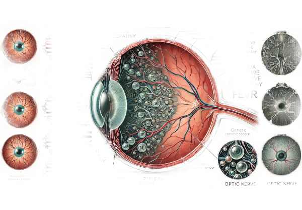
What is Familial Exudative Vitreoretinopathy?
Familial exudative vitreoretinopathy (FEVR) is a rare genetic disorder that affects the formation and function of blood vessels in the retina, the light-sensitive tissue at the back of the eye. This condition can cause vision problems ranging from mild impairment to total blindness. FEVR is frequently inherited and can manifest in a variety of ways, including within the same family. Symptoms can appear at any age, but they are most commonly identified during childhood. The severity and progression of FEVR varies greatly between individuals.
Detailed Investigation of Familial Exudative Vitreoretinopathy
Familial exudative vitreoretinopathy (FEVR) is a complex retinal vascular disorder characterized by peripheral retinal vascularization deficiencies. The disease is genetically heterogeneous, with multiple genes involved in its pathogenesis, resulting in a variety of inheritance patterns, including autosomal dominant, autosomal recessive, and X-linked recessive forms.
Gene Basis and Inheritance Patterns
FEVR is typically inherited in an autosomal dominant pattern, which means that only one copy of the mutated gene is required to cause the disorder. However, autosomal recessive and X-linked recessive forms do exist. The key genes associated with FEVR include:
- FZD4: This gene encodes a receptor for the Wnt signaling pathway, which is essential for retinal vascular development. Mutations in FZD4 prevent normal blood vessel formation in the retina.
- LRP5: This gene also functions in the Wnt signaling pathway. Mutations in LRP5 can impair retinal vascularization and cause bone density anomalies.
- NDP: Mutations in this gene, which encodes the protein Norrin, are the primary cause of the X-linked form of FEVR. Norrin is necessary for retinal and cochlear vascular development.
- TSPAN12: This gene regulates the Wnt signaling pathway and retinal angiogenesis. Mutations can impair the formation of normal retinal blood vessels.
Pathophysiology
The primary defect in FEVR is abnormal development of the retinal vasculature. During fetal development, blood vessels grow from the optic nerve head to the peripheral retina. FEVR disrupts this process, resulting in incomplete vascularization and peripheral avascular zones. The ischemic retina responds by producing vascular endothelial growth factor (VEGF), which promotes the development of abnormal, leaky blood vessels.
These abnormal vessels can cause a variety of complications:
- Exudation: Fluid leakage from these vessels can cause swelling of the retina and the accumulation of exudate.
- Hemorrhage: Fragile, abnormal vessels are more likely to bleed, causing additional retinal tissue damage.
- Fibrosis: The proliferation of fibrovascular tissue can cause retinal traction and the formation of folds or detachments.
Clinical Presentation
FEVR’s clinical presentation varies greatly, even among people who share the same genetic mutation. Some patients may be asymptomatic, while others develop severe visual impairment. Common symptoms include:
- Reduced Visual Acuity: This can range from minor blurring to complete vision loss.
- Floaters are small, dark shapes that move across the field of view.
- Photophobia: High sensitivity to light.
- Strabismus: Eye misalignment, which is commonly seen in children.
- Leukocoria: A white reflection of the retina seen in photographs.
Stages of FEVR
FEVR is classified into different stages depending on the severity of retinal changes:
- Stage 1: Peripheral avascularity with no additional complications. Often asymptomatic and detected during routine eye exams.
- Stage 2: Peripheral avascularity with extraretinal fibrovascular proliferation causes mild visual symptoms.
- Stage 3: Extraretinal fibrovascular proliferation occurs with traction, resulting in retinal folds or partial detachment.
- Stage 4: Partial retinal detachment affecting the macula, resulting in significant visual impairment.
- Stage 5: Total retinal detachment, which leads to severe vision loss or blindness.
Complications
FEVR can cause several serious complications that endanger vision.
- Retinal Detachment: One of the most serious complications, in which the retina peels away from the underlying support tissue, causing vision loss if not treated immediately.
- Vitreous Hemorrhage: Bleeding into the vitreous humor (the gel-like substance inside the eye) can impair vision and complicate surgical procedures.
- Neovascular Glaucoma: Abnormal blood vessels can form on the iris and obstruct the drainage of intraocular fluid, causing increased eye pressure and potential optic nerve damage.
- Macular Ectopia: Retinal traction can cause displacement of the macula, which is the central part of the retina responsible for detailed vision.
Epidemiology
FEVR is a rare condition, and its exact prevalence is unknown due to its variable clinical presentation and risk of underdiagnosis. It affects people of all ethnicities and genders equally. The onset can occur at any age, but it is most commonly discovered in childhood during routine eye exams or after the appearance of symptoms.
Differential Diagnosis
FEVR shares clinical features with several other retinal disorders, so differential diagnosis is essential.
- Retinopathy of Prematurity (ROP): Affects premature infants and causes similar retinal vascular abnormalities. However, ROP is linked to a history of prematurity and low birth weight.
- Coats’ Disease: A unilateral condition characterized by retinal telangiectasia and exudation, similar to FEVR but presenting unilaterally and sporadically.
- Persistent Fetal Vasculature (PFV): This congenital condition is characterized by the persistence of fetal vasculature, which results in similar retinal findings but is typically unilateral and associated with anterior segment anomalies.
- Norrie Disease: An X-linked disorder that causes retinal detachment and blindness. It frequently presents with similar retinal vascular changes to FEVR but is also accompanied by other systemic symptoms such as hearing loss and intellectual disability.
Prognosis
The prognosis for people with FEVR is determined by the severity of the disease at the time of diagnosis and the effectiveness of early intervention. Regular monitoring and timely treatment can help manage complications and save vision. However, advanced stages with significant retinal detachment or extensive fibrovascular proliferation are linked to a poorer visual outcome.
Diagnostic Approaches for Familial Exudative Vitreoretinopathy
To accurately diagnose Familial Exudative Vitreoretinopathy, a combination of clinical evaluations and advanced imaging techniques are used.
Clinical Examination
A comprehensive clinical examination by an ophthalmologist is the first step in diagnosing FEVR. This includes:
- Visual Acuity Testing: Evaluates the patient’s vision to determine the level of visual impairment.
- Fundus Examination: An ophthalmoscopic examination of the retina and vitreous. This may reveal peripheral avascular zones, retinal folds, and exudative changes.
- Family History: Because FEVR is inherited, detailed information about any family members with similar ocular conditions should be gathered.
Imaging Techniques
Several imaging modalities are used to provide detailed views of the retina and its vasculature.
- Fluorescein Angiography: In this technique, a fluorescent dye is injected into the bloodstream and images are taken as it travels through the retinal blood vessels. It aids in the detection of non-perfusion, abnormal vessel growth, and leakage.
Optical Coherence Tomography (OCT) is a non-invasive imaging test that produces high-resolution cross-sectional images of the retina. OCT is useful for detecting macular involvement, retinal thickening, tractional changes, and detachment. - Ultrasound B-Scan: When vitreous hemorrhage or dense cataracts obscure the retinal view, B-scan ultrasonography can reveal the posterior segment and detect retinal detachment or other structural abnormalities.
Genetic Testing
Genetic testing is critical in confirming the diagnosis of FEVR and determining the specific genetic mutation involved. This includes:
- Molecular Genetic Testing: Mutations in genes associated with FEVR, such as FZD4, LRP5, NDP, and TSPAN12, can help confirm the diagnosis. Affected families should seek genetic counseling to better understand their inheritance patterns and the implications for other family members.
Differential Diagnosis
Given the clinical similarities with other retinal conditions, differential diagnosis is critical for ruling out other potential causes. Additional testing and clinical evaluation may be required to distinguish FEVR from conditions such as retinopathy of prematurity, Coats’ disease, and persistent fetal vasculature.
Healthcare providers can treat patients using these diagnostic methods.
Advanced Therapies for Familial Exudative Vitreoretinopathy
Treatment for Familial Exudative Vitreoretinopathy (FEVR) focuses on symptom management, avoiding complications, and preserving vision. Given the variability in clinical presentation, treatment plans are frequently tailored to each patient’s specific needs.
- Observation: In mild or early stages (Stage 1 and some Stage 2 cases), where there are no significant symptoms or risk of progression, close observation and regular follow-ups may be advised.
- Laser Photocoagulation is a common treatment for avascular retina. The laser helps to seal off leaking blood vessels and prevent the formation of new abnormal vessels, lowering the risk of retinal detachment and other complications.
- Cryotherapy: In cases where laser treatment is not possible, cryotherapy (freezing therapy) can be used to treat peripheral retinal avascularity.
- Intravitreal Injections: Anti-VEGF (vascular endothelial growth factor) injections, such as bevacizumab (Avastin) or ranibizumab (Lucentis), are used to decrease neovascularization and exudation. These injections help to stabilize the retina, lowering the risk of further damage.
- Scleral Buckling: This surgical procedure repairs retinal detachments by indenting the eye’s wall, bringing it closer to the detached retina and allowing it to reattach.
- Vitrectomy: In advanced cases, vitrectomy surgery may be required. This involves removing the vitreous gel and any fibrovascular tissue that is causing traction on the retina. It is frequently paired with laser therapy or cryotherapy.
Innovative and Emerging Therapies
- Gene Therapy: Research into gene therapy seeks to correct the underlying genetic defects that cause FEVR. Although still in the experimental stage, early studies show promise for future treatment options.
- Stem Cell Therapy: Stem cell therapy is under investigation as a potential treatment for retinal diseases. The goal is to regenerate or replace damaged retinal cells, which could restore vision.
- New Drug Developments: Current research into new pharmacological treatments focuses on multiple pathways involved in retinal vascular development and maintenance. These include new anti-VEGF agents and other molecular targets.
- Retinal Implants and Prosthetics: Technological advancements have resulted in the development of retinal implants and prosthetics that can restore vision in people who have suffered severe retinal damage.
Best Practices to Prevent Familial Exudative Vitreoretinopathy
While Familial Exudative Vitreoretinopathy (FEVR) is a genetic condition that cannot be completely avoided, certain precautions can help manage the risk and detect early symptoms to avoid complications.
- Genetic Counseling: Families with a history of FEVR should seek genetic counseling to better understand their risk and explore potential genetic testing options.
- Regular Eye Examinations: Early detection through regular comprehensive eye exams, particularly for people with a family history of FEVR, can help manage the condition more effectively.
- Monitor for Symptoms: Be aware of any early symptoms, such as visual changes, floaters, or light sensitivity, and seek immediate medical attention if they occur.
- Healthy Lifestyle: Maintain a healthy lifestyle to support overall eye health, such as eating a balanced diet rich in antioxidants, exercising regularly, and not smoking.
- Protective Eyewear: Wear protective eyewear during activities that could cause eye injury, as trauma can aggravate retinal conditions.
- Education and Awareness: Inform family members about the symptoms of FEVR and the significance of early intervention.
- Adherence to Treatment Plans: Comply with prescribed treatment plans and attend all scheduled follow-up appointments to closely monitor the condition.
- Genetic Testing for At-Risk Individuals: Consider genetic testing for children and other at-risk family members to aid in early detection and treatment.
Individuals who follow these practices can help manage the risks associated with FEVR while also maintaining good ocular health.
Trusted Resources
Books
- “Inherited Retinal Disease: Diagnosis and Management” by Stephen H. Tsang
- “Retinal Vascular Disease” by A.M. Joussen, T.W. Gardner, B. Kirchhof, S.J. Ryan
Online Resources
- American Academy of Ophthalmology: AAO
- Retina International: Retina International
- National Eye Institute: NEI
- Genetic and Rare Diseases Information Center: GARD


