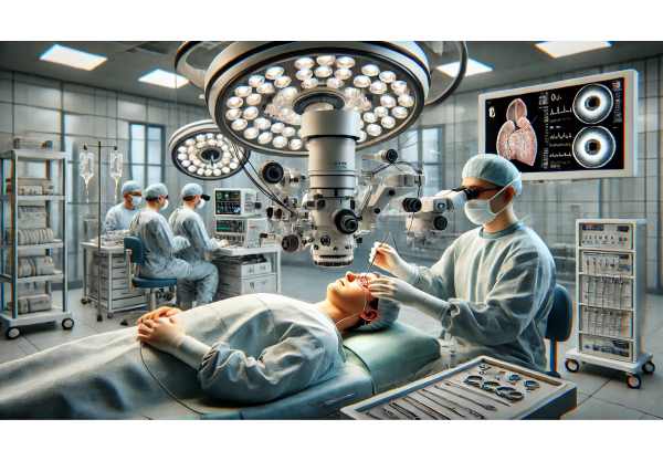
Globe rupture is among the most severe ocular emergencies, resulting from a full-thickness injury to the eye wall that threatens sight and, in some cases, the eye itself. Often caused by blunt or penetrating trauma, globe rupture requires urgent recognition, rapid stabilization, and specialized care to maximize the chance of saving vision. Understanding the mechanisms, identifying symptoms, and acting promptly can make the difference between recovery and permanent vision loss. This comprehensive guide explores the latest in globe rupture management, from time-tested treatments and surgical repair to new technologies and clinical trials reshaping ocular trauma care.
Table of Contents
- Introduction to Globe Rupture and Epidemiology
- Standard Medical and Supportive Therapies
- Surgical Techniques and Interventional Management
- Recent Innovations and Technological Advances
- Research Pipeline and Future Directions
- Frequently Asked Questions
- Disclaimer
Introduction to Globe Rupture and Epidemiology
Globe rupture refers to a full-thickness break in the outer walls of the eye (cornea and/or sclera), typically resulting from significant trauma. Unlike a superficial injury, a globe rupture means the eye’s structural integrity is lost, allowing internal contents (like vitreous, lens, or even retina) to potentially escape. Recognizing this vision-threatening emergency and seeking immediate care is vital.
Key Pathophysiology and Causes
- Mechanisms: Blunt trauma (e.g., sports injuries, falls, assaults) or penetrating objects (metal shards, wood, glass, etc.).
- Predisposing Factors: Previous eye surgery, thin corneal tissue, elderly (more fragile sclera), children (accidents).
- Where Ruptures Occur: Often at the weakest areas—behind muscle insertions, old surgical sites, or thinned tissue.
Epidemiology
- Incidence is higher in males and working-age adults due to occupational and sports injuries.
- Children and older adults are also vulnerable (play accidents, falls).
- Major cause of monocular blindness globally, especially in regions with limited eye trauma care.
Risk Factors
- Unprotected work in construction, agriculture, or metalwork.
- Lack of eye protection during risky activities.
- Underlying eye disease (degeneration, previous surgery).
Warning Signs & Symptoms
- Sudden pain and loss of vision.
- Misshapen (“collapsed”) eye or irregular pupil.
- Extrusion of eye contents, blood in the eye (hyphema), subconjunctival hemorrhage.
- Nausea/vomiting may occur with severe pain.
- Sometimes subtle—never try to manipulate an injured eye.
Prevention Strategies
- Always wear protective eyewear during high-risk activities.
- Educate children and workers about ocular safety.
- Address home hazards (sharp objects, tools) to protect vulnerable individuals.
Standard Medical and Supportive Therapies
Immediate medical attention is crucial in the management of globe rupture. While surgical intervention is almost always necessary, medical and supportive therapies help stabilize the eye and prevent further damage prior to surgery.
First Aid and Emergency Response
- Do NOT apply pressure: Never press on the injured eye.
- Protect the eye: Place a rigid shield or cup (not gauze) over the eye to prevent further trauma.
- Avoid manipulation: No attempts to remove foreign bodies or fluids.
- NPO Status: No food or drink in anticipation of urgent surgery.
- Immediate transfer: Expedite transport to an emergency room or eye trauma center.
Medical Stabilization
- Pain Control: Administer oral or IV analgesics as needed.
- Antiemetics: Prevent vomiting to avoid increased intraocular pressure.
- Tetanus Prophylaxis: Update if necessary.
- Antibiotics: Broad-spectrum intravenous antibiotics to reduce infection risk (endophthalmitis).
- Common regimens: vancomycin + ceftazidime.
- Sedation: Sometimes required for agitated patients or children.
Imaging and Assessment
- CT scan of orbits: Gold standard for detecting globe rupture and locating foreign bodies.
- Avoid ocular ultrasound: May worsen injury.
Special Considerations
- No eye drops or ointments prior to surgical assessment.
- Do not check eye pressure: This can force contents out.
Patient and Family Support
- Reassure patient and family, explaining each step.
- Offer practical advice: avoid coughing, bending, or straining.
- Prepare for surgical intervention and possible hospital stay.
Practical Prehospital Tips
- If unsure, treat any significant eye injury as a possible rupture and seek help immediately.
- Early action can be sight-saving.
Surgical Techniques and Interventional Management
Definitive treatment for globe rupture is surgical repair, aiming to restore the globe’s integrity, minimize infection risk, and preserve vision whenever possible.
Timing of Surgery
- Urgent surgery within hours is ideal.
- Delays increase risk of infection (endophthalmitis), loss of intraocular tissue, and worse outcomes.
Core Surgical Steps
- Anesthesia: General anesthesia is standard for pain control and safety.
- Wound Exploration: Precise identification of entry/exit wounds and extent of tissue damage.
- Debridement: Removal of devitalized tissue and foreign material.
- Closure: Layered suturing of cornea and/or sclera with fine, non-reactive sutures.
- Intraocular Content Management: Replace/reshape prolapsed tissue if viable, excise if not.
- Anterior Segment Repair: Address lens, iris, and anterior chamber structures.
- Posterior Segment Access: For injuries extending to retina/choroid, retina specialists may assist.
- Antibiotic Irrigation: May be used to further reduce infection risk.
Dealing with Complications
- Endophthalmitis: Aggressive intravitreal antibiotics.
- Retinal Detachment: Vitrectomy may be required.
- Sympathetic Ophthalmia: Rare but serious; may require steroids or, rarely, enucleation.
Secondary and Staged Procedures
- Further surgeries may be needed for lens replacement, scar revision, or retinal detachment.
Ocular Prosthesis
- If the eye cannot be saved, removal (enucleation) and later fitting with a prosthetic eye may be considered.
Postoperative Care
- Intensive antibiotic therapy.
- Pain and inflammation control (oral and topical medications).
- Protective eye shielding.
- Strict follow-up for visual rehabilitation and monitoring complications.
Rehabilitation
- Early vision support, low vision aids, and psychological counseling as needed.
Patient and Family Guidance
- Discuss realistic visual outcomes and need for possible further interventions.
- Support coping with trauma and visual changes.
Recent Innovations and Technological Advances
New developments are improving both surgical techniques and overall care in globe rupture management, promising better outcomes and safer interventions.
Diagnostic and Imaging Advances
- High-Resolution CT and MRI: Allow better visualization of tiny foreign bodies and subtle injuries.
- 3D Surgical Planning: Software assists in mapping complex wounds before surgery.
Bioengineered Materials
- Tissue Adhesives: New glues may temporarily seal wounds in field or military settings.
- Artificial Corneas and Scleral Patches: Bioengineered grafts can replace or reinforce damaged tissue.
- Stem Cell-Enhanced Patches: In experimental stages, these promote healing and reduce scarring.
Infection Prevention
- Antimicrobial Sutures: Release antibiotics to prevent infection.
- Advanced Intravitreal Antibiotics: Deliver longer-lasting infection protection.
Micro-Surgical Tools and Techniques
- Femtosecond Lasers: Enable ultra-precise corneal wound repairs (currently mostly experimental).
- Minimally Invasive Vitrectomy: Tiny instruments allow early intervention for posterior injuries.
Regenerative Medicine
- Stem Cell Therapies: Investigated for retinal and optic nerve regeneration after severe injury.
Telemedicine and Remote Consultation
- Mobile platforms help rural and military medics transmit images and receive expert advice rapidly.
Digital Patient Monitoring
- Wearable devices track healing, warn of infection, and alert doctors remotely.
Patient-Focused Innovations
- VR-based visual rehabilitation and counseling programs.
- Smartphone-based reminders and support for medication adherence.
Practical Advice for Patients and Families
- Ask your team about eligibility for new therapies or trials.
- Use digital tools for medication reminders and vision rehabilitation.
Research Pipeline and Future Directions
Current research is focused on not only improving globe repair but also on restoring vision and reducing trauma-related complications.
Key Areas of Research
- Retinal and Optic Nerve Regeneration: Stem cell and gene therapies to restore vision.
- Smart Prosthetics: Ocular implants with sensors for partial vision or eye movement.
- Anti-Fibrotic Agents: Medications to reduce scarring after surgery.
- Improved Biomaterials: Scleral and corneal substitutes closer to natural tissue.
- Real-Time Biosensors: Alert to infection, healing issues, or recurrent trauma.
Clinical Trials
- Ongoing studies are testing new surgical materials, infection prevention protocols, and rehabilitation strategies.
- Several international collaborations focus on telemedicine support and rapid triage in remote trauma settings.
Global Health Initiatives
- Expanding trauma centers and outreach to underserved regions.
- Education campaigns for prevention and rapid care-seeking.
Vision for the Future
- Personalized surgical planning using genetic and imaging data.
- Integration of artificial intelligence in trauma triage and surgical navigation.
- Hope for not only globe preservation but meaningful vision restoration after even severe injuries.
Staying Informed
- Patients and families should engage with support organizations and follow reputable sources for research updates.
Frequently Asked Questions
What is a globe rupture and how serious is it?
A globe rupture is a full-thickness wound of the eyeball, usually from trauma. It is a vision-threatening emergency requiring urgent surgery and expert care.
What are the first steps if globe rupture is suspected?
Do not touch or apply pressure to the eye. Shield it with a rigid cup, avoid food and drink, and seek immediate emergency medical care.
Can vision be saved after a globe rupture?
Timely surgical repair and medical care may preserve vision, but the outcome depends on injury severity and speed of treatment. Ongoing advances are improving success rates.
What complications can occur after globe rupture?
Infection, retinal detachment, sympathetic ophthalmia, and loss of the eye are possible. Close follow-up and prompt management of complications are essential.
How can globe rupture be prevented?
Wear protective eyewear during risky activities, educate children and workers, and remove hazards in the environment.
What new treatments or technology exist for globe rupture?
Innovations include tissue adhesives, bioengineered grafts, antimicrobial sutures, high-resolution imaging, and telemedicine for remote support.
Is globe rupture more common in certain populations?
Yes, it’s more common in males, the elderly, children, and those in high-risk occupations. Previous eye surgery or disease may also increase risk.
Disclaimer
This article is intended for educational purposes only and should not replace direct consultation with a medical professional. If you suspect an eye injury, seek immediate medical attention. For more information and updates, follow us on social media and please share this article to support our mission of providing trusted, expert eye health content.


