A foreign body in the eye can happen to anyone—whether you’re working outdoors, playing sports, or simply dusting your home. Even tiny particles like dust, metal, or wood fragments can cause discomfort, pain, and potentially serious complications if not managed promptly and correctly. Understanding the full spectrum of treatment and management—from first aid to surgical removal and the latest innovations—empowers you to act swiftly and seek proper care. In this comprehensive guide, we’ll explore the epidemiology, therapies, surgical solutions, emerging research, and practical advice for both patients and caregivers, ensuring clarity at every step.
Table of Contents
- Condition Basics and Epidemiology
- Non-Surgical and Medical Treatment Approaches
- Surgical Removal and Advanced Procedures
- Cutting-Edge Technologies and Innovative Solutions
- Ongoing Trials and Future Prospects
- Frequently Asked Questions
- Disclaimer
Condition Basics and Epidemiology
Foreign bodies in the eye are among the most frequent ocular emergencies, affecting all age groups and backgrounds. They can range from minor nuisances, like eyelashes or dust, to severe injuries caused by glass, metal, or plant material.
Definition and Types
- A foreign body refers to any object or material that becomes lodged on the surface of the eye (conjunctiva, cornea) or inside the eye (intraocular).
- Common examples: dust, sand, metal shavings, wood splinters, insects, and even contact lens fragments.
Pathophysiology
- The presence of a foreign body can trigger inflammation, cause corneal abrasions, introduce infection, or—if deeply embedded—result in scarring or vision loss.
- Superficial objects usually rest on the conjunctiva or cornea, while penetrating foreign bodies breach the globe (eyeball), requiring urgent intervention.
Epidemiology
- Incidence: Ocular foreign bodies are estimated to make up 35–45% of all eye-related emergency visits worldwide.
- Demographics: More common in males, often due to workplace accidents, especially in construction, metalworking, or agricultural settings.
- Children: Frequently affected by plant matter, sand, or craft materials during play.
Risk Factors
- Occupations involving grinding, hammering, or drilling without eye protection.
- Sports, cycling, and outdoor activities.
- Windy or dusty environments.
- Pre-existing dry eye or conditions that impair blink reflex.
- Inadequate use or absence of protective eyewear.
Prevention and Safety Tips
- Always wear approved eye protection during hazardous work or sports.
- Use safety goggles, face shields, or wraparound glasses.
- Keep work and play areas clean to minimize airborne debris.
- Teach children to avoid rubbing their eyes and supervise play with small, sharp objects.
Clinical Presentation
- Symptoms: Sudden eye pain, redness, tearing, light sensitivity, blurred vision, and the sensation of something “in the eye.”
- Signs: Visible object on the eye surface, scratch marks, eyelid swelling, or even visible bleeding if trauma is severe.
Early Recognition
- If you notice any persistent discomfort, do not ignore it. Even a tiny particle can lead to complications like infection or corneal ulcer if left untreated.
- Avoid rubbing the eye, as this may embed the foreign body deeper or cause additional injury.
Non-Surgical and Medical Treatment Approaches
Many cases of superficial foreign bodies in the eye can be managed safely and effectively without surgery, especially if addressed early.
First Aid and Immediate Steps
- Do not rub your eye.
- Wash hands thoroughly before attempting any intervention.
- Inspect the eye: In good lighting, gently pull down the lower lid and ask the person to look up, then up and look down.
- Rinse with saline: Use sterile saline or clean water to flush out loose particles. A gentle stream can dislodge many superficial objects.
- Blink repeatedly: Sometimes, the natural tear film can wash away small debris.
Do’s and Don’ts
- Do not attempt to remove embedded or sharp objects on your own.
- Do not use unsterilized objects (cotton swab, tissue) to dig out foreign bodies.
- Seek immediate medical attention if there’s persistent pain, reduced vision, or bleeding.
Medical Office Management
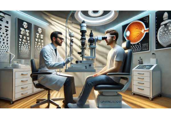
- Slit lamp examination: Ophthalmologists use specialized microscopes to identify and localize the foreign body.
- Fluorescein dye test: A special dye reveals abrasions and highlights the location.
- Removal techniques:
- Moistened cotton-tipped applicator: For superficial particles not embedded in the cornea.
- Eye irrigation: A steady flow of sterile saline.
- Eye spud or needle: For stubborn corneal foreign bodies—performed only by trained professionals.
Pharmacological Support
- Topical anesthetics: Used for pain control during examination or removal.
- Antibiotic eye drops/ointment: Prevents secondary infection, especially if the cornea is scratched.
- Lubricating drops: Promotes healing and reduces irritation.
- Tetanus prophylaxis: Considered if the object is metallic, organic, or if the vaccination is outdated.
Aftercare and Monitoring
- Follow-up visits to monitor healing, especially in corneal injuries.
- Watch for warning signs: increased redness, pain, discharge, or changes in vision.
Practical Advice
- Keep an emergency saline solution handy if you work in risky environments.
- Avoid using tap water for rinsing unless no other clean option is available.
- Educate children and coworkers about eye safety and immediate reporting of any injury.
Surgical Removal and Advanced Procedures
When a foreign body is deeply embedded, intraocular, or has caused significant trauma, surgical intervention becomes necessary.
Indications for Surgery
- Penetrating injuries or intraocular foreign bodies.
- Objects embedded in the cornea or sclera.
- Suspected plant material, as it increases risk of fungal infection.
- Associated complications: hyphema (blood in the anterior chamber), lens damage, or retinal involvement.
Types of Surgical Procedures
- Foreign body extraction under anesthesia: Local or general anesthesia is used, especially in children or anxious patients.
- Corneal/scleral repair: If the globe is ruptured, layered closure and repair of all involved tissues are critical.
- Anterior chamber washout: For cases where the foreign body or debris enters the front of the eye.
- Vitrectomy: A highly specialized surgery to remove foreign bodies from the vitreous or retina, often using microsurgical instruments and visualization.
Laser-Assisted Removal
- Nd\:YAG laser: Sometimes used to break up or dislodge small metallic particles embedded in the cornea.
- Femtosecond laser: Used in rare cases for precise, controlled removal in delicate areas.
Implantation and Repair
- Intraocular lens implants: In cases where the natural lens is damaged.
- Suturing: For full-thickness lacerations or to close the wound after removal.
Post-Surgical Care
- Eye patching or shield: To protect the healing eye and prevent accidental rubbing or trauma.
- Topical and systemic antibiotics: Reduce infection risk after surgery.
- Steroid drops: Control post-operative inflammation as prescribed.
- Regular follow-ups: To monitor healing, intraocular pressure, and vision.
Rehabilitation and Vision Recovery
- In some cases, vision therapy or low vision aids may be recommended during recovery.
- Early rehabilitation and adherence to post-op instructions dramatically improve outcomes.
Tips for a Smooth Recovery
- Avoid strenuous activities and swimming until cleared by your doctor.
- Use all prescribed medications exactly as directed.
- Attend every scheduled follow-up—even if you feel fine.
Cutting-Edge Technologies and Innovative Solutions
Technological progress is transforming the way ocular foreign bodies are managed, reducing risks and improving patient outcomes.
Modern Diagnostic Tools
- High-definition optical coherence tomography (OCT): Allows for precise localization of foreign bodies and assessment of eye structures.
- Confocal microscopy: Provides detailed imaging of corneal layers for complex injuries.
- Portable slit lamps: Used for field diagnosis in emergencies or remote settings.
Minimally Invasive Devices
- Micro-forceps and magnets: Specialized tools now allow for less traumatic removal of metallic objects.
- Disposable removal kits: Sterile, single-use kits are increasingly available for use in primary care and field settings.
AI-Powered Triage and Virtual Assessment
- Telemedicine: Smartphone cameras and AI-assisted platforms now allow rapid assessment and triage, guiding whether immediate ER referral or local management is needed.
- Automated detection algorithms: Ongoing research aims to train AI models to identify foreign bodies and complications in real-time using images.
Next-Generation Therapeutics
- Bioactive ocular dressings: Dressings that release antibiotics or healing factors to reduce infection and speed up recovery.
- Antimicrobial coatings: Development of antimicrobial surface coatings for contact lenses and surgical instruments reduces post-removal infections.
Smart Aftercare
- Digital medication reminders: Apps that alert you to administer drops on time.
- Remote monitoring: Wearable eye patches that track temperature or pressure and send updates to your healthcare team.
Practical Innovations
- Seek out clinics that offer digital follow-up for convenience.
- Ask about new technology if your case is complex or you have a history of poor healing.
Ongoing Trials and Future Prospects
Research continues to expand the frontiers of ocular foreign body management, promising safer, more effective solutions in the years ahead.
Current Clinical Trials
- Antimicrobial eye drops: Investigating new agents to better prevent post-removal infection.
- Biodegradable materials: Development of new sutureless patches and sealants for corneal repair.
- Gene therapy: Early research into gene-based approaches for promoting corneal regeneration after trauma.
- AI decision support: Multi-center studies evaluating the accuracy of AI-assisted triage and treatment recommendations.
Emerging Research Themes
- Nanotechnology: Nanoparticles designed to deliver antibiotics directly to the injury site.
- Regenerative medicine: Stem cell therapies aiming to heal severe corneal abrasions and restore clarity.
- Sensor technology: Contact lenses with embedded sensors to detect infection or elevated pressure after injury.
Future Directions
- Integration of AI and telehealth for real-time injury triage globally.
- Broader adoption of minimally invasive extraction techniques in rural and under-resourced settings.
- Continued innovation in patient self-monitoring and home-based recovery protocols.
How to Stay Informed
- Ask your ophthalmologist about ongoing clinical trials relevant to your injury or risk profile.
- Join patient education programs or online communities focused on eye safety and injury recovery.
Frequently Asked Questions
What should I do first if I get something in my eye?
Immediately rinse your eye with clean water or saline and avoid rubbing. If the object doesn’t come out or you have pain or blurred vision, seek professional help.
How is a foreign body in the eye removed?
Superficial objects can often be flushed out with saline or removed using a moistened cotton tip. Embedded or intraocular foreign bodies require removal by an eye specialist using specialized tools.
When should I seek emergency care for a foreign body in the eye?
Go to the emergency room if you have severe pain, vision loss, bleeding, or if the foreign body is metallic, sharp, or deeply embedded.
Can a foreign body in the eye cause long-term damage?
Yes, if not promptly removed, it can cause infection, scarring, corneal ulcer, or even vision loss. Early intervention is crucial for the best outcome.
What treatments help healing after foreign body removal?
Antibiotic drops, lubricating ointments, and sometimes steroid eye drops help prevent infection and promote healing. Always follow your doctor’s advice on medication use and follow-up care.
Are there any new treatments for eye injuries caused by foreign bodies?
Recent advances include bioactive dressings, antimicrobial agents, minimally invasive removal devices, and even AI-powered virtual triage systems for rapid assessment and referral.
Disclaimer
This article is provided for educational purposes only and is not a substitute for professional medical advice. Always consult an eye care professional or physician if you have questions about eye injuries, treatments, or if you suspect a foreign body in your eye.
If you found this guide useful, please share it with others on Facebook, X (formerly Twitter), or any platform you prefer. Support us by spreading reliable health information—your encouragement helps us create more expert resources for the community.


