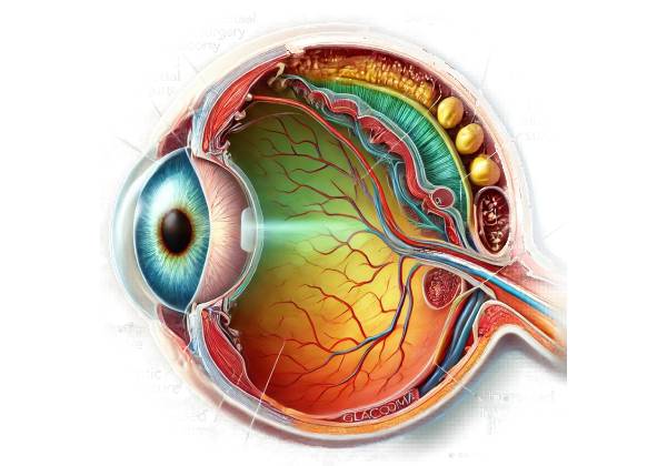
Post-vitreoretinal surgery glaucoma, also known as secondary glaucoma, is a condition characterized by high intraocular pressure (IOP) following vitreoretinal surgery. Vitreoretinal surgeries are a class of surgical procedures used to treat retina and vitreous humor disorders such as retinal detachment, macular holes, epiretinal membranes, and vitreous hemorrhage. While these surgeries are frequently successful in treating the underlying retinal issues, they can occasionally result in complications, such as the development of glaucoma.
Understanding Vitreoretinal Surgery
Vitreoretinal surgery includes a variety of procedures aimed at restoring or improving retinal function as well as addressing issues within the vitreous cavity. Common vitreoretinal surgeries include the following:
- Pars Plana Vitrectomy (PPV): This procedure removes the vitreous humor to improve access to the retina. Retinal detachments, vitreous hemorrhage, and macular holes are common treatments.
- Scleral Buckling is a technique for treating retinal detachments that involves placing a silicone band around the eye to indent the sclera and relieve traction on the retina.
- Pneumatic Retinopexy: This procedure involves injecting a gas bubble into the vitreous cavity to reattach the retina by pressing it against the eye’s wall.
- Laser Photocoagulation: Used to close retinal tears or destroy abnormal blood vessels in conditions such as diabetic retinopathy.
- Endolaser Photocoagulation: A laser treatment for retinal tears or proliferative diabetic retinopathy performed inside the eye during vitrectomy.
The Pathophysiology of Glaucoma Following Vitreoretinal Surgery
Glaucoma develops after vitreoretinal surgery through a variety of mechanisms that are influenced by the type of surgery performed, the presence of pre-existing ocular conditions, and the patient’s overall health. The key mechanisms include:
- Inflammation: Surgical trauma causes an inflammatory response, resulting in the release of inflammatory mediators, which can cause trabecular meshwork swelling and impaired aqueous humor outflow.
- Mechanical Obstruction: Surgical instruments, silicone oil, or gas bubbles used during surgery can obstruct the trabecular meshwork or the angle of the anterior chamber, resulting in elevated IOP.
- Scarring and Fibrosis: Postoperative healing can cause scarring and fibrosis around the trabecular meshwork, reducing its effectiveness in draining aqueous humor.
- Hemorrhage: Intraoperative or postoperative bleeding can obstruct the trabecular meshwork or Schlemm’s canal, reducing fluid outflow and increasing IOP.
- Gas and Silicone Oil Tamponade: The introduction of gas or silicone oil during procedures such as pneumatic retinopexy or vitrectomy can physically obstruct the aqueous outflow pathways.
Risk Factors
Several factors can increase the risk of developing glaucoma after vitreoretinal surgery:
- Pre-existing Glaucoma: Patients with a history of glaucoma are more vulnerable because of pre-existing damage to the trabecular meshwork and compromised outflow pathways.
- Inflammatory Ocular Conditions: Conditions such as uveitis can worsen postoperative inflammation, increasing the risk of glaucoma.
- Complex Surgeries: Longer or more complicated vitreoretinal surgeries are more likely to cause postoperative glaucoma.
- Age: Older patients are more vulnerable due to decreased regenerative capacity and the presence of other ocular comorbidities.
- Systemic Conditions: Diabetes and hypertension can slow healing and raise the risk of postoperative complications, such as glaucoma.
Clinical Presentation
The symptoms of post-vitreoretinal surgery glaucoma can differ depending on the severity and timing of the condition.
- Acute Postoperative Glaucoma: This type develops within a few days to weeks of surgery and can cause:
- Severe Eye Pain: Acute and sudden pain in the affected eye.
- Redness: Significant redness caused by inflammation or hemorrhage.
- Blurred Vision: Rapid loss of vision.
- Halos Around Lights: The perception of halos, particularly at nighttime.
- Nausea and Vomiting: These symptoms can occur in conjunction with severe IOP increases.
- Chronic Postoperative Glaucoma: This type develops slowly, over months or years, and may present with:
- Gradual Vision Loss: A progressive loss of peripheral vision.
- Mild to Moderate Eye Pain: Persistent discomfort, but less severe than in acute cases.
- Ocular Hypertension: Increased IOP detected during routine check-ups.
- No Symptoms: Many patients may remain asymptomatic until significant vision loss occurs.
Complications
If left untreated, post-vitreoretinal surgery glaucoma can cause a number of serious complications.
- Optic Nerve Damage: Prolonged elevated IOP can irreversibly damage the optic nerve, resulting in permanent vision loss.
- Corneal Edema: High IOP can swell the cornea, causing pain and blurred vision.
- Secondary Infections: Inflammation and mechanical blockages can make the eye vulnerable to secondary infections.
- Retinal Detachment: Severe inflammation or hemorrhage can cause retinal detachment, which is a vision-threatening condition.
Differential Diagnosis
To diagnose post-vitreoretinal surgery glaucoma, distinguish it from other causes of elevated IOP and similar ocular conditions:
- Primary Open-Angle Glaucoma is chronic and progressive, with no recent surgical history.
- Primary Angle-Closure Glaucoma: Acute onset, typically unrelated to recent surgery.
- Uveitis: Inflammation of the uveal tract can cause elevated IOP, but the causes vary.
- Ocular Hypertension: Elevated intraocular pressure (IOP) without optic nerve damage, usually without symptoms.
- Infectious Endophthalmitis: A post-surgical infection that causes pain, redness, and vision loss, but often includes signs of severe infection.
Epidemiology
The risk of post-vitreoretinal surgery glaucoma varies according to the type of surgery and the patient population. According to studies, it may occur in 5% to 15% of patients undergoing vitreoretinal surgery. It is more common in patients undergoing complex procedures, those with pre-existing glaucoma, and certain high-risk groups, such as the elderly or those with inflammatory eye conditions.
Prognosis
The prognosis for glaucoma following vitreoretinal surgery is determined by the timing of diagnosis and the efficacy of treatment. Early detection and treatment can prevent serious vision loss and improve outcomes. However, delayed diagnosis or inadequate treatment can result in permanent optic nerve damage and blindness.
Effects on Quality of Life
Glaucoma that develops after vitreoretinal surgery can have a significant impact on a patient’s quality of life. Acute cases can cause severe pain and immediate vision loss, whereas chronic cases can result in gradual but irreversible vision loss. The need for ongoing monitoring, as well as the possibility of multiple treatments or surgeries, can place a financial and emotional burden on patients.
Diagnostic methods
To confirm the diagnosis and understand the underlying cause, glaucoma after vitreoretinal surgery is diagnosed using a combination of clinical examination, imaging studies, and, in some cases, laboratory tests.
Clinical Examination
A comprehensive clinical examination is the first step in diagnosing post-vitreoretinal surgery glaucoma.
- Visual Acuity Test: This test determines how well a patient sees at different distances. It aids in determining how elevated IOP affects vision.
- Intraocular Pressure (IOP) Measurement: The eye care professional uses tonometry to measure the pressure within the eye. Elevated IOP is a strong indicator of glaucoma.
- Slit-Lamp Examination: This procedure uses a specialized microscope to examine the anterior and posterior segments of the eye. It can detect inflammation, hemorrhage, lens position, and other abnormalities.
- Gonioscopy: This procedure uses a special lens to examine the angle between the iris and the cornea, allowing for the detection of angle closure or drainage system abnormalities.
Imaging Studies
Imaging studies provide detailed views of the eye’s internal structures and aid in diagnosing and monitoring post-surgical glaucoma.
- Optical Coherence Tomography (OCT): OCT can produce high-resolution cross-sectional images of the retina and optic nerve. It is useful for detecting optic nerve damage and retinal changes caused by glaucoma.
- Ultrasound Biomicroscopy: This imaging technique uses high-frequency sound waves to visualize the anterior segment of the eye. It is especially useful for examining the ciliary body, angle structures, and any postoperative changes.
- Anterior Segment Optical Coherence Tomography (AS-OCT): This technique evaluates anterior segment structures, such as the angle and positioning of intraocular lenses.
Lab Tests
In some cases, laboratory tests may be required to identify underlying causes or associated systemic conditions.
- Blood Tests: Routine blood tests can detect systemic conditions such as diabetes or inflammatory diseases, which can lead to postoperative complications.
- Aqueous Humor Analysis: In cases of suspected infection or inflammation, a sample of aqueous humor may be tested for pathogens or inflammatory markers.
Treatment of Glaucoma Following Vitreoretinal Surgery
Managing post-vitreoretinal surgery glaucoma requires a multifaceted approach that includes medical treatments, laser procedures, and surgical interventions to control intraocular pressure (IOP) and prevent optic nerve damage. The severity of the condition, the underlying cause, and the patient’s overall health all influence the management strategy chosen.
Medical Management
- Topical Medications: The first line of treatment for post-vitreoretinal surgery glaucoma is usually eye drops to reduce IOP. These medications fall into several categories:
- Prostaglandin Analogues: Drugs such as latanoprost and bimatoprost increase the flow of aqueous humor, lowering IOP.
- Beta-Blockers: Drugs like timolol and betaxolol reduce the production of aqueous humor, which helps to lower IOP.
- Alpha Agonists: Brimonidine and apraclonidine decrease aqueous humor production while increasing its outflow.
- Carbonic Anhydrase Inhibitors: These drugs, available as eye drops (e.g., dorzolamide, brinzolamide) or oral medications (e.g., acetazolamide), reduce the production of aqueous humor.
- Rho Kinase Inhibitors: Netarsudil promotes aqueous humor outflow via the trabecular meshwork.
- Oral Medications: If topical medications are ineffective, oral carbonic anhydrase inhibitors such as acetazolamide may be prescribed to further reduce IOP.
- Anti-Inflammatory Agents: Nonsteroidal anti-inflammatory drugs (NSAIDs) or corticosteroids can be used to treat inflammation, which is a common cause of high IOP after surgery.
Laser Therapy
Laser procedures can be extremely effective in treating post-vitreoretinal surgery glaucoma.
- Laser Trabeculoplasty: This procedure utilizes a laser to improve aqueous humor drainage through the trabecular meshwork.
- Argon Laser Trabeculoplasty (ALT): This procedure uses an argon laser to create small burns in the trabecular meshwork, which improves fluid outflow.
- Selective Laser Trabeculoplasty (SLT): A low-energy laser that targets pigmented cells in the trabecular meshwork while minimizing damage to surrounding tissue.
- Laser Iridotomy: Used to treat angle-closure glaucoma, this procedure creates a small hole in the iris to improve aqueous fluid flow.
Surgical Management
Surgery is frequently required when medical and laser treatments fail to control IOP effectively:
- Trabeculectomy: This common surgical procedure creates a new drainage pathway by removing a portion of the trabecular meshwork. It allows aqueous humor to drain into the space beneath the conjunctiva, resulting in a bleb. Regular monitoring is required to ensure that the bleb remains operational.
- Glaucoma Drainage Devices: These devices, also known as tube shunts, help drain excess aqueous humor from the eye and into an external reservoir. Examples include the Ahmed valve, the Baerveldt implant, and the Molteno implant.
- Minimally Invasive Glaucoma Surgery (MIGS): MIGS procedures are less invasive than traditional surgeries and frequently used in conjunction with vitreoretinal surgery. Examples include the iStent, Hydrus Microstent, and Xen Gel Stent. These devices improve aqueous outflow by reducing complications and shortening recovery time.
- Cyclophotocoagulation: This procedure employs a laser to inhibit the ciliary body’s ability to produce aqueous humor. It is possible to perform it either externally (transscleral cyclophotocoagulation) or internally (endocyclophotocoagulation).
Post-operative Care and Monitoring
Effective postoperative care is critical for ensuring successful outcomes and avoiding complications.
- Medications: Typically, patients are given antibiotic and anti-inflammatory eye drops to prevent infection and reduce inflammation. It is critical to adhere to the ophthalmologist’s medication regimen.
- Follow-Up Visits: Regular follow-up visits are scheduled to monitor the healing process, check the IOL’s positioning, and evaluate visual acuity. These visits aid in the early detection and treatment of any complications, such as infection, elevated intraocular pressure, or lens displacement.
- Activity Restrictions: Patients should avoid strenuous activities, heavy lifting, and rubbing their eyes during the initial healing period. Protective eyewear may be prescribed to protect the eye from injury and bright light.
Lifestyle and Supportive Measures
- Diet and Exercise: A healthy lifestyle, which includes a well-balanced diet and regular exercise, can help to improve overall eye health. Patients should avoid activities that cause significant increases in IOP, such as heavy lifting and certain yoga positions.
- Protective Eyewear: Wearing protective eyewear during activities that could injure the eye is critical, especially after surgery.
Trusted Resources and Support
Books
- “Cataract Surgery: A Patient’s Guide to Treatment” by Robert S. Feder, MD
- This book provides comprehensive information on cataract surgery, postoperative care, and managing complications such as glaucoma.
- “Glaucoma: A Patient’s Guide to the Disease” by Graham E. Trope, MD
- An informative resource that covers various types of glaucoma, including secondary glaucoma, and offers insights into diagnosis and treatment options.
Organizations
- American Academy of Ophthalmology (AAO)
- Website: www.aao.org
- The AAO provides extensive resources on glaucoma, including patient education materials, research updates, and professional guidelines.
- Glaucoma Research Foundation (GRF)
- Website: www.glaucoma.org
- The GRF offers valuable information on glaucoma types, treatment options, and ongoing research, along with support resources for patients and caregivers.


