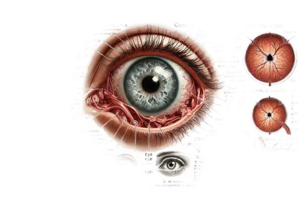
What is microphthalmia?
Microphthalmia is a congenital ocular condition in which one or both eyes are abnormally small and malformed. This condition can range in severity from slightly smaller-than-normal eyes to severely underdeveloped eyes, and it is sometimes associated with other ocular anomalies. Microphthalmia can occur alone or in conjunction with other systemic abnormalities. It is a significant cause of visual impairment and blindness in children, necessitating early detection and intervention to ensure optimal management and support.
Detailed Examination of Microphthalmia
Microphthalmia is a complex condition with a wide range of clinical manifestations and underlying causes. Understanding microphthalmia necessitates a thorough examination of its causes, genetic factors, associated syndromes, clinical manifestations, and implications for vision and overall health.
Etiology and Pathogenesis
Microphthalmia is caused by disruptions in the eye’s normal development during embryogenesis. Genetic mutations, chromosomal abnormalities, and environmental factors that disrupt ocular development can all contribute to the condition. Key points in the pathogenesis of microphthalmia are:
- Genetic Mutations: Microphthalmia has been associated with mutations in several genes. PAX6, SOX2, OTX2, and CHX10 are key genes for early eye development. Mutations in these genes can cause abnormal eye development and growth.
- Chromosomal Abnormalities: Microphthalmia can be caused by chromosomal anomalies such as trisomy 13, trisomy 18, or chromosome segment deletions or duplications. These chromosomal abnormalities frequently result in a variety of developmental issues, including microphthalmia.
- Environmental Factors: Certain environmental factors during pregnancy, such as infections (rubella, toxoplasmosis, cytomegalovirus), drugs, alcohol, or radiation, can disrupt normal eye development and result in microphthalmia.
Genetic Factors and Inheritance
Microphthalmia can be sporadic or inherited in a variety of ways, including autosomal dominant, autosomal recessive, or X-linked inheritance. Understanding the genetic basis is critical for making an accurate diagnosis, providing genetic counseling, and assessing future pregnancy risks.
- Autosomal Dominant Microphthalmia: In this type of inheritance, a single copy of the mutated gene from an affected parent is sufficient to cause the condition. Each pregnancy carries a 50% risk of passing on the condition to offspring.
- Autosomal Recessive Microphthalmia: The condition can only manifest in a child if both parents carry a copy of the mutated gene. Each pregnancy increases the risk of having an affected child by 25%, while the child has a 50% chance of becoming a carrier.
- X-Linked Microphthalmia: Caused by X chromosome mutations. Males are more severely affected because they only have one X chromosome, whereas females can be carriers with milder or no symptoms at all.
Associated syndromes
Microphthalmia may be part of a larger syndrome with systemic involvement. Common syndromes associated with microphthalmia are:
- CHARGE Syndrome: This syndrome encompasses a variety of congenital anomalies, such as coloboma (an eye structural defect), heart defects, choanal atresia, growth retardation, genital abnormalities, and ear anomalies.
- Lenz Microphthalmia Syndrome is a rare genetic disorder that causes severe microphthalmia or anophthalmia (the absence of one or both eyes), as well as other physical and developmental abnormalities.
- Aicardi Syndrome: This rare syndrome, which primarily affects females, is characterized by microphthalmia, brain abnormalities (agenesis of the corpus callosum), and seizures.
Clinical Manifestations
The clinical presentation of microphthalmia varies greatly according to its severity and associated anomalies. Key clinical characteristics include:
- Ocular Features: The affected eye(s) look smaller than usual. Other possible ocular abnormalities include coloboma, cataract, glaucoma, and retinal dysplasia. These anomalies can have a significant impact on vision.
- Visual Impairment: The degree of visual impairment varies from mild to severe, depending on the size and structure of the affected eye(s) and any associated ocular anomalies.
- Facial Asymmetry: Unilateral microphthalmia can cause the face to appear asymmetrical due to the size difference between the eyes.
- Systemic Features: Syndromic patients may also have systemic abnormalities affecting the heart, kidneys, central nervous system, and other organs.
Impact on Vision and Development
Microphthalmia has a significant impact on visual development and overall health. Children with this condition frequently encounter challenges related to:
- Visual Acuity: Reduced visual acuity is common, and severe cases may result in complete blindness in the affected eye(s).
- Binocular Vision: Unilateral microphthalmia can impair binocular vision, which affects depth perception and coordination.
- Psychosocial Development: Children with visible ocular anomalies may face social difficulties and require assistance to develop self-esteem and social skills.
- Educational Needs: Early intervention with visual aids, special education services, and supportive therapies is critical for improving learning and development.
Diagnostic methods
Accurate diagnosis of microphthalmia requires a combination of clinical examination, imaging studies, genetic testing, and consultation with various specialists. Early diagnosis is critical for effective treatment and intervention.
Clinical Examination
Diagnosing microphthalmia begins with a thorough clinical examination by an ophthalmologist. This includes:
- Visual Inspection: Examine the size and shape of the eyes, eyelids, and other facial structures. Asymmetry or obvious ocular abnormalities are present.
- Ophthalmoscopy: A thorough examination of the eye’s internal structures, such as the retina and optic nerve, for the presence of coloboma or other structural defects.
- Visual Acuity Testing: An evaluation of visual function to determine the degree of visual impairment.
Imaging Studies
Imaging studies can provide detailed information about the structure and development of the eyes.
- Ultrasound: Ocular ultrasound measures the axial length of the eye and evaluates internal structures. It is especially useful for determining the severity of microphthalmia and detecting associated anomalies.
- MRI and CT Scans: Magnetic resonance imaging (MRI) and computed tomography (CT) scans produce high-resolution images of the eye and its surrounding structures. These imaging techniques aid in detecting abnormalities in the orbit, optic nerve, and brain.
Genetic Testing
Genetic testing is critical in determining the underlying cause of microphthalmia:
- Karyotyping: Chromosomal analysis to detect large chromosomal abnormalities, such as trisomies or deletions, which may be associated with microphthalmia.
- Molecular Genetic Testing: Look for specific gene mutations that are known to cause microphthalmia. This includes the sequencing of genes like PAX6, SOX2, and OTX2, among others. Complex cases may warrant whole-exome or whole-genome sequencing.
- Family history and genetic counseling: A detailed family history can reveal information about the inheritance pattern and risk to other family members. Genetic counseling is essential for discussing the implications of genetic discoveries and providing assistance to affected families.
Multidisciplinary Evaluation
A comprehensive diagnosis frequently requires consultation with a variety of specialists, including:
- Pediatricians and Geneticists: To assess the child’s overall health and check for any associated systemic abnormalities.
- Neuroimaging Specialists: To look for brain abnormalities that could be associated with syndromic forms of microphthalmia.
- Oculoplastic Surgeons: For surgical evaluation and planning in cases where reconstructive surgery is required.
Effective Therapies for Microphthalmia
Microphthalmia treatment is multifaceted and tailored to the condition’s severity as well as any associated abnormalities. Treatment objectives include maximizing visual potential, addressing cosmetic concerns, and managing any underlying systemic conditions.
Surgical Interventions
- Orbital Expansion Surgery: In severe microphthalmia, especially when the eye socket is underdeveloped, orbital expansion surgery may be required. This involves using orbital expanders or other surgical techniques to stimulate eye socket growth and development.
- Enucleation and Prosthesis: If the affected eye is non-functional and causes significant cosmetic concerns or discomfort, enucleation (eye removal) and placement of an ocular prosthesis may be considered. This enhances facial symmetry and appearance.
- Reconstructive Surgery: If a patient has associated eyelid or facial abnormalities, reconstructive surgery may be required to correct the defects and improve function and appearance.
Visual Rehabilitation
- Corrective Lenses: Corrective lenses, such as glasses or contact lenses, can help people with partial vision reach their full visual potential. It is possible to use specialized lenses, such as those for correcting refractive errors or providing magnification.
- Low Vision Aids: Magnifiers, telescopic lenses, and electronic visual aids can help people with severe visual impairment perform daily tasks and improve their quality of life.
- Vision Therapy: Vision therapy consists of exercises and activities that aim to improve visual skills and processing. This can be especially beneficial for children with microphthalmia by improving their visual development and coordination.
Medical Management
- Glaucoma Treatment: Microphthalmia is frequently associated with glaucoma, a condition marked by high intraocular pressure that can harm the optic nerve. Medication to lower intraocular pressure may be used, as well as surgical interventions to improve fluid drainage.
- Cataract Management: Congenital cataracts are common in microphthalmia and may necessitate surgical intervention to improve vision. Early intervention is critical for preventing amblyopia (lazy eye).
- Anti-Inflammatory Medications: In cases of associated ocular inflammation, anti-inflammatory medications such as corticosteroids may be prescribed to alleviate symptoms and prevent complications.
Innovative and Emerging Therapies
- Gene Therapy: Research into gene therapy for microphthalmia is still ongoing. This novel approach aims to correct the underlying genetic defects that cause the condition. Gene therapy has the potential to help affected people regain normal eye development and function.
- Stem Cell Therapy: Stem cells are being studied as a possible treatment for microphthalmia. This approach uses stem cells to regenerate damaged ocular tissues and promote normal eye development.
- Regenerative Medicine: Advances in regenerative medicine are investigating the use of tissue engineering and biomaterials to reconstruct and repair ocular tissues damaged by microphthalmia. These therapies show potential for improving visual outcomes and cosmetic appearance.
Effective Ways to Improve and Avoid Microphthalmia
- Prenatal Care: Regular prenatal care is necessary to monitor the health of both the mother and the developing fetus. Prenatal screening can help identify potential risk factors for microphthalmia.
- Avoid Teratogenic Substances: Pregnant women should stay away from substances that are known to cause birth defects, such as alcohol, certain medications, and environmental toxins. Before taking any medications during pregnancy, consult a healthcare provider.
- Vaccination: Ensure that women of childbearing age receive vaccines against infections that can cause congenital anomalies, such as rubella. Vaccination helps prevent maternal infections that can cause microphthalmia in the developing fetus.
- Genetic Counseling: Families with a history of microphthalmia or other genetic disorders should consult a genetic counselor. This can aid in determining the risk of recurrence in subsequent pregnancies and providing information on available testing options.
- Healthy Lifestyle: Maintaining a healthy lifestyle, which includes a well-balanced diet high in essential nutrients, regular exercise, and quitting smoking, can benefit both maternal and fetal health.
- Regular Eye Examinations: Infants and children benefit from early and regular eye examinations to detect and manage microphthalmia and other ocular conditions. Early intervention is critical for achieving optimal visual outcomes.
- Awareness and Education: Raising awareness and providing education about microphthalmia and its risk factors can help prevent the condition while also ensuring timely diagnosis and treatment.
- Prenatal Screening: Prenatal testing for genetic and chromosomal abnormalities can aid in the identification of pregnancies at risk for microphthalmia. Early detection enables appropriate monitoring and intervention.
- Avoid Infections During Pregnancy: Pregnant women should take precautions to avoid infections that can harm fetal development, such as staying away from sick people and practicing good hygiene.
Trusted Resources
Books
- “Genetics and Eye Diseases: An Overview” by Robert W. Massof and G. E. Leguire
- “Ocular Development and Genetics” by Ann T. Gronowski and E. Y. Song
- “Congenital Anomalies of the Eye” by Zeynel A. Karcioglu


