Definition of Secondary Acquired Lacrimal Duct Obstruction
Secondary acquired lacrimal duct obstruction (SALDO) is a condition in which the tear drainage system of the eye becomes clogged due to an external factor or underlying disease, causing excessive tearing (epiphora) and recurrent eye infections. This condition is distinct from primary acquired nasolacrimal duct obstruction, which occurs without a known external cause. SALDO can have a significant impact on quality of life, causing discomfort, blurred vision, and potential complications like chronic dacryocystitis (lacrimal sac infection).
The lacrimal system consists of the lacrimal glands, which produce tears, and the lacrimal drainage pathways, which include the puncta, canaliculi, lacrimal sac, and nasolacrimal duct, which transport tears from the eye surface to the nasal cavity. The obstruction in SALDO typically occurs in the nasolacrimal duct, but it can affect any part of the drainage system.
Trauma, inflammation, infections, tumors, and iatrogenic factors like complications from surgery or radiation therapy are all common causes of SALDO. Chronic conditions such as sarcoidosis, Wegener’s granulomatosis, and other systemic inflammatory diseases can also result in secondary obstruction. Symptoms frequently include persistent tearing, recurring eye infections, pain, swelling around the eye, and discharge.
A detailed patient history, clinical examination, and imaging studies are all required to diagnose SALDO. Techniques like dacryocystography, dacryoscintigraphy, and nasal endoscopy aid in determining the location and cause of the obstruction, allowing for appropriate treatment.
Traditional Management and treatment of secondary acquired lacrimal duct obstruction
Secondary acquired lacrimal duct obstruction requires a multifaceted approach based on the underlying cause and severity of the obstruction. The treatment aims to restore normal tear drainage, alleviate symptoms, and prevent complications.
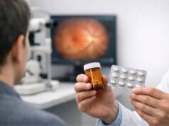
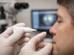
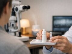
- Conservative Management: For mild cases, initial treatment may include warm compresses, massage, and topical antibiotics or anti-inflammatory medications to control infection and inflammation. These methods can alleviate symptoms and clear minor obstructions.
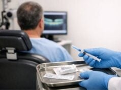
- Probing and Irrigation: The lacrimal drainage system is routinely probed and irrigated for diagnostic and therapeutic purposes. Probing entails inserting a thin, flexible instrument through the punctum and canaliculi to clear the blockage, followed by saline or antibiotic irrigation. This procedure may be effective for partial obstructions or early-stage disease.
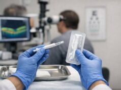
- Dilation and Stenting: Dilation of the nasolacrimal duct with increasingly larger probes or balloons can aid in the opening of narrowed passages. Stenting is the process of inserting a small tube or stent into the lacrimal system to keep it open and prevent further obstruction. Silicone stents are frequently used and kept in place for several months to allow the tissues around them to heal.
- Dacryocystorhinostomy (DCR): DCR is the preferred surgical procedure for treating nasolacrimal duct obstructions that do not respond to conservative treatment. This surgery opens up a new drainage pathway between the lacrimal sac and the nasal cavity, bypassing the obstructed nasolacrimal duct. DCR can be performed using an external approach, which involves making an incision on the side of the nose, or an endonasal approach, which employs nasal endoscopy. Endonasal DCR is a less invasive procedure with shorter recovery times and no visible scars.
- Conjunctivodacryocystorhinostomy (CDCR): CDCR is a specialized procedure performed when the canaliculi are severely scarred or obstructed. It entails creating a new drainage pathway from the conjunctiva (the membrane that lines the eyelids) to the nasal cavity, which is typically done with a glass tube known as a Jones tube. This procedure is usually reserved for complex or recurring cases.
- Adjunctive Therapies: Depending on the root cause of the obstruction, additional treatments may be required. For example, treating systemic inflammatory diseases with corticosteroids or immunosuppressive agents can help reduce inflammation and prevent recurrence. Antifungal or antiviral medications may be required to treat infections caused by specific pathogens.
- Regular Monitoring and Follow-Up: Continuous monitoring by an ophthalmologist or oculoplastic surgeon is required for patients with SALDO. Regular follow-up visits enable the evaluation of treatment efficacy, early detection of complications, and timely changes to the management plan.
Innovative Strategies for Treating Secondary Acquired Lacrimal Duct Obstructions
Recent advances in the understanding and treatment of secondary acquired lacrimal duct obstruction have resulted in novel therapies and diagnostic tools that are transforming the condition’s management. These cutting-edge innovations aim to improve patient outcomes, shorten recovery times, and increase treatment precision and effectiveness.
Advanced Imaging and Diagnostic Techniques
SALDO management requires accurate and early diagnosis. Imaging technology advancements enhance the ability to visualize and assess the extent of lacrimal duct obstructions, allowing for more precise treatment planning.
- High-Resolution Dacryocystography: High-resolution dacryocystography employs contrast media and advanced imaging techniques to produce detailed images of the lacrimal drainage system. This enables precise localization of the obstruction and evaluation of the surrounding anatomical structures, guiding targeted interventions.
- Optical Coherence Tomography (OCT): OCT is a non-invasive imaging technique for obtaining high-resolution cross-sectional images of the lacrimal system. Enhanced depth imaging OCT provides detailed visualization of the lacrimal sac and nasolacrimal duct, enabling precise assessment of obstructions and tissue changes. OCT can track treatment progress and detect early signs of recurrence.
- Ultrasound Biomicroscopy (UBM): UBM uses high-frequency ultrasound to create detailed images of the eye’s anterior segment, including the lacrimal drainage system. This technique is especially useful for assessing the canaliculi and proximal nasolacrimal duct, which provides important information for surgical planning.
Minimal Invasive Surgical Techniques
Surgeons are developing less invasive techniques for treating SALDO, reducing recovery times and improving patient comfort.
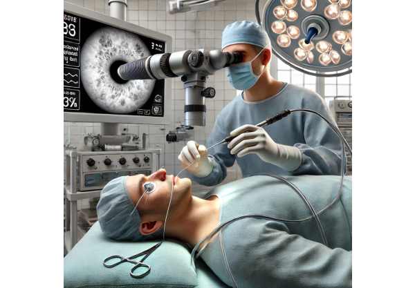
- Endoscopic Dacryocystorhinostomy (Endo-DCR): Endo-DCR is a minimally invasive procedure that employs nasal endoscopy to establish a new drainage pathway between the lacrimal sac and the nasal cavity. This method avoids external incisions, resulting in no visible scars and a faster recovery. Advances in endoscopic instruments and imaging technologies are improving Endo-DCR precision and success rates.
- Balloon Dacryoplasty: A catheter containing an inflatable balloon is used to dilate the obstructed nasolacrimal duct. This technique, which can be performed with local anesthesia, is less invasive than traditional surgical methods. Balloon dacryoplasty is especially useful for partial obstructions or in situations where traditional surgery is not an option.
- Microendoscopic Surgery: Microendoscopic techniques use miniature endoscopes and instruments to diagnose and treat lacrimal drainage system obstructions. These techniques enable the precise removal of obstructions and placement of stents, reducing tissue trauma and improving results.
Advanced Stenting Technology
Advanced stenting technologies are opening up new options for keeping the lacrimal drainage system open and preventing re-obstruction.
- Drug-Eluting Stents: Drug-eluting stents deliver medications, such as anti-inflammatory or antifibrotic agents, directly into the tissues. These stents reduce inflammation and scar tissue formation, increasing the long-term success rate of stenting procedures. Drug-eluting stents are especially beneficial for patients with a high risk of recurrence.
2) Bioabsorbable Stents: Bioabsorbable stents are made of materials that dissolve slowly and are absorbed by the body over time. These stents provide temporary support for the lacrimal drainage system, lowering the risk of long-term complications that come with permanent stents. Bioabsorbable stents are useful for patients who need temporary stenting to allow for tissue healing.
Regenerative Medicine and Tissue Engineering
Advances in regenerative medicine and tissue engineering are providing innovative solutions for repairing and regenerating damaged lacrimal tissues in SALDO patients.
- Stem Cell Therapy: Stem cells have the ability to regenerate damaged lacrimal duct tissues and restore normal function. Mesenchymal stem cells (MSCs) possess immunomodulatory and regenerative properties, making them a promising treatment for lacrimal duct obstructions. Preclinical studies have demonstrated that MSCs can improve tissue repair and reduce inflammation. Clinical trials are currently underway to determine the safety and efficacy of stem cell therapy in SALDO patients.
- Bioengineered Tissues: Tissue engineering techniques are being used to create bioengineered lacrimal duct tissues that have the same structural and functional properties as natural tissues. These bioengineered tissues have the potential to repair or replace damaged sections of the lacrimal drainage system, providing a long-term and effective solution for patients with severe obstructions.

