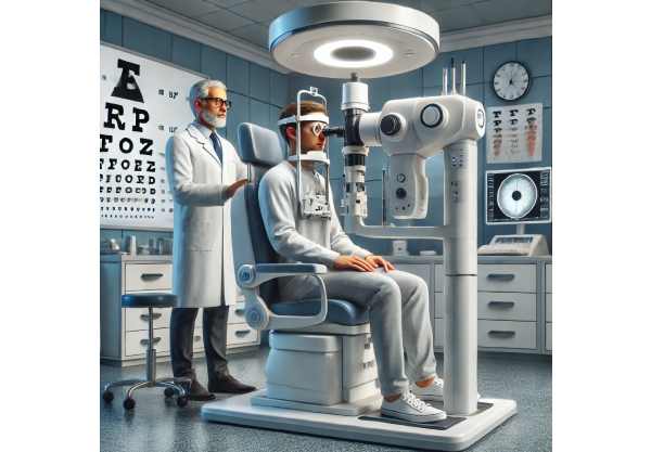
Optic disc pallor is a pale appearance of the optic disc, the visible portion of the optic nerve located at the back of the eye. This condition indicates optic nerve damage or atrophy, which can be caused by a variety of underlying conditions including optic neuritis, ischemic optic neuropathy, glaucoma, or trauma. The optic disc is typically pinkish-orange due to its abundant blood supply. However, when nerve fibers degenerate, the disc loses color and appears pale, which is a significant clinical finding during an eye exam.
The presentation of optic disc pallor can differ, with patients experiencing a variety of symptoms depending on the severity and underlying cause. Common symptoms include decreased visual acuity, visual field defects, and difficulties with color perception. A comprehensive eye examination is usually required to diagnose, which includes fundoscopy to directly observe the optic disc, visual field testing to assess the extent of vision loss, and imaging studies such as optical coherence tomography (OCT) and magnetic resonance imaging (MRI) to evaluate the optic nerve and surrounding structures. Understanding the underlying cause of optic disc pallor is critical for effective treatment and management, as it can have a significant impact on the patient’s visual outcome.
Standard Optic Disc Pallor Management
The primary goals of managing and treating optic disc pallor are to address the underlying cause of the condition, prevent further optic nerve damage, and improve the patient’s remaining vision. The following are the standard treatment methods for managing optic disc pallor:
- Addressing the Underlying Cause: The first step in treating optic disc pallor is to identify and treat the underlying condition that caused the optic nerve injury. For example, if optic neuritis is the cause, corticosteroids or immunosuppressive medications may be prescribed to reduce inflammation. To avoid further vascular damage in cases of ischemic optic neuropathy, systemic conditions such as hypertension and diabetes must be managed.
- Visual Rehabilitation: Patients who have significant vision loss due to optic disc pallor may benefit from visual rehabilitation services. These services include training in the use of low-vision aids, adaptive technologies, and strategies for maximizing residual vision. Rehabilitation programs are tailored to each patient’s specific needs, allowing them to maintain independence and improve their quality of life.
- Glaucoma Management: If glaucoma is the underlying cause of optic disc pallor, lowering intraocular pressure is critical to preventing additional optic nerve damage. Glaucoma treatment options include medications (prostaglandin analogs and beta-blockers), laser therapy (selective laser trabeculoplasty), and surgical interventions (trabeculectomy or glaucoma drainage implants).
- Neuroprotective Therapies: Neuroprotective agents are intended to protect the optic nerve from further damage and promote its survival. While these treatments are still being investigated, some studies indicate that medications such as brimonidine or citicoline may provide neuroprotective benefits in optic neuropathies.
- Monitoring and Follow-Up: Patients with optic disc pallor should schedule regular follow-up appointments to monitor the condition’s progression and treatment effectiveness. Visual field testing, OCT, and other imaging studies are frequently repeated to monitor changes in the optic nerve and adjust treatment plans accordingly.
- Lifestyle Modifications: Encouraging patients to lead a healthy lifestyle can benefit their overall ocular health. Maintaining a balanced, antioxidant-rich diet, getting regular exercise, quitting smoking, and managing systemic health conditions like hypertension and diabetes are all recommended.
Advanced Treatments for Optic Disc Pallor
Recent advances in the understanding and treatment of optic disc pallor have resulted in novel approaches that provide new hope to patients. These cutting-edge innovations include advanced imaging techniques, genetic research, neuroprotective therapies, regenerative medicine, and integrated care models. Each of these innovations provides distinct advantages and potential for improving the management of optic disc pallor.
Advanced Imaging Techniques
Imaging technology advances have made it much easier to diagnose and monitor optic disc pallor. High-resolution imaging modalities enable detailed visualization of the optic nerve and its surrounding structures, allowing for early detection and precise assessment of nerve damage.
Optical Coherence Tomography Angiogram (OCTA): OCTA is a non-invasive imaging technique that produces detailed images of the retina and choroidal vasculature. It aids in detecting vascular changes associated with optic nerve damage, such as decreased blood flow or neovascularization. OCTA improves the ability to track the progression of optic disc pallor and identify complications early on.
Enhanced Depth Imaging-OCT(EDI-OCT): EDI-OCT provides better visualization of deeper structures within the optic nerve head, making it especially useful for detecting optic nerve atrophy. This advanced imaging method allows for a better understanding of the extent of nerve damage, resulting in more accurate diagnosis and management.
Magnetic Resonance Imaging (MRI): MRI is useful for evaluating the optic nerve and brain, particularly when optic disc pallor is suspected to be caused by neurological conditions like multiple sclerosis or tumors. High-resolution MRI can detect subtle changes in the optic nerve and surrounding tissues, helping to identify the underlying cause.
Genetic Research and Therapy
Genetic research has shed light on the hereditary nature of certain conditions that cause optic disc pallor. Understanding the genetic basis opens up new avenues for targeted therapies and personalized medicine.
Genetic Testing: Identifying specific genetic mutations linked to optic neuropathies can aid in predicting the likelihood of developing optic disc pallor and understanding its progression. Genetic testing allows for early diagnosis in family members and guides personalized monitoring and treatment plans.
Gene Therapy: Gene therapy shows promise in treating genetic optic neuropathies that cause optic disc pallor. Gene therapy aims to halt or reverse the pathological processes that cause optic nerve damage by addressing the underlying genetic defects. Ongoing research in this field may eventually lead to viable treatment options for patients who are genetically predisposed to optic disc pallor.
Neuroprotective Therapies
Neuroprotective therapies aim to preserve the function of the optic nerve and retinal ganglion cells, potentially slowing the progression of vision loss caused by optic disc pallor.
Neurotrophic Factors: Neurotrophic factors, including BDNF and CNTF, are essential for neural cell survival and function. The goal of studying how to administer these factors is to protect the optic nerve from further damage caused by various insults. Experimental treatments involving neurotrophic factors are being investigated for their ability to preserve vision in patients with optic disc pallor.
Antioxidant Therapies: Oxidative stress is linked to the progression of optic nerve damage. Antioxidant therapies are intended to reduce oxidative stress and protect neural tissues. Vitamin E, vitamin C, and alpha-lipoic acid are being investigated for their neuroprotective properties in patients with optic disc pallor.
Regenerative Medicine
Regenerative medicine provides novel approaches to repairing and restoring damaged optic nerve tissues, opening up new avenues for patients with optic disc pallor.
Stem Cell Therapy: Stem cells are used to regenerate damaged or lost tissue in the optic nerve. Recent advances in stem cell technology have allowed for the creation of induced pluripotent stem cells (iPSCs), which can be generated from the patient’s own cells, lowering the likelihood of immune rejection. Researchers are investigating the potential of iPSCs in regenerating optic nerve tissues and restoring vision in patients with optic disc pallor.
Optic Nerve Regeneration: Researchers are looking into different ways to promote optic nerve regeneration, such as the use of growth factors, scaffolds, and gene editing techniques. These approaches aim to stimulate the growth of new nerve fibers while also repairing damaged ones, providing hope for reversing optic nerve atrophy.
Integrative and Complementary Approaches
Integrative approaches combine traditional medical treatments with complementary therapies to provide comprehensive care for patients with optic disc pallor.
Acupuncture: Acupuncture is being investigated for its ability to increase blood flow to the optic nerve and lower intraocular pressure. According to some studies, acupuncture can help manage symptoms and improve overall eye health, making it a valuable addition to traditional treatments.
Herbal Medicine: Some herbal remedies, such as ginkgo biloba and bilberry, have been studied for their potential benefits to eye health. These herbs are thought to improve blood circulation and provide antioxidant protection, potentially alleviating the symptoms of optic disc pallor. While more research is needed, herbal medicine provides a complementary approach to conventional treatments.
Personalized Medicine
Personalized medicine tailors treatment plans to each patient’s unique characteristics, including genetics, lifestyle, and disease manifestations.
Precision Medicine: Advances in genetic testing and molecular diagnostics have enabled the development of precision medicine approaches to optic disc pallor. Understanding the genetic and molecular underpinnings of the condition allows clinicians to create personalized treatment plans that target the specific pathways involved in optic nerve damage and progression.
Lifestyle and Nutritional Interventions: Personalized medicine emphasizes the importance of lifestyle and nutrition in treating optic disc pallor. Patients can benefit from personalized dietary recommendations, exercise plans, and stress management techniques that are tailored to their specific needs and health profiles.


